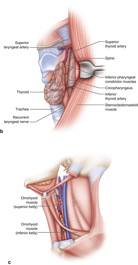
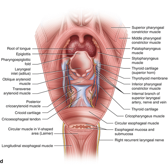
Fig. 1.1
Cervical esophagus. (a) The upper esophagus is best exposed through a left oblique incision on the neck. (b) From this incision, the anterior border of the sternocleidomastoid muscle is retracted laterally and, with careful dissection, the structures in this figure may be exposed. The left recurrent laryngeal nerve typically is identified in the tracheoesophageal groove, inferior to the inferior thyroid artery. Posterior to the esophagus, the spine and prevertebral fascia allow easy passage of a finger during dissection to begin mobilization of the esophagus. (c) The omohyoid muscle, also referred to as the “doorway to the esophagus,” typically is divided to gain access to the esophagus during dissection. (d) The upper esophagus from a posterior view. The relationship of the cervical esophagus to the cricoid cartilage, the superior laryngeal and recurrent nerves, and adjacent structures may be seen more clearly from a posterior view. This view is more commonly seen when performing endoscopy and looking for the esophageal lumen. It is important to be aware of adjacent structures and to see how an esophageal diverticulum may develop when the cricopharyngeus muscle is too tight. Learning to distinguish between cricopharyngeus, strap muscles of the neck, and omohyoid is critical to successfully performing surgery of the cervical esphagus
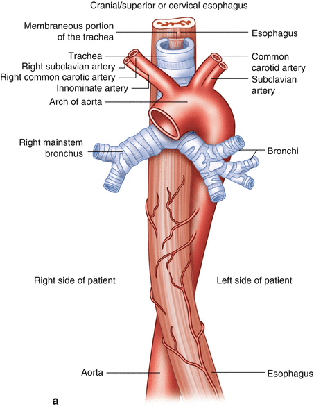
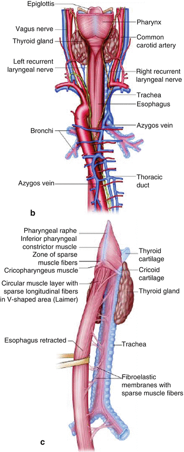
Fig. 1.2
Thoracic esophagus. The upper thoracic esophagus is best exposed through either a right thoracoscopic approach or a right posterolateral thoracotomy. The esophagus lies behind the trachea and the left atrium of the heart. The azygos vein arches over the esophagus just above the tracheal bifurcation and descends along the side of the esophagus on the right side of the chest, parallel to the course of the esophagus. An unnamed vein that runs directly into the esophagus from the azygos vein may be a source of bleeding when blunt dissection is used. On either side of the esophagus, the vagus nerves are found, with branches exiting their parallel pathway, extending to the bronchial branches of the lungs. In most circumstances, the aorta descends parallel to the esophagus in the left chest but may be identified easily when dissecting the esophagus from the right. The close relationship of the aorta and esophagus is best appreciated with esophageal ultrasound. The thoracic duct courses between the aorta, esophagus, and azygos vein. During esophageal dissection, special attention should be paid to the lymphatics low in the chest and to the close proximity of the left and right bronchi. The lower thoracic esophagus is best approached from the left chest, as the esophagus courses to the left in the lower thorax. A left thoracotomy also gives a good view of the esophageal hiatus from above. (a) Anterior view. (b) Posterior view. (c) Relationship of the thoracic esophagus to the trachea
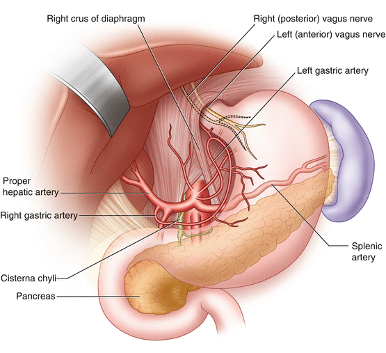
Fig. 1.3


Abdominal esophagus. The abdominal esophagus is best exposed from either a chevron, laparoscopic, or upper midline abdominal incision. With the left lateral lobe of the liver retracted, the phrenoesophageal membrane, hepatogastric ligament, and crura are seen. The aorta is located behind the crural decussation, and just to the right of the aorta, in front of the crura, lies the cisterna chyli. The left vagus nerve takes an anterior course, and the right vagus nerve travels posteriorly as it descends and innervates the stomach. The celiac trunk and its branches are exposed. The pancreas lies just posterior to the stomach and in front of the splenic vessels
Stay updated, free articles. Join our Telegram channel

Full access? Get Clinical Tree


