Fig. 6.1
Rigid esophagoscopy. The rigid esophagoscope is carefully inserted, taking care to protect the patient’s teeth. This procedure allows therapeutic endoscopy of the esophagus with larger instruments than with a flexible endoscope
Endoscopic Biopsy of the Esophagus
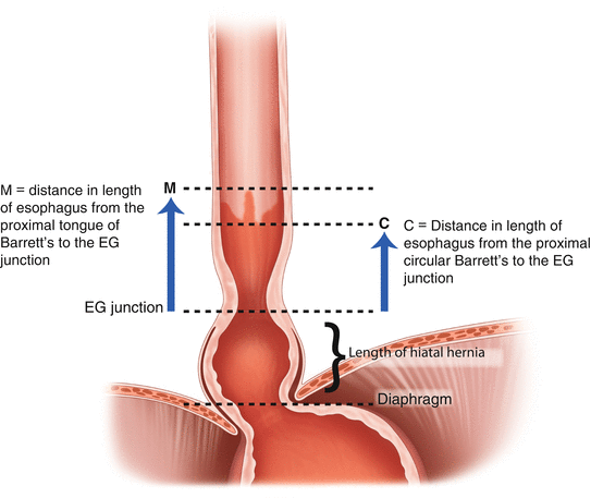
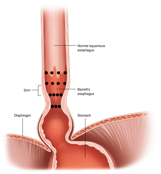
Esophageal biopsies are taken when pathology is seen. A number of forceps are available for these biopsies, and the use of cautery usually is not recommended for a diagnostic esophageal biopsy. The Prague classification (Fig. 6.2 and Table 6.1) is used to describe the Barrett’s segment macroscopically. With regard to esophageal biopsies for Barrett’s surveillance, the Seattle protocol of four-quadrant biopsies must be observed. This allows histological assessment of any Barrett’s segment (Fig. 6.3).


Fig. 6.2
The Prague classification of Barrett’s esophagus. This schematic describes the steps for performing an esophagogastroduodenoscopy and assessing the Barrett’s epithelium according to the Prague criteria [2] (Adapted from Sharma et al. [2]; with permission from Elsevier.) Table 6.1 further outlines the six numbered steps shown in this figure
Table 6.1
Steps of the esophagogastroduodenoscopy (EGD) examination in a patient with Barrett’s esophagus
Steps | Action points |
|---|---|
1 | Full EGD examination. Particularly note the presence of any hiatus hernia. Observe the position of the gastroesophageal junction (GEJ) in relation to the hiatal impression of the diaphragm |
2 | Note the location (in centimeters from the incisors) of the GEJ. Anatomical clues: Top of gastric mucosal folds The “pinch” of the lower esophageal sphincter |
3 | Note whether the squamocolumnar junction (or Z line) is above the GEJ |
4 | Note the location (in centimeters from the incisors) of the most proximal Circumferential extent of the suspected Barrett’s epithelium. This measurement = C |
5 | Note the location (in centimeters from the incisors) at the Maximum extent (i.e., most proximal extension) of suspected Barrett’s epithelium. This measurement = M |
6 | Subtract the C and M values noted in Step 4 and Step 5 from the depth of endoscope insertion noted in Step 2 (i.e., the GEJ). This is the relative position of the circumferential and maximal Barrett’s in relation to the GEJ |
7 | Report these values as Prague C and M [2]. For example, e.g., Barrett’s with a circumferential length of 4 cm but a maximum length of 8 cm would be C4M8 |

Fig. 6.3
The Seattle protocol of four-quadrant biopsies for histologic assessment of Barrett’s epithelium. When Barrett’s esophagus is seen, or at surveillance endoscopy, biopsies from all four quadrants of the circumference of the esophagus should be performed every 2 cm to clearly document the length of the Barrett’s epithelium
Dilation of Esophageal Strictures
Esophageal strictures may be benign or malignant. At times, the distinction is unclear, so biopsy should be performed on all strictures of the esophagus. Dilation of the stricture will frequently facilitate biopsy and cytologic brushings and will also relieve dysphagia. Dilation should be undertaken with caution because of the risk of esophageal perforation.
Strictures vary in their diameter and in their response to dilation. Some are treated easily by the passage of the scope itself or by a single balloon dilation. Other strictures, such as those resulting from caustic ingestion, are inelastic, demonstrate great resistance to dilation, and frequently are long. The endoscopist should assess the most likely etiology for the stricture and proceed with caution. The use of intraoperative fluoroscopy is often advisable, and excessive force should be avoided. If the patient complains of pain, the procedure should be promptly terminated and a contrast study should be performed to rule out esophageal perforation.
Strictures following operations such as esophagectomy are usually very amenable to endoscopic dilation. Early dilation may result in a more effective and long-lasting result. The gastroscope is positioned immediately above the narrowing and the characteristics of the stricture are noted. Biopsy and cytologic specimens of the edges of the stricture may be obtained at this time and again after dilation.
Strictures of the esophagus may present initially with bolus obstruction. Urgent endoscopy must be undertaken to remove the bolus, usually with a snare or grasping forceps, but the endoscopist must not ignore the stricture that caused the event. It may be dealt with shortly after removal of the food bolus.
Balloon Dilation
To perform a balloon dilation of an esophageal stricture, the endoscope is positioned above the stricture and an appropriate balloon is selected. The normal human esophagus is 2 cm wide, and a 20-mm balloon is usually the maximum dilation that would be attempted. The flaccid balloon is passed through the biopsy channel of the scope and positioned within the stricture. If the stricture is complex and the lumen is narrow, contrast can be injected to allow fluoroscopic guidance of a wire over which the balloon is passed. This technique prevents inadvertent perforation and allows safe passage of the sheathed balloon into the lumen. The balloon should be long enough to traverse the entire length of the stricture. After positioning, the balloon is inflated using radiopaque contrast material under fluoroscopic monitoring. This step may be accomplished without fluoroscopic guidance, using saline to fill the balloon, but without fluoroscopy there can be no assurance that the maximum diameter of the balloon has been achieved.
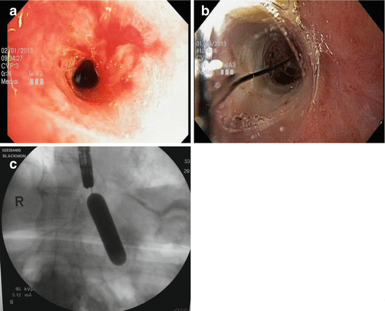
Using fluoroscopic guidance and a manometer, the balloon is insufflated with contrast agent until the waist is obliterated (Fig. 6.4a–c). The manometer prevents balloon rupture. The balloon should be inflated for a minute to ensure adequate dilation. After the balloon is deflated and removed, the scope may be passed through the stricture, or a larger dilator may be used if necessary. Once the scope is passed through the narrowing, it is important to obtain tissue samples from the stricture’s lower extent.


Fig. 6.4
(a) Endoscopic appearances of a benign esophageal stricture, pre-balloon dilatation. (b) Endoscopic appearances during balloon dilatation of a benign esophageal stricture. (c) Fluoroscopy demonstrating balloon dilation of an esophageal stricture. The balloon is filled with contrast to enable the endoscopist to observe adequate dilation of the stricture and obliteration of the “waist.”
Balloon dilation of esophageal strictures allows rapid clinical improvement in a patient’s dysphagia, but the strictures often recur and require repeated dilations.
Savary Dilation
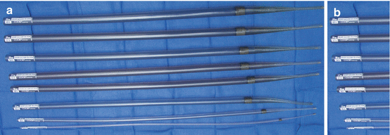
An alternative to using balloon dilation is wire-guided rigid Savary dilation of esophageal strictures. The Savary dilators are usually more successful in dilating recalcitrant strictures but are often more uncomfortable for the patient. One must use fluoroscopy when using these dilators. Savary dilators, which range in size from 15 Fr to 60 Fr (5–20 mm) in diameter, are passed over a guidewire in progressive size increments to dilate the stricture safely (Fig. 6.5). The endoscope should always be passed back into the esophagus after dilation to assess the esophageal mucosa.


Fig. 6.5


Savary esophageal dilators. (a) Savary esophageal dilators have a narrow tip inserted over a guidewire that has been placed endoscopically. This wire allows the dilator to be passed safely even though the endoscope is removed during dilation. (b) Savary dilators are available in a range of sizes from 15 to 60 Fr (5–20 mm) and are passed sequentially to dilate the stricture
Stay updated, free articles. Join our Telegram channel

Full access? Get Clinical Tree


