(1)
Division of General Thoracic Surgery, Mayo Clinic, 200 First Street SW, Rochester, MN 55905, USA
Keywords
AchalasiaBird’s beak esophagusApple core lesionDiverticulumDiverticulaParaesophageal herniaHiatal herniaEsophageal cancerLeiomyomaRadiation strictureEsophageal strictureImaging of the esophagus is best approached in a multidisciplinary fashion. The surgeon and radiologist bring different attributes to the interpretation of the images, and a dialogue between all members of the team caring for the patient is essential. Preoperative discussion of a patient’s images can allow appropriate staging of any tumor and allow selection of the most appropriate neoadjuvant therapy and operative approach. Further discussion regarding postoperative anatomy is essential for accurate interpretation of the images; it is especially important for those patients in whom a complication such as an anastomotic leak or obstruction is encountered. This chapter focuses on imaging the esophagus using a variety of techniques to image benign and malignant pathology. The upper gastrointestinal (GI) contrast swallow examination, plain x-ray, CT imaging, MR imaging, and positron emission tomography (PET) imaging are reviewed (Figs. 4.1–4.21). Ultrasound imaging of the esophagus is reviewed in Chap. 5.
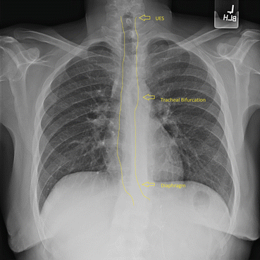
Fig. 4.1
Normal esophageal images seen on plain film. In this normal chest x-ray, the esophagus has been highlighted for better identification. The esophagus extends from C6 to T10 and is approximately 25–30 cm long. Cephalad, the esophagus is posterior to the trachea and subsequently is posterior to the left atrium. The esophagus deviates to the left of the midline caudad to the left mainstem bronchus. UES—upper esophageal sphincter
Upper Gastrointestinal Contrast Swallow Examination
Barium swallow, esophagram, or upper GI study involves the patient swallowing a contrast agent to identify structural and functional abnormalities of the esophagus. In the postoperative setting, if a leak is suspected, the test will start with a water-soluble agent like iohexol (Omnipaque™) or diatrizoic acid (Gastrografin®); if no leak is detected, barium may be used. The ordering practitioner should be aware of the densities of materials used for upper GI studies. Barium will preclude good imaging if a CT scan of the chest to view the esophagus is required later or if a leak is present, as extravasation of the barium remains within the leak. Adequate CT images cannot be obtained until enough time has passed for the contrast to leave the esophagus or the cavity formed by the leak, so it is common to image first with a CT scan and later with a swallow if a leak is suspected. Gastrografin pneumonitis and pneumonia can be fatal, and many institutions have changed to using vascular contrast agents like Omnipaque or iodixanol (Visipaque™). Omnipaque dissolves quickly after extravasation, is less caustic if aspirated, and is less expensive than Visipaque, which was originally developed for patients with renal failure. Barium remains the gold standard for imaging subtle mucosal changes and anatomic abnormalities of the esophagus. The individual images that are recorded during these swallow examinations are only a representative selection of the study obtained, however. It is best for the surgeon to attend the examination and watch the real-time pictures of the study, to correlate his or her understanding of the postoperative anatomy with the images obtained.
Thick barium (high density and high viscosity) and thin barium (lower density, low viscosity) may be used for examinations. Thick barium is useful when evaluating for aspiration during a modified barium swallow evaluation performed by a radiologist and speech pathologist. Other densities of food (e.g., applesauce, marshmallows, or pudding) may also be mixed with barium to assess swallowing with different textures. In the outpatient setting, barium swallows may be used to assess strictures (benign or malignant) in patients with dysphagia. A barium tablet also may be swallowed to see if there is any delay in passage through the esophagus, indicating a stricture.
CT Scans
CT images of the esophagus are usually obtained in order to assess esophageal perforation and local contamination and for the staging of esophageal tumors. For tumor staging, CT will not reliably determine T stage, but it sometimes can help to determine whether there is a fat plane between the esophageal lesion and local structures. If there is doubt with regard to invasion of local structures, endoscopic ultrasound (EUS), intravascular ultrasound (IVUS), and MRI may help to differentiate which lesions are growing into the aorta, lung, mediastinal structures, or other great vessels. CT images are also important for determining evidence of metastatic spread. If esophageal perforation has occurred, pneumomediastinum, free fluid or fluid collections, and a left pleural effusion may commonly be seen on CT scans.
When evaluating the esophagus using CT scanning, if no lumen can be seen, half of the maximum thickness of the esophagus is a surrogate for the diameter of the esophagus. More recently, three-dimensional CT models can be printed from two-dimensional CT images, and with the administration of an oral effervescent agent, the esophagus can be distended and relationships to other mediastinal structures can be accurately delineated. A positive enteric contrast agent is given to the patient prior to administration of effervescent granules. Positive contrast agents used in our practice include an iso-osmotic iodinated agent diluted with water for postoperative patients (Omnipaque 300; GE Healthcare, Cork, Ireland; 15 mL added to a 500-mL bottle of water) or barium sulfate suspension for preoperative patients (Berry Smoothie Readi-Cat two barium sulfate suspension 2.1 % w/v; E-Z-EM Inc., Lake Success, NY). The patient is then given a packet of effervescent granules containing sodium bicarbonate (E-Z-GAS II; E-Z-EM Inc., Lake Success, NY) to release carbon dioxide within the esophagus. The effervescent granules are mixed with approximately 20 mL of water and swallowed, or injected via a tube if the patient cannot swallow. After the patient is placed in the decubitus position on the scanner table, several more swallows or injections of positive enteric contrast agent are given to fill a redundant or patulous esophagus. Images are generally acquired in two positions using automatic exposure control (AEC) to ensure similar image noise across the acquired volume. Standard low-dose techniques such as kV selection, AEC, and iterative reconstruction are used to create high contrast differences between the esophageal wall, air, periesophageal fat, and positive enteric contrast. Routine multiplanar images used for diagnostic purposes are obtained. From these data, additional thin (1-mm) images are reconstructed in order to minimize stair-step artifacts in 3D printing. The imaging data, stored in Digital Imaging and Communication in Medicine (DICOM) format, is imported into proprietary software (Materialise, Belgium). The imaging data are then segmented using Hounsfield units (as well as hand segmented) to provide greater accuracy of the critical structures involved. (Hounsfield units are an indicator of radiodensity on CT scanning [air, −1000; water, 0; blood, 30–40; soft tissue, 100–300] and may help to determine if duplication cysts near the esophagus are hemorrhagic or are filled with thin fluid.) The segmented data are converted into a virtual 3D anatomic model, which is then exported into an STL (STereoLithography) file format. The final STL file is reimported into the source imaging data to ensure that its outline accurately matches what was initially segmented. The STL files are imported into Mimics software (Materialise, Belgium) for printing (Objet350 Connex multi-material 3D printer; Stratasys, Rehovot, Israel). Using the 3D printing software, different colors are assigned to the various anatomic structures, and several materials, both rigid and flexible, are selected. Life-size models are then printed using liquid photopolymers on the polyjet 3D printer. The material is printed with surrounding support material, which is washed off after the model is created. These life-size anatomic models can be used for multidisciplinary preoperative discussions, surgical planning, and as part of the patient education and consent process.
Related posts:


Stay updated, free articles. Join our Telegram channel

Full access? Get Clinical Tree


