, André Smout1 and Jan Tack2
(1)
Gastroenterology and Hepatology, Academic Medical Centre, Amsterdam, The Netherlands
(2)
Gastroenterology and Hepatology, UZ Leuven, Leuven, Belgium
1.1 Introduction
The gastrointestinal tract can be regarded as a hollow tube divided into several compartments. Each compartment has its own structure, related to its function. We distinguish the esophagus, stomach, small bowel, and large bowel or colon. Glands produce juices that play an important role in digestion, the salivary glands produce saliva, the stomach secretes hydrochloric acid and pepsin, the liver produces bile, and the pancreas produces amylase, lipase, and tryptase. The various compartments differ in diameter and are delimited by sphincters that open and close at the correct moments (Fig. 1.1).
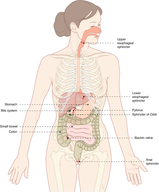
Fig. 1.1
Schematic display of the different compartments and sphincters of the gastrointestinal tract. Each compartment is divided from the previous and following compartment by sphincters (Published with kind permission of © Rogier Trompert Medical Art 2015)
This allows the partly digested food or feces to be expelled in the right direction. The wall of the compartments of the gastrointestinal tract is different for each compartment; however, the basic structure is similar.
1.2 The Basic Structure
The wall consists of several layers (Fig. 1.2). From the inner to the outer layers, there is the mucosa, submucosa, muscularis propria, and serosa (Fig. 1.3).
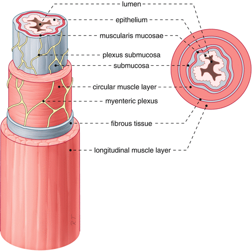
Fig. 1.2
Schematic figure of the different layers of the gastrointestinal tract (Published with kind permission of © Rogier Trompert Medical Art 2015)
Fig. 1.3
Variations in wall structure of the gastrointestinal tract (Published with kind permission of © Rogier Trompert Medical Art 2015)
1.2.1 The Mucosa
The mucosa forms the barrier between the luminal content and the internal environment. The mucosa consists of the epithelium, a thin muscular layer of smooth muscle (muscularis mucosae), and in between the connective tissue of the lamina propria.
1.2.2 The Submucosa
The submucosa mainly consists of fibrous tissue. In this layer a network of nerve cells is found, the submucosal plexus.
1.2.3 The Muscularis Propria
The muscularis propria surrounds the submucosa. The muscularis propria consists of an inner longitudinal muscular layer and an outer circular muscular layer. In between the two muscular layers, a nervous network exists called the myenteric plexus that plays an important role in regulation and coordination of the contractions of these muscular layers.
1.2.4 The Serosa
The serosa is the outer layer of the gastrointestinal tract and consists of fibrous tissue. In the serosa blood vessels, lymph vessels, and nerve fibers are present. In contrast to the other parts of the digestive canal, the esophagus does not have a serosa.
1.3 The Esophagus
The esophagus is a muscular tube that connects the pharynx with the stomach. Its function is to transport food and saliva from the mouth to the stomach and to prevent reflux of gastric contents. The structure and function of the esophagus are closely connected.
The mucosa of the esophagus consists of multiple layers of squamous epithelium. The transition from esophageal to gastric mucosa is normally in the distal esophagus. It is sometimes called the z-line because of its z-shaped appearance that one can find during endoscopy. The esophagus does not play a role in absorption of food and liquids and its lining is relatively smooth.
The upper and lower esophageal sphincters seal off the esophagus from the pharynx and stomach. The upper esophageal sphincter consists of striated muscle, and the lower esophageal sphincter consists of smooth muscle. In the tubular esophagus, between the two sphincters, one can find these two kinds of muscle fibers. The proximal third consists of striated muscle and the distal two-thirds consist of smooth muscle.
1.4 The Stomach
The stomach is a bag-shaped organ bordered by the lower esophageal sphincter and the pylorus. The pylorus connects the stomach with the small bowel. A distinction in function can be made between the fundus and antrum and in between the body of the stomach. The fundus serves as a temporary storage for food until it can be grinded in the antrum.
The gastric mucosa has many folds, in order to create a large surface. The gastric mucosa consists of a singular layer of cylindrical epithelial cells. In the epithelium mucus is produced that protects the cells against the acidic gastric contents. In the body of the stomach, there are gland tubes with parietal cells and gastric chief cells. Parietal cells produce acid and intrinsic factor, and chief cells produce pepsinogen, the precursor of the protein-digesting enzyme pepsin.
The gastric muscle layer is rather thick, in particular in the antrum. Proximally an extra muscle layer is present below the circular muscle layers. This extra muscle layer consists of diagonal muscle fibers and allows creation of a tonical pressure in the stomach.
1.5 The Small Bowel
The small bowel is the longest part of the gastrointestinal tract and measures about 5 m in total. The duodenum is about 25 cm long and is followed by 2 m of jejunum and 3 m of ileum. The small bowel starts at the pylorus and ends at the ileocecal valve (valve of Bauhin), where the small bowel empties in the colon.
The small bowel mucosa consists of a single layer of cylindrical epithelium and is even much more folded than in the stomach. The folds and villi increase the surface of the 5 m long small bowel up to the surface of a soccer field, which allows intensive transportation of nutrients through and along epithelial cells. The most important function of the small bowel is the uptake of these nutrients. The muscle layers of the small bowel are much thinner than the muscle layers of the distal stomach. The serosa of the small bowel is attached to the mesentery. The position of the small bowel is very flexible, and only the duodenum and ileocolonic valve are fixated.
1.6 The Colon
The colon is about one-and-a-half meter long and is also called large bowel because of its diameter. The colon starts at the ileocolonic valve and ends at the anal canal. We differentiate the cecum, ascending colon, transverse colon, descending colon, sigmoid, and rectum. Also in the colon, a single layer of cylindrical epithelium is found. The colonic wall is not folded and is relatively flat, and surface enhancement is realized with crypts. The organization of the muscle layers of the colon is a variation of the overall structure of the gastrointestinal tract. The inner circular muscle layer contracts at intervals, and these are called haustrations and give the colon its typical shape. The outer longitudinal muscle layer is organized into three bundles called taeniae. Between the taeniae there are virtually no longitudinal fibers present. The function of the colon is absorption of electrolytes and water which results in the feces becoming more solid.
1.7 The Anorectum
The rectum is the continuation of the colon but differs from it considerably in structure and function. The longitudinal muscle layers of the rectum are not organized in bundles but are continuous, and the rectum is covered by the peritoneum. The function of the rectum is storage of feces until it is defecated. The rectum ends at the anal canal. The anal canal is 2–4 cm long and is lined proximally by a single layer of cylindrical epithelium and distally by anoderm. The transition from cylindrical epithelium to anoderm is zipper or teeth shaped and is therefore sometimes called dentate line or linea dentata. The most important function of the anus is to maintain continence, and this is brought about by the smooth muscles of the internal anal sphincter and the striated muscles of the external anal sphincter. In addition to this, the muscles of the pelvic floor play an important role in continence as well.
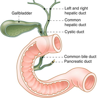
1.8 The Gallbladder and Bile Ducts
The production of bile from the liver flows through the bile ducts to the intestine (Fig. 1.4).
 < div class='tao-gold-member'>


Only gold members can continue reading. Log In or Register to continue
Related posts:


Stay updated, free articles. Join our Telegram channel
Full access? Get Clinical Tree
