, André Smout1 and Jan Tack2
(1)
Gastroenterology and Hepatology, Academic Medical Centre, Amsterdam, The Netherlands
(2)
Gastroenterology and Hepatology, UZ Leuven, Leuven, Belgium
2.1 Introduction
The diagnosis of disorders of gastrointestinal motility and functional disorders of the gastrointestinal tract can be facilitated by various investigational techniques. The most important of these will be discussed in this chapter.
2.2 Manometry
Measurement of the pressure (manometry) in the lumen of the gastrointestinal canal is an important aid in the study of the motor patterns of the canal. The most commonly used types of manometry allow the detection and quantification of the phasic contractions of the wall of the digestive tube. In addition, specialized forms of manometry can be utilized to record the tonic tone in sphincters and relaxations of sphincters.
An important principle is that a contraction of the gut wall is associated with a measurable increase in intraluminal pressure only if the contraction leads to a closed compartment in which a pressure rise can occur. Contractions that do not occlude the lumen do not lead to a measurable pressure increase. Thus, manometry can fail to lead to meaningful results when it is applied to organs that are wide, such as the proximal part of the stomach or the colon, or in a part of the digestive tract that has become distended during the course of a disease process (such as intestinal pseudo-obstruction). In fact, the majority of phasic contractions of the digestive tract will not lead to compartmentalization and thus not to a measurable pressure rise.
2.2.1 Perfused versus Solid-State Manometry
Basically, there are two different ways of measuring pressures in the lumen of the gastrointestinal canal (Fig. 2.1). With the first of these, a multi-lumen catheter is used, the channels of which are each perfused with water (water-perfused manometry). The water that runs through the individual water channels exits the catheter through side holes. Thus, perfused manometric systems measure pressure in one direction of the circumference. In water perfusion manometry, the pressure in the lumen of the gut is transferred, via the water in the individual channels, to pressure transducers outside the human body. The conversion of the pressure in each of the water-perfused channels to electrical signal takes place outside the body. For the perfusion of the water channels in the catheter, specialized systems are used that ensure a low and constant perfusion speed (between 0.08 and 0.3 ml/min). These so-called minimally compliant perfusion systems make it possible that the perfusion speed is not affected by pressure changes. This allows reliable pressure recording.
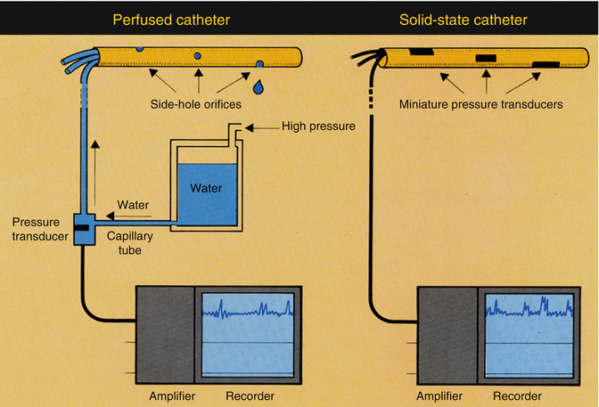
Fig. 2.1
Schematic representation of manometry with a perfused system (left) and with a solid-state catheter (right)
The recording of pressures exerted at side holes in a perfused manometric catheter assembly is useful for the study of peristaltic sequences in tubular parts of the digestive tract (e.g., esophageal peristalsis) but has clear limitations when the motor functions of a sphincter are to be studied. Movements, such as caused by respiration or shortening, make the pressure sensor slip in and out of the sphincter, leading to a pressure signal that is difficult to interpret. In order to solve this problem, the Australian gastroenterologist John Dent developed the so-called sleeve sensor. The sleeve sensor consists of a 5–6-cm-long membrane mounted on the manometric catheter. Water runs underneath the sleeve. The highest pressure exerted at any point on the sleeve is recorded. The Dent sleeve is particularly useful for pressure recording from the lower esophageal sphincter (LES). With the advent of high-resolution manometry, the sleeve sensor has become less indispensable, since high-resolution manometry also makes it possible to record sphincter pressures in a reliable way. This will be discussed below (Fig. 2.2).
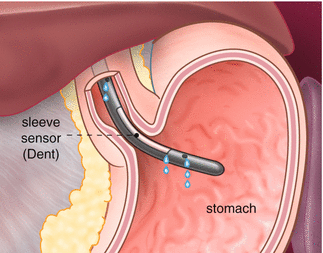
Fig. 2.2
Sleeve catheter, positioned in the lower esophageal sphincter (Published with kind permission of © Rogier Trompert Medical Art 2015)
Basal pressure in a sphincter (tone) can also be measured by pulling the manometric catheter through the sphincter while continuously recording the pressure (“pull-through technique”).
The second method for manometry in the gastrointestinal canal uses miniature pressure sensors mounted in or on a catheter. No water perfusion is used with this so-called “solid-state” method. Each of the pressure sensors in the catheter converts the pressure to which it is exposed to an electrical signal. Because water perfusion is not needed, the solid-state technique can be used much more easily for pressure recording in ambulatory subjects. This offers advantages when prolonged manometry is to be carried out (e.g., to detect esophageal spasm in patients with noncardiac chest pain or to study small bowel dysmotility in patients with (suspected) intestinal pseudo-obstruction).
The advantages of water-perfused manometry are that the catheters are relatively cheap and very durable. A disadvantage is that it can be time-consuming to make the manometric system ready for use, because the water flow through all perfused channels has to be constant and there may be no air bubbles in the system.
Disadvantages of the solid-state manometry are the high price of the catheters and their vulnerability. Its enormous advantage is the ease of performance of the manometric investigation.
2.2.2 High-Resolution Manometry
The development of high-resolution manometry has revolutionized manometric practice, in particular that of esophageal manometry. In high-resolution manometry, pressures are measured at closely spaced sites along the manometric catheter. The distance between the sensors typically is 1 cm but can even be smaller. High-resolution manometry can be carried out with a perfused multichannel catheter (and an array of perfused external pressure transducers) or with a solid-state catheter. The latter has gained enormous popularity, in particular in the form of 36-channel esophageal manometry. A 96-channel manometric technique using fiber-optic technology has also been developed but is as yet not commercially available. When a 36-channel catheter with sensors at 1-cm intervals is used, the catheter simultaneously “sees” all relevant structures, namely, the UES, the esophageal body, and the LES. This makes correct positioning of the catheter easy, allowing the manometric procedure to be performed by a less experienced person.
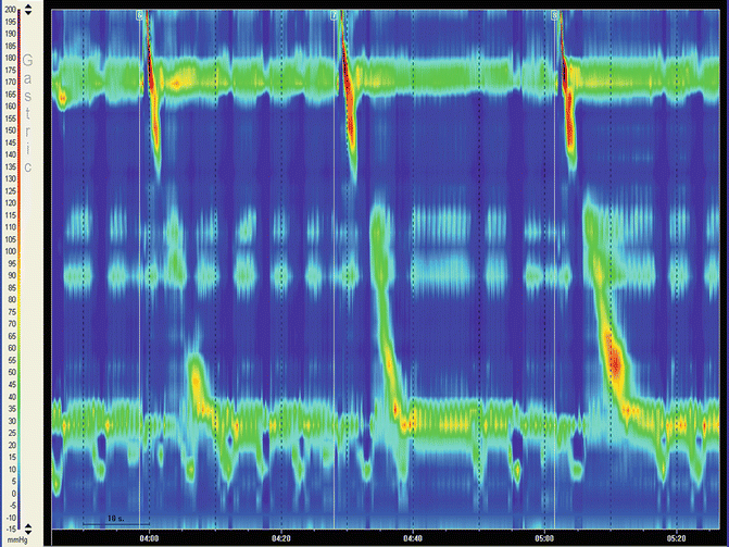
When 36 or more pressure signals are recorded, it becomes difficult to overview all signals. Conventional line plots (with time on the horizontal axis and amplitude on the vertical) fall short in providing a clear overview (Fig. 2.3). Therefore, other ways of representing the data were sought for. Universally, color plots are utilized in which amplitudes are expressed as color (Fig. 2.4). This type of representation is also referred to as “pressure topography” or as “Clouse plots,” named after the investigator who pioneered this, the late Ray Clouse.
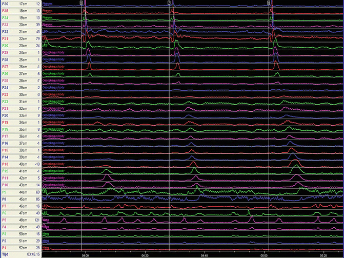
Fig. 2.3
36-channel recording of pressures in the pharynx, UES, esophagus, LES, and proximal stomach
Obviously, high-resolution manometry can be applied to all parts of the digestive canal. In clinical practice, application in the esophagus and its sphincters has gained most popularity, but high-resolution anorectal manometry has also become standard in many centers (Fig. 2.5).
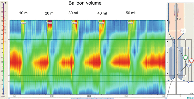
Fig. 2.5
High-resolution manometry of anorectum showing the recto-anal inhibition reflex (relaxation of internal anal sphincter at filling of rectal balloon with 10–50 ml of air)
2.3 pH Recording
Recording of intraluminal pH is mostly done in the esophageal body and less frequently in the stomach. In most parts of the world, esophageal pH monitoring is done most often with a miniature pH electrode mounted on a flexible catheter. Such a catheter is introduced via the nose and positioned at a predefined position. For esophageal pH monitoring, the electrode is usually placed at 5 cm above the upper border of the LES. The catheter is taped to the nose and attached to a portable digital data recorder (Fig. 2.6). The recorder usually is worn at a belt or on a shoulder strap.
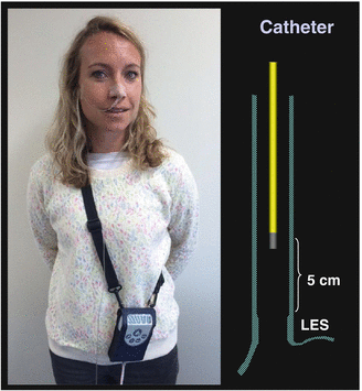
Fig. 2.6
Ambulatory esophageal pH monitoring with a transnasal pH catheter
In most cases, the measurement is carried out as an outpatient procedure and lasts 24 h or more. It is essential that the patient records all symptom episodes (e.g., heartburn) that occur during the measurement, by pressing an event marker button on the portable data recorder and also by filling in a paper symptom diary.
Catheters for pH recording come into three types. Firstly, a glass pH electrode can be used. There is no doubt that glass electrodes yield the best quality, but they are more bulky than the other types and cannot be sterilized. Secondly, catheters with an antimony pH electrode are available. The technical specifications of antimony electrodes are clearly inferior to those of pH electrodes. An antimony electrode is sensitive not only to the pH of the refluxate but also to other constituents, such as food components. However, antimony electrodes can be produced at low cost, making disposable pH catheters economically viable. Lastly, pH can be measured with an ISFET (ion-sensitive field effective transistor) electrode. The performance of ISFET pH electrodes is almost as good as that of glass pH electrodes. ISFET pH electrodes can also be marketed at prices compatible with disposable use.
It is also possible to measure esophageal pH without a transnasal catheter. Catheter-free pH monitoring employs a capsule containing a pH electrode (antimony) and a radio transmitter. The capsule is placed in the esophagus using a specialized introducer and attached to the esophageal mucosa by penetrating it with a metal pin (Fig. 2.7). The signals transmitted by the capsule are received by an external receiver-storage device, which needs to be in close proximity of the body. The system, consisting of capsule, receiver, and signal analysis software, is marketed as Bravo. The most recent versions of the system allow recording for at least 48 h, which has been shown to be advantageous for diagnosis. The advantage of the pH capsule is that transnasal intubation is avoided. However, some patients experience intolerable retrosternal discomfort or pain once the capsule is placed. The costs associated with capsule pH monitoring are higher than those associated with catheter-based pH monitoring.
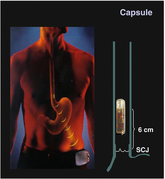
Fig. 2.7
Ambulatory esophageal pH monitoring with a telemetric capsule (SCJ squamocolumnar junction, also known as z-line)
2.4 Impedance Monitoring
Electrical impedance is the resistance encountered by an alternating electrical current. The well-known Ohm’s law describes resistance to a direct electrical current as the voltage difference across the resistance divided by the current running through it. The resistance is expressed in Ohm (Ω). In the case of an alternating current, the resistance, still expressed in Ω, is composed of not only a resistive but also a capacitive and inductive component, making it dependent of the frequency of the current.
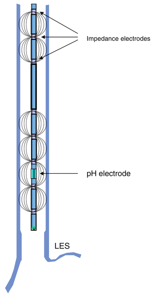
There are several applications of the impedance measurement technique in the human body. One of these is measurement of intraluminal impedance in the digestive canal. For this application a catheter is used on which an array of circular electrodes is mounted (Fig. 2.8). An alternating current that runs from one electrode to the next experiences a resistance (impedance) that depends on what is between the two electrodes. When the current goes through air, it will experience an almost infinitely high impedance. In contrast, when a well-conducting fluid, such as saliva or gastric juice, is between the electrodes, the impedance is low. Using these principles, intraluminal impedance measurement can be used to study the transit of air and fluid through the gastrointestinal canal. The most frequently used application of intraluminal impedance monitoring is in the esophagus.
 < div class='tao-gold-member'>


Only gold members can continue reading. Log In or Register to continue
Related posts:


Stay updated, free articles. Join our Telegram channel
Full access? Get Clinical Tree
