, André Smout1 and Jan Tack2
(1)
Gastroenterology and Hepatology, Academic Medical Centre, Amsterdam, The Netherlands
(2)
Gastroenterology and Hepatology, UZ Leuven, Leuven, Belgium
7.1 Introduction
The most important function of the colon is to reabsorb water and electrolytes from the liquid chyme that arrives from the small bowel. Under normal circumstances approximately 1500 ml of chyme is delivered to the cecum per day. After reabsorption only 150 ml remains (Fig. 7.1).
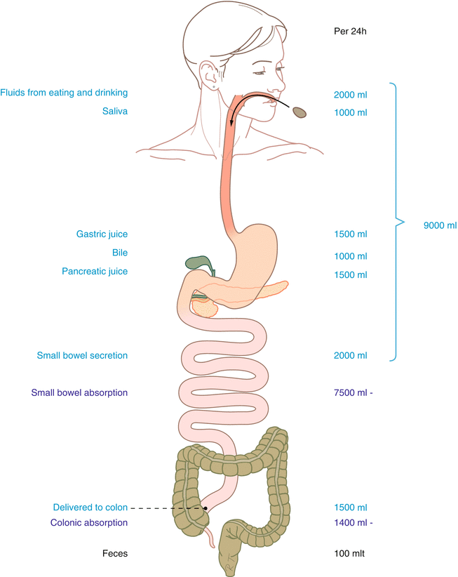

Fig. 7.1
Fluid secretion and absorption in the gastrointestinal canal (Published with kind permission of © Rogier Trompert Medical Art 2015)
In addition to the reabsorption function, the colon also has an important storage function. The feces that is produced during the course of one or several days can remain stored in the colon until a suitable moment for defecation occurs.
For the execution of the abovementioned tasks, the colon not only needs a sufficiently large absorbing surface, but also movements of the wall of the organ. Moreover, specialized functions of the pelvic floor, the anal sphincters, and the rectum are required, but these will be discussed in Chap. 8 of this book.
7.2 Anatomy and Innervation
The human colon is 1–1.5 m long. The organ usually has more curves and bends than are depicted in the anatomy books. From proximal to distal, the cecum, the ascending colon, the transverse colon, the sigmoid, and the rectum are distinguished (Fig. 7.2).
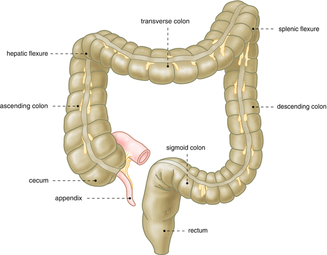

Fig. 7.2
Anatomy of the colon (Published with kind permission of © Rogier Trompert Medical Art 2015)
Like the other parts of the digestive canal, the colon has an outer longitudinal and an inner circular muscle layer. However, in the colon the longitudinal layer is not equally distributed over the circumference. Most longitudinal muscle fibers are found in three longitudinal bands known as taeniae (singular: taenia). Along the length of the colon, with the exception of the rectum, the colon has circular indentations with saccular pouches in between. These are called haustra (singular: haustrum). As in the other parts of the canal the wall of the colon contains neuronal plexuses, such as the myenteric and the submucosal plexus. The parasympathetic fibers that reach the right part of the colon are branches of the vagus nerve. The left part of the colon receives its parasympathetic innervation via the pelvic splanchnic nerves. The orthosympathetic innervation of the colon is supplied via perivascular plexuses.
The motility of the colon is regulated not only neurally but also hormonally. The most important hormones that stimulate colonic motility are gastrin and cholecystokinin. The most important inhibitory hormones are glucagon and vasoactive intestinal polypeptide (VIP).
7.3 Colonic Motility
Two types of colonic contractile patterns can be distinguished. These are haustrating (segmenting) contractions and mass contractions, also known as “high-amplitude propagated contractions” (HAPCs) (Fig. 7.3).
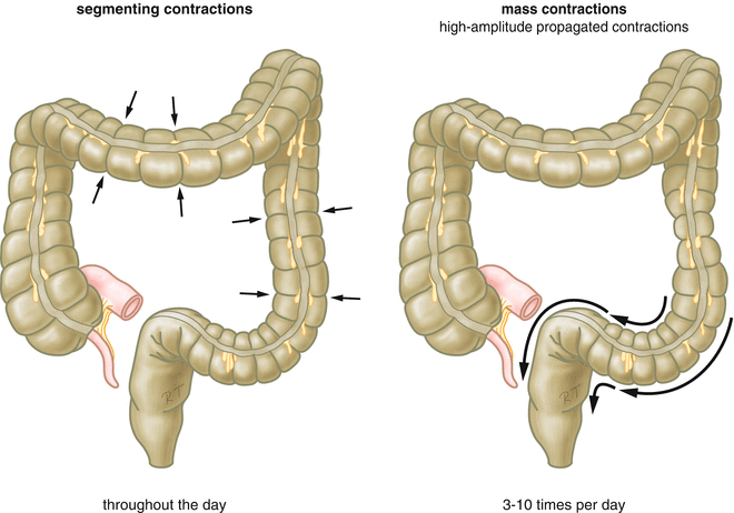

Fig. 7.3
Schematic representation of the two types of colonic contractions (Published with kind permission of © Rogier Trompert Medical Art 2015)
7.3.1 Haustrating Contractions
The haustrating contractions are brought about by local contractions of the circular muscle layer. Many of the haustrating contractions are stationary or almost stationary. They give the colon its typical haustrated appearance. The movements of these contractions are so slow that during short observations one gets the impression that the haustrated appearance of the colon is based on preformed anatomical structures. Prolonged observations of colonic appearance on X-ray have made clear however that the characteristic haustration pattern changes continuously.
Using intraluminal manometry the haustrating contractions can be recognized as pressure waves of variable duration and amplitude, without a clearly recognizable rhythm (Fig. 7.4). Electromyographic studies of the colon have shown that electrical slow waves are present, but, unlike the situation in the small bowel, these lack regularity. Often multiple slow wave frequencies coexist in the colon.
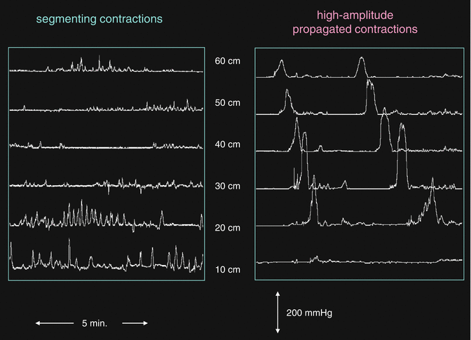

Fig. 7.4
Manometric recordings of the two types of colonic contractions. Left segmenting contractions, right high-amplitude peristaltic contractions
The haustrating contractions of the colon do not lead to significant displacements of colonic contents. Their primary function is to mix and triturate the contents, enabling optimal contact with the mucosa. This ensures that the resorption of water and salts is optimal. The frequency and amplitude of haustrating contractions increase after a meal, and haustrating contractions almost cease during the night.
7.3.2 Mass Contractions
Mass contractions are powerful, prolonged circular contractions that travel in the direction of the anus. When studied manometrically, the pressure in the lumen of the colon is found to reach values of several hundreds of mmHg during these contractions. Hence, the alternative name high-amplitude peristaltic contractions (HAPCs). The propagation speed of the contractions is about 1 cm per second. HAPCs do not occur frequently; in the human colon about 6 HAPCs contractions can be seen per day. They occur preferentially in the morning after awakening, after breakfast, and after meals. HAPCs effectively propel colonic contents in the direction of the anus. When feces arrives in the rectum, a sensation of urgency can arise and defecation may follow.
7.4 Postprandial versus Interdigestive Activity
The interdigestive migrating motor complex (MMC) that is the hallmark of fasting state motility in the stomach and small intestine is absent in the colon. Nevertheless, there are differences between interdigestive and postprandial motor activities in the colon. Within minutes after ingestion of a meal the motility of the colon increases significantly. This so-called gastrocolonic response lasts 30–60 min. During the gastrocolonic response the incidence and amplitude of the haustrating contractions is increased. In addition, the meal may elicit one or more mass contractions. The gastrocolonic response is mediated by release of the hormones cholecystokinin and gastrin, but an increased activation via the autonomic nervous system also plays a role.
7.5 Symptoms of Disordered Colonic Motility and Perception
Disordered motility of the colon can lead to constipation, diarrhea, and abdominal pain. When colonic visceral sensitivity is increased, pain is the predominant symptom, but the feeling of a distended abdomen (bloating) may also arise.
7.5.1 Constipation
It is difficult to define constipation. In fact, constipation comprises a variable assortment of symptoms. In the past often a low defecation frequency (fewer than three defecations per week) was used to define constipation. We now accept that constipation can also be present when defecation frequency is in the normal range. It has also been proposed to use fecal weight (in grams per 24 h) as an objective criterion for constipation. A production of less than 100 g would be abnormal. However, this criterion is difficult to use in daily practice. Many patients with constipation do not complain of a low frequency of defecation, and hardly any patient would report a low fecal weight. Rather, the patients’ presenting symptoms are hard stools, the feeling of incomplete evacuation, or the necessity to use straining (Table 7.1). In the Rome III classification all of these symptoms are taken into account for the diagnosis of functional constipation.
Table 7.1
Characteristics of constipation
Low frequency of defecation (<3 week) |
Necessity to strain excessively during defecation |
Hard stools |
Feeling of incomplete evacuation |
Feeling of anorectal obstruction or blockade |
Need for manual assistance during defecation |
7.5.2 Diarrhea
It is also difficult to define diarrhea. In addition to a higher frequency of defecation and an increased fecal output (more than 200 g per day), the Bristol stool form scale is used. In this scale consistency and shape of the stools are used to categorize the stools into one of seven types (Fig. 7.5).
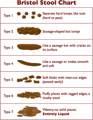

Fig. 7.5
Bristol stool scale. Stools are categorized based on consistency and form
Whereas constipation most often is caused by disordered motor function (of colon or pelvic floor), diarrhea is often the consequence of diminished resorption of water by the colonic mucosa. This can be caused by a variety of disorders such as inflammatory bowel disease (Crohn’s disease and ulcerative colitis). Diarrhea can also be the consequence of malabsorption at the level of the small intestine, as for instance in celiac disease. Both in cases with acute and chronic diarrhea motility disorders should only be considered when all other causes of diarrhea have been excluded (Table 7.2).
Table 7.2
Causes of diarrhea
Osmotic diarrhea |
Carbohydrate malabsorption |
Lactase deficiency |
Excessive intake of poorly absorbable carbohydrates (fructose, sorbitol) |
Use or abuse of osmotic laxatives |
Fat maldigestion and fat malabsorption |
Other malabsorption syndromes |
Bacterial overgrowth of small bowel |
Secretory diarrhea |
Infections |
Bacterial |
Parasites |
Viral |
Neuroendocrine tumors |
Carcinoid |
Medullary thyroid carcinoma |
VIPoma |
Inflammation of mucosa |
Crohn’s disease |
Collagenous colitis |
Ulcerative colitis |
Celiac disease |
Short bowel syndrome |
Diarrhea mediated by abnormal motility |
Hyperthyroidism |
Carcinoid |
Post-vagotomy |
Irritable bowel syndrome |
Diabetes mellitus |
Intestinal pseudo-obstruction syndromes |
Stimulant laxatives |
7.5.3 Distended Abdomen
Not infrequently the gastroenterologist sees patients whose predominant complaint is that they have a distended abdomen. Some of these patients do not really have abdominal distension but they perceive a feeling of bloating. In others the abdomen is visibly distended. This condition can be caused by serious conditions such as ascites, hepatosplenomegaly, and large tumors or cysts in the abdomen, but often the distension is brought about by accumulation of gas, fluid, or feces in the gastrointestinal tract, in particular in the colon. Such accumulation of liquid, gaseous, or solid material in the colon can take place when the colonic transit is slow.
It is also possible however to have a visibly distended abdomen in the absence of an increase of the contents of the abdomen. This is brought about by a combination of a low position of the diaphragm, which is abnormally contracted, and relaxation of the abdominal wall muscles. Especially in patients with functional bowel disorders (such as irritable bowel syndrome) who complain of intermittent abdominal distension, this mechanism is frequently present.
< div class='tao-gold-member'>
Only gold members can continue reading. Log In or Register to continue
Stay updated, free articles. Join our Telegram channel

Full access? Get Clinical Tree


