, André Smout1 and Jan Tack2
(1)
Gastroenterology and Hepatology, Academic Medical Centre, Amsterdam, The Netherlands
(2)
Gastroenterology and Hepatology, UZ Leuven, Leuven, Belgium
8.1 Introduction
The last parts of the gastrointestinal canal, the rectum and the anus, take care of a number of specialized functions. These are to retain the produced feces until a suitable moment for defecation has arrived and, subsequently, to achieve effective expulsion of the fecal mass. The rectum and the anal sphincter exert these functions in close collaboration with the pelvic floor. Thus, smooth muscle (rectal muscles and internal anal sphincter [IAS]) and striated muscle (pelvic floor and external anal sphincter [EAS]) work closely together in this area. Disordered functioning of the anus, rectum, and pelvic floor can result in two groups of functional problems: impaired defecation and fecal incontinence.
8.2 Anatomy
The rectum, 10–15 cm in length, is the most distal part of the colon. This is an extraperitoneally located part of the gastrointestinal canal. The longitudinal muscles of the rectum are continuous with the longitudinal muscles of the colon. At the transition from the sigmoid colon to the rectum, the three longitudinal taeniae join to form the continuous longitudinal layer of the rectum. The internal anal sphincter (IAS), 2–3 cm long, is continuous with the circular muscles of the rectum. Its smooth muscle is not under voluntary control.
The external anal sphincter (EAS), 3–4 cm long, surrounds the IAS. The EAS is a striated muscle sphincter that is under voluntary control. This sphincter collaborates closely with the pelvic floor, a structure composed of number of striated muscles. In the direct surroundings of the anus, the most important muscles of the pelvic floor are the puborectalis muscle and the levator ani muscle. The puborectalis muscle is U-shaped and forms a sling around the distal part of the rectum. Its two anterior ends are inserted into the pubic bone. The puborectalis continuously pulls the rectum forward and creates a sharp angle between the rectal and the anal canal (Fig. 8.1). The tonic contraction of the levator ani muscle maintains an elevated position of the pelvic floor and the anus. During defecation the levator ani muscle and the puborectalis muscle relax, leading to a downward displacement of the pelvic floor, as well as to a more obtuse anorectal angle.
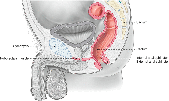
Fig. 8.1
Anatomy of the pelvic floor and surrounding structures (Published with kind permission of © Rogier Trompert Medical Art 2015)
The innervation of anus and rectum is complex. Like elsewhere in the gastrointestinal canal, the rectum has a myenteric and submucosal plexus. The extrinsic innervation of the IAS is supplied by the autonomic nervous system (sympathetic and parasympathetic). The sympathetic fibers originate in the L5 segment of the spinal cord and reach the IAS via the hypogastric plexus and the pelvic plexus. The parasympathetic fibers leave the spinal cord in the segments S2 to S4 and they reach the IAS via the pelvic plexus. The innervation of the EAS is via the pudendal nerve that comes from the sacral part of the spinal cord. The pelvic floor muscles also receive neuronal input via this nerve (Fig. 8.2).
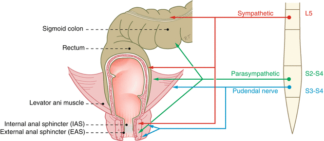
Fig. 8.2
Nerve supply to the rectum, anus, and pelvic floor (Published with kind permission of © Rogier Trompert Medical Art 2015)
In addition to this efferent, motility-driving innervation, there is an important afferent neuronal circuitry in the rectoanal area. Its function is to relay information from the sensory elements in the wall of the rectum and anus to the enteric nervous system but also to the brain. The system makes it possible to feel the urge to defecate, and the combination of sensory information from the anal canal and the rectum even makes it possible to perceive whether the contents of the rectum are solid, liquid, or gaseous.
8.3 Anorectal Constipation
As discussed in Chap. 7, constipation is often caused by disordered colon function. However, in a subset of patients with constipation, the cause of the constipation lies in abnormal function or an anatomical abnormality in the anorectal area, including the pelvic floor.
8.3.1 How to Recognize Constipation Caused by an Anorectal Disorder
The patient’s history can provide important information that may help to distinguish between constipation caused by disordered colonic function and constipation caused by an abnormality in the anorectal area. When the patient reports that he or she rarely experiences an urge to defecate but that when the urge occurs it is not a major problem to empty the bowel, it is not likely that the cause of the constipation can be found in the anorectum or the pelvic floor. On the other hand, if the patient regularly experiences the urge to defecate but is then unable to expel the feces, the possibility of an anorectal disorder must be considered. Furthermore, a sudden onset of the constipation and a young age at the onset of the constipation point in the direction of an anorectal disorder.
However, it can be difficult to differentiate between the two types of defecation on the basis of the history. Many patients with colonic constipation have hard stools, which can be difficult to evacuate, even when the pelvic floor and the anorectum function normally.
8.3.1.1 Colonic Transit Tests
Some clinicians use a colonic transit test to distinguish constipation based on an anorectal disorder from constipation caused by a colonic function disorder. When the holdup of the markers (radioopaque pellets or radioactivity) is in the proximal colon, this would favor a diagnosis of a colonic dysfunction, whereas stasis in the rectosigmoid area would support a diagnosis of dyssynergic defecation. However, there is no evidence that corroborates this view. On the other hand, a study in healthy subjects has shown that voluntary suppression of defecation, which mimics a defecation disorder, leads to stasis of the markers in the right colon in a proportion of the individuals. This suggests that colonic transit tests do not provide reliable information as to the mechanism of the constipation. A colonic transit test is thus not recommended for distinguishing between functional constipation and dyssynergic defecation.
8.3.2 Functional and Structural Causes
Defecation disorders can be caused by various abnormalities. Two groups of causes can be distinguished. Firstly, there may be abnormal function of the anal sphincter or the pelvic floor muscles. This can occur in a behavioral abnormality known as dyssynergic defecation and in Hirschsprung’s disease. On the other hand, disordered rectal evacuation can also be caused by a structural abnormality that leads to obstruction. In the next paragraphs the functional and structural abnormalities that can lead disordered defecation will be discussed.
8.4 Constipation Caused by Disordered Anorectal Function
8.4.1 Dyssynergic Defecation
In patients with dyssynergic defecation, the pelvic floor and the EAS do not relax sufficiently during attempts to defecate. In many cases, these muscles even contract paradoxically. Other names that have been given to this disorder are spastic pelvic floor syndrome and anismus. Dyssynergic defecation is an acquired condition that can have its debut at any age but often starts in adolescence. Not infrequently, psychological factors play a role. Dyssynergic defecation has been shown to be associated with sexual abuse.
8.4.2 Diagnosis
The diagnosis of dyssynergic defecation requires specialized tests. Some claim that the diagnosis can be made with digital examination, but this view is not shared by many experts in the field.
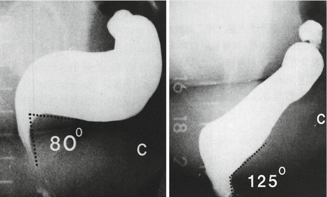
A useful technique for assessment of pelvic floor function is defecography. In this procedure videofluorography is used to visualize the defecation process. The rectum is filled with a thickened barium suspension, the vagina is filled with contrast, and the small bowel is made radioopaque with orally ingested contrast. Using laterolateral X-ray, moving images of the pelvic region of the patient – who is seated on a toilet – are obtained. After having acquired images in rest and during straining, the patient is instructed to empty the rectum. In the analysis of the acquired images, attention is paid to the patient’s ability to empty the rectum, to the widening of the rectoanal angle, and to anatomical abnormalities that can occur during straining (Fig. 8.3). Nowadays, defecography can also be carried out with magnetic resonance imaging (MRI) (Fig. 8.4). The advantage over radiography is that no ionizing radiation is applied. The disadvantage is that the investigation can only be carried out with the patient in a supine position.
 < div class='tao-gold-member'>


Only gold members can continue reading. Log In or Register to continue
Related posts:


Stay updated, free articles. Join our Telegram channel
Full access? Get Clinical Tree
