Contrast-Induced Acute Kidney Injury


Contrast-Induced Acute Kidney Injury


Steven D. Weisbord

Paul M. Palevsky


Acute kidney injury is a well-recognized complication of intravascular iodinated contrast administration.1,2,3,4 Epidemiologic trends and clinical factors suggest that contrast-induced acute kidney injury (CIAKI), which is associated with serious adverse short- and long-term outcomes, will continue to be an important clinical entity for the foreseeable future. First, patients are living longer with a greater burden of chronic illness, suggesting that there will be an increasing demand for radiographic procedures that use intravascular (IV) radiocontrast. Second, chronic kidney disease (CKD), which is the principal risk factor for CIAKI, and diabetes mellitus, which amplifies the risk for CIAKI in patients with baseline renal impairment, are increasing in prevalence.5 Third, recent studies linking gadolinium-containing contrast agents used to enhance the diagnostic accuracy of magnetic resonance imaging (MRI) studies with nephrogenic systemic fibrosis, a potentially severe fibrosing disorder in patients with advanced CKD, has led to the performance of fewer contrast-enhanced MRI procedures in patients with kidney disease and is likely to result in a greater reliance on imaging modalities that use iodinated radiocontrast in patients at risk for CIAKI.6,7,8 Lastly, advancements in modern imaging technology have led to an increasing array of diagnostic and therapeutic procedures that employ iodinated contrast. As a result of these factors, CIAKI is likely to remain a common iatrogenic complication.

This chapter reviews the pathophysiology, risk factors, clinical presentation, and incidence of CIAKI; discusses the adverse outcomes associated with the development of CIAKI; and summarizes the data regarding interventions to prevent this iatrogenic condition.


THE PATHOPHYSIOLOGY OF CONTRAST-INDUCED ACUTE KIDNEY INJURY

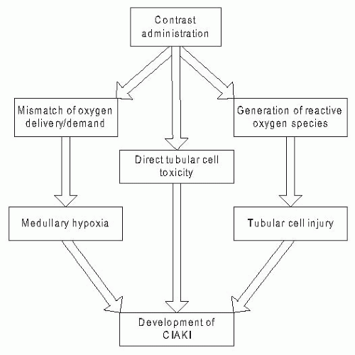
Our current understanding of the pathophysiology of CIAKI principally derives from animal studies examining the effect of contrast media on renal hemodynamics, oxygen delivery, and kidney function. These studies suggest that multiple overlapping pathophysiologic processes triggered by contrast administration collectively contribute to renal injury. These include mismatched oxygen supply and demand resulting in medullary hypoxia, direct toxic effects of contrast on tubular epithelial cells, and the generation of cytotoxic oxygen free radicals within the kidney that intensify renal injury (Fig. 33.1). Animal models used to investigate the pathophysiology of CIAKI have relied upon additional nephrotoxic insults such as volume depletion or concomitant nonsteroidal anti-inflammatory administration to enhance the susceptibility of the kidneys to contrast-induced injury. For example, Heyman et al.9 compared the effect of contrast media on renal vasoconstriction in intact rats and uninephrectomized, salt-depleted rats that were injected with intravenous indomethacin. Renal blood flow dropped to a substantially greater degree following contrast administration in the preconditioned rats. Similarly, Agmon and colleagues10 pretreated rats with indomethacin and nitro-L-arginine methyl ester (L-NAME), an inhibitor of nitric oxide synthesis to examine the effect of iodinated contrast on renal medullary blood flow. Whether such additional nephrotoxic insults in animals accurately reflect the mechanisms underlying the increased susceptibility of the human kidney to iodinated contrast from renal impairment is not clear. Nonetheless, such studies form much of the basis for our current understanding of the pathophysiology of CIAKI.


Vasoconstriction and Medullary Hypoxia

Multiple studies have demonstrated that the administration of iodinated contrast media induces vasoconstriction of the renal vasculature.11,12,13 However, this effect is not uniform throughout the kidney but rather regional because blood flow to the cortex appears to be maintained but decreases significantly in the renal medulla. Oxygen delivery to the renal cortex is high, yet delivery to the medulla is considerably lower, resulting in low medullary tissue oxygen tension, with values as low as 30 mm Hg detected under normal physiologic conditions in rats, dogs, and humans. At baseline, oxygen extraction by the renal medulla is near maximal, reaching 79% of regional oxygen delivery. Thus, medullary oxygen
reserve is marginal under normal conditions.14,15 In the intact kidney, medullary blood flow is usually maintained, even in the presence of systemic and local vasoconstrictive stimuli, by the combined effects of vasodilators including nitric oxide and prostaglandin E2, as well as the unique regional vasodilatory effects of renal vasoconstrictors.16 However, in certain settings, including altered renal reserve, vasoconstriction can overwhelm the kidney’s capacity to preserve oxygen delivery, leading to medullary hypoxia. In particular, the vasoconstrictors endothelin and adenosine have been implicated in the reduction in outer medullary blood flow following the administration of iodinated contrast.17,18,19,20 However, the precise role of and degree to which renal injury from contrast is directly attributable to these vasoconstrictors is unknown. Nonetheless, a series of studies have examined the role of medullary hypoxia in the pathogenesis of CIAKI.21,22 Liss and colleagues22 demonstrated a fall in medullary oxygen tension from approximately 30 mm Hg to 15 mm Hg after the administration of low-osmolal and iso-osmolal contrast agents. The contrast-induced exacerbation of medullary hypoxemia has also been suggested by noninvasive blood-oxygen level dependent magnetic resonance imaging (BOLD MRI), which detects an increased unsaturated hemoglobin concentration within the renal medulla and by the detection of increased levels of hypoxia-inducible factors (HIF) shortly after contrast administration.23,24 Systemic effects of iodinated contrast media may also contribute to the decline in renal medullary oxygen tension. Contrast administration is associated with the induction of pulmonary ventilation-perfusion mismatch, reduced cardiac output with a secondary decrease in renal perfusion pressure, rheologic alterations of blood, and a leftward shift of the oxygen-hemoglobin dissociation curve.25,26,27,28,29 However, these systemic effects appear to play a less prominent role in the reduction in medullary oxygen tension than the intrarenal mechanisms.


FIGURE 33.1 Pathophysiology of contrast-induced acute kidney injury.

Simultaneous with its effect on medullary oxygen tension, iodinated contrast increases oxygen demand within the kidney. The administration of contrast media induces an abrupt and transient natriuresis and increases glomerular filtration and urinary output, effects that are mediated, at least in part, by an increase in plasma volume and the release of natriuretic peptides.17,30,31 These effects, along with the increased osmotic load following contrast media administration, lead to enhanced solute delivery to the distal nephron. The increased active transport in the distal nephron increases oxygen demand. Thus, the contrast-induced decrease in medullary oxygen tension is accompanied by a concomitant increase in oxygen demand, particularly at the corticomedullary junction, resulting in a mismatch between oxygen supply and demand, tissue hypoxia, and cellular injury.


Direct Tubular Toxicity and Generation of Reactive Oxygen Species

Radiocontrast agents result in tubular cell injury directly through direct cytotoxicity and indirectly through the generation of reactive oxygen species (ROS).32,33,34,35,36,37 Following filtration at the glomerulus, iodinated contrast enters the urinary space and increases the viscosity of the tubular fluid. The increased urine viscosity combined with intratubular cast formation by cellular debris and the precipitation of contrast medium with urinary proteins may increase exposure of the tubules to the contrast medium, thus increasing the risk for direct cytotoxicity. Evidence supporting the role of direct tubular cell toxicity in the pathogenesis of CIAKI derives from renal biopsy specimens demonstrating vacuolization and necrosis of tubular cells in patients who recently underwent contrast-enhanced procedures.38 Additionally, data from in vitro studies of renal epithelial cells demonstrate contrast-associated DNA fragmentation, altered cell polarity, inhibition of mitochondrial function, increased brush border marker enzyme activity, and apoptosis—effects that support the direct nephrotoxic effects of contrast.34,35,36

In vitro studies demonstrate that iodinated contrast agents also induce oxygen free radical mediated injury through the oxidation of cell membranes, cellular proteins, and nucleic acids.39,40,41,42,43 Secondary activation of reparative processes, such as the DNA mending poly-(ADP-ribose) polymerase (PARP) may, in turn, precipitate additional depletion of intracellular energy stores and tubular damage.44,45 Endothelial cells may also be injured by the evolving hypoxic stress. Following exposure to contrast media, HIF-2α accumulates in medullary endothelial capillaries.46 Resultant endothelial damage, induced by reactive oxygen species and energy consuming reparative mechanisms, such as PARP, leads to endothelial dysfunction, further exacerbating regional tissue hypoxia.40,47,48,49 Thus, the hemodynamic and toxic effects of
iodinated contrast media are synergistic, leading to the amplification of kidney injury.


RISK FACTORS FOR CONTRAST-INDUCED ACUTE KIDNEY INJURY


Patient-Related Risk Factors

Risk factors for CIAKI can be categorized as patient related or procedure related (Table 33.1). CIAKI rarely develops in the absence of patient-related risk factors, which are collectively characterized by functional and structural changes impairing the capacity of the kidneys to adequately compensate for the hemodynamic and microcirculatory stresses caused by iodinated contrast media. Preexisting disease of the renal parenchyma (i.e., CKD) is characterized by an abnormal medullary microcirculation and a diminished capacity to compensate for hemodynamic perturbation. Clinical studies confirm that underlying renal impairment is the major patient-related risk factor for CIAKI, with increasing levels of dysfunction associated with escalating levels of risk.50 In a study of 378 hospitalized patients undergoing nonrenal angiography, D’Elia et al.51 found that preexisting renal insufficiency was the only risk factor predisposing to CIAKI. In an analysis of nearly 3,700 patients, McCullough and colleagues52 found a strong inverse relationship between the baseline kidney function and a risk of both CIAKI and the need for acute dialysis. Schemata outlining the risk associated with varying levels of CKD have been proposed. For patients with mild to moderate underlying renal insufficiency, the incidence of CIAKI is approximately 5% to 10%. Superimposition of diabetes mellitus on mild to moderate renal insufficiency heightens this risk, whereas the incidence of CIAKI increases to as much as 50% or more in patients with very advanced CKD.


TABLE 33.1 Risk Factors for Contrast-Induced Acute Kidney Injury


Patient Related


Procedure Related


Renal impairment


High-osmolal contrast media


Diabetes mellitusa


Large contrast volume


Absolute intravascular volume depletion


Multiple sequential procedures


Effective intravascular volume depletion


Intra-arterial administration


Concomitant nephrotoxic medication use


a Amplifies risk in the setting of renal impairment; not a strong independent risk factor.


Although diabetes mellitus substantially amplifies the risk of CIAKI in patients with underlying renal impairment, the presence of diabetes in the setting of intact kidney function does not appear to be associated with an increased risk of CKAKI.51,52,53,54,55 For example, in a study by Rudnick et al.53 that compared the effects of high- and low-osmolal contrast agents in 1,196 patients undergoing coronary angiography, CIAKI—defined by a postprocedure increase in serum creatinine (SCr) of ≥ 1.0 mg per deciliter—occurred in none of 359 patients without diabetes or underlying CKD and in just 2 of 315 (0.6%) patients with diabetes but no underlying CKD. However, in study participants with baseline renal impairment, 17 of 296 nondiabetics (6%) and 42 of 213 diabetics (20%) developed CIAKI. Thus, diabetes substantially amplifies the risk of CIAKI in patients with impaired kidney function, but does not appear to represent a notable risk factor in the setting of intact kidney function.

Patients with absolute or effective intravascular volume depletion have increased susceptibility to renal injury from iodinated radiocontrast media.56 Both clinical states are associated with reduced renal blood flow, which can exacerbate the impact of renal vasoconstriction following intravascular radiocontrast administration. Absolute extracellular volume depletion due to gastrointestinal (GI) losses or diuresis and effective intravascular volume depletion due to advanced heart failure or end-stage liver disease, which are associated with an increased reliance on the vasodilatory effects of prostaglandins to maintain renal microperfusion, also augment the risk for CIAKI.56,57 Similarly, nephrotoxic medications, specifically nonselective and cyclo-oxygenase-2 selective nonsteroidal anti-inflammatory medications, which inhibit vasodilatory prostaglandins in the kidney, are associated with an increased risk of CIAKI.58 Other medications that may also increase the likelihood of contrast-associated renal injury include aminoglycosides and calcineurin inhibitors.

Additional factors that have been reported to increase the risk of CIAKI include older age, hypertension, and anemia.59,60 However, the independent impact of these factors on the risk for CIAKI is uncertain because each is strongly correlated with the presence of underlying CKD. Recent studies suggest that an elevated serum glucose concentration at the time of contrast administration may confer an added risk of CIAKI, particularly among nondiabetics.61 Likewise, elevated urinary protein excretion has been shown to be associated with increased risk of AKI in several clinical settings, although its role in the context of contrast administration is currently unknown.62 In animal studies, intratubular light chains, particularly if accompanied by intravascular volume depletion, augment the nephrotoxic potential of radiocontrast media.34 However, more recent studies in humans do not support an association of multiple myeloma with an increased risk for CIAKI.63 An analysis of 476 patients with multiple myeloma who received iodinated contrast revealed
an incidence of CIAKI of just 0.6% to 1.25%.64 Early reports of CIAKI following contrast exposure in patients with multiple myeloma may not have fully accounted for other comorbid factors such as sepsis and volume depletion.

In summary, the administration of iodinated contrast media leads to hemodynamic and toxic effects that, in healthy subjects, are balanced by protective regulatory systems that maintain renal parenchymal oxygenation, function, and integrity. These protective regulatory systems are impaired in the setting of patient-related risk factors, particularly preexisting CKD, leading to an increased susceptibility to renal injury and the development of CIAKI.


Procedure-Related Risk Factors

A series of procedure-related factors have been identified that increase the risk for CIAKI. The dose of contrast has been the subject of considerable attention, with some studies demonstrating an association of higher volumes of contrast with greater risk and other studies showing no such association.65,66,67 Miller and colleagues67 prospectively evaluated 200 patients undergoing procedures with intravenous or intra-arterial contrast and reported no consistent change in renal function with increasing doses of contrast media. Conversely, Cigarroa et al.65 demonstrated that decreasing the volume of contrast administered during coronary angiography was associated with a reduction in the incidence of CIAKI. Although a specific threshold volume of contrast above which the risk for CIAKI increases substantially has not been definitively determined, multiple sequential procedures or procedures employing larger volumes of contrast appear to pose a greater risk. Similarly, higher doses of iodine have also been associated with a greater risk for CIAKI, which has led to the development of formulas that incorporate the dose of iodine to estimate risk.68,69 However, the role and importance of an iodine dose relative to the overall volume of contrast requires further study.

The type of contrast agent has also been strongly associated with risk for CIAKI. The first iodinated contrast media widely used in clinical practice were high-osmolal ionic derivatives of triiodobenzene, such as diatrizoate, meglumine, or metrizoate.70 These high-osmolal contrast media (HOCM) were characterized by osmolalities that were five to eight times greater than blood (approximately 1,500 to 2,000 mOsm per kilogram of water). A series of studies in the early 1990s demonstrated that contrast agents with osmolalities of approximately 600 to 850 mOsm per kilogram (low-osmolal contrast media [LOCM]) were less nephrotoxic than conventional HOCM.53,71 A third-generation iso-osmolal agent (approximately 300 mOsm per kilogram), iodixanol, was introduced in the late 1990s. Several clinical trials and meta-analyses have compared the nephrotoxicity of this agent with various LOCM, with conflicting results (see section on prevention).72,73,74,75

Iodinated contrast media are administered intra-arterially in the setting of an angiography and intravenously with computed tomography (CT). Direct comparisons of the incidence of CIAKI across different procedure types have been scarce. In a study of 660 patients with CKD, Weisbord et al.76 demonstrated that the incidence of CIAKI, defined by an increase in SCr ≥25%, was higher following noncoronary angiography (13.2%) than both coronary angiography (8.5%) and CT imaging (6.5%). However, these analyses did not account for variation in baseline clinical risk factors such as diabetes mellitus, heart failure, and the severity of CKD or in differential application of preventive interventions such as IV fluids. Differences in such factors may account for the higher observed rate of CIAKI following angiography in this study and may also underlie the perception that procedures involving intra-arterial contrast administration are associated with a higher risk of CIAKI than procedures that use IV injection.


THE CLINICAL PRESENTATION OF CONTRAST-INDUCED ACUTE KIDNEY INJURY

CIAKI presents as an acute decline in renal function that characteristically develops within 72 hours following contrast administration. Serum creatinine typically peaks within 3 to 5 days and returns toward baseline levels within 7 to 10 days. Most patients with CIAKI remain nonoliguric, although oliguric acute kidney injury (AKI) may occur. CIAKI is a form of acute tubular necrosis (ATN), for which the principal differential diagnoses include ischemic ATN and renal atheroembolic disease.77 Differentiation from ischemic ATN is generally based on the clinical setting, although distinguishing between these two forms of AKI may be difficult in hemodynamically unstable patients who sustain episodes of hypotension contemporaneous with contrast administration. The urine sediment in CIAKI commonly demonstrates coarsely granular “muddy brown” casts, as is seen in other etiologies of ATN. The fractional excretion of sodium in CIAKI is often less than 1%, although this does not have sufficient diagnostic reliability to definitively differentiate CIAKI from ischemic or other forms of ATN.

Renal atheroembolic disease, which is considerably less common than CIAKI and ischemic ATN, results from the release of cholesterol crystals and other atheromatous debris into the systemic circulation from ulcerated atherosclerotic plaques.78 Although renal atheroembolic disease can occur spontaneously, it is more common following angiographic procedures.78 The time course of angiography-associated atheroembolic disease and CIAKI differ; although acute atheroembolism can occur immediately following vascular catheterization, more commonly it is delayed, typically developing days to weeks following vascular instrumentation.78 Unlike CIAKI, atheroembolic disease is generally associated with specific systemic manifestations including mesenteric ischemia, digital ischemia (“blue toe” syndrome) and livedo reticularis, and laboratory abnormalities that include eosinophilia, eosinophiluria, and hypocomplementemia.79


In clinical practice, the identification of patients with CIAKI is based on observing a rise in SCr that occurs in the characteristic time frame following contrast administration. Because clinically detectable elevations in SCr are not evident for many hours to days after renal injury, the diagnosis and implementation of supportive care are typically delayed. Earlier identification of renal injury in the incipient stage of AKI could lead to more immediate implementation of supportive care in the postprocedure period. A series of candidate urinary and serum biomarkers have been identified for the early diagnosis of AKI, including neutrophil gelatinaseassociated lipocalin (NGAL), interleukin-18 (IL-18), and kidney injury molecule-1 (KIM-1).80,81 In addition, these biomarkers have the potential to differentiate volume responsive AKI (prerenal azotemia) from intrinsic AKI. Given their early expression in the urine following tubular injury, these or other biomarkers may ultimately assist in the diagnosis of CIAKI prior to a change in SCr.


THE INCIDENCE OF CONTRAST-INDUCED ACUTE KIDNEY INJURY

The reported incidence of CIAKI is highly dependent on the patient population studied and on the criteria employed to define renal injury. Serologic criteria that have been used in past studies to define CIAKI include an increase in SCr of at least 0.3 mg per deciliter, 0.5 mg per deciliter, 1.0 mg per deciliter, or 2.0 mg per deciliter, or a relative increase of at least 10%, 25%, or 50% within 5 days following contrast administration.51,65,72,82,83,84,85,86,87,88,89,90,91 D’Elia and coworkers51 reported that 0.68% of nonazotemic patients and 17.4% of azotemic patients experienced a 1.0 mg per deciliter rise in SCr following nonrenal angiography. A 12% incidence of CIAKI was reported in seriously ill, hospitalized patients, using an elevation in SCr of ≥1 mg per deciliter within 48 hours as the criterion for nephrotoxicity.92 A study of 537 patients undergoing angiography that defined CIAKI as an increase in SCr of at least 1.0 mg per deciliter within 24 hours demonstrated no episodes of CIAKI.93 More recently, an observational cohort study by Weisbord and colleagues76 enrolled patients with baseline estimated glomerular filtration rate (eGFR) less than 60 mL/min/1.73m2 who were undergoing nonurgent coronary or noncoronary angiography or CT, and reported on the rates of CIAKI with these procedures. The incidence of CIAKI, defined by an increase in SCr of ≥25%, was 13.2% following noncoronary angiography, 8.5% following coronary angiography, and 6.5% following CT scans.76 Using more robust increments in SCr to define CIAKI resulted in considerably lower rates of renal injury with less than 1% of patients overall experiencing a rise in SCr of ≥ 1.0 mg per deciliter. In a recent observational study of 1,111 hospitalized patients who underwent procedures with intravascular contrast, the incidence of CIAKI, defined as an increase in SCr of ≥0.5 mg per deciliter within 1 to 5 days, was as high as 44% among patients with baseline renal insufficiency and concomitant diabetes.94

The substantial variability in the reported rates of CIAKI across these and other studies highlights the important impact that patient population, procedure type, and criteria used to define renal injury have on the reported disease incidence. Determining accurate estimates of the incidence of CIAKI, particularly if defined by diminutive increments in SCr, is further confounded by the underlying fluctuation in SCr that occurs independently of iodinated contrast administration. Bruce et al.95 studied the incidence of AKI, defined by an increase in SCr ≥0.5 mg per deciliter or a decrease in eGFR ≥25%, among 11,588 patients who underwent a total of 13,274 CT scans either with iodinated contrast (n = 5,790) or without iodinated contrast (n = 7,484). Among patients with baseline CKD, the overall incidence of renal injury in patients who did not receive iodinated contrast (8.8%) was comparable to that of patients who received iodinated contrast (9.7% with iso-osmolal iodixanol and 9.9% with low-osmolal iohexol). Thus, baseline variability in SCr and factors other than iodinated contrast should be considered when estimating the incidence of acute kidney injury from contrast administration.


OUTCOMES ASSOCIATED WITH CONTRAST-INDUCED ACUTE KIDNEY INJURY


Short-Term Outcomes Associated with Contrast-Induced Acute Kidney Injury

Several studies have demonstrated that CIAKI is associated with increased short-term mortality (Table 33.2).52,96,97,98,99,100 In a retrospective study of 183 hospitalized patients, Levy et al.96 demonstrated that CIAKI was associated with an increased risk of in-hospital mortality after adjustment for underlying level of comorbid illness (odds ratio [OR] = 5.5, P< .001). McCullough et al.52 evaluated 1,826 patients who had undergone percutaneous coronary intervention and documented an in-hospital mortality rate of 7.1% among patients with CIAKI (defined by an increase in SCr of >25%) compared to 1.1% in those without CIAKI (P< .0001). Patients who developed CIAKI that required renal replacement therapy experienced an in-hospital mortality rate greater than 35%. In a retrospective study, Rihal et al.100 examined outcomes in 7,586 patients undergoing percutaneous coronary intervention and reported that patients who developed CIAKI had a marked increased risk for in-hospital mortality (OR = 10.8, P< .0001). Among more than 20,000 patients who underwent percutaneous coronary intervention, Bartholomew and colleagues97 demonstrated that CIAKI, defined by increases in SCr ≥1.0 mg per deciliter, was associated with a striking increase in in-hospital mortality (OR = 22, 95% confidence interval [CI]: 16 to 31). In a recent analysis, Shema et al.94 demonstrated that among over 1,100 hospitalized patients who underwent contrast-enhanced radiographic procedures, CIAKI was independently associated with a nearly 10-fold increase in in-hospital mortality (OR = 9.8, 95% CI: 4.4 to 22.0).
Finally, in a retrospective analysis of over 27,000 patients who underwent coronary angiography, Weisbord et al.99 found that postangiography increases in SCr greater than 0.25 mg per deciliter but no higher than 0.5 mg per deciliter were independently associated with increased in-hospital mortality (OR = 1.83, 95% CI: 1.35 to 2.49).


TABLE 33.2 Association of Contrast-Induced Acute Kidney Injury with Short-Term Risk of Mortality


Study Authors


# Study Patients


Definition of CIAKI


Adjusted OR for Death


95% CI


Levy et al.


357


↑SCr ≥25% to ≥2.0 mg/dL


5.5


2.9-13.2


Gruberg et al.


439


↑SCr >25%


3.9


2.0-7.6


Shema et al.


1,111


↑SCr ≥50% or ↓eGFR ≥25%


3.9


1.2-12.0


McCullough et al.


1,826


↑SCr >25%


6.6


3.3-12.9


From et al.


3,236


↑SCr ≥25% or ≥0.5 mg/dL


3.4


2.6-4.4


Rihal et al.


7,586


↑SCr >0.5 mg/dL


10.8


6.9-17.0


Bartholomew et al.


20,479


↑SCr ≥1.0 mg/dL


22


16-31


Weisbord et al.


27,608


↑SCr 0.25-0.5 mg/dL


1.8


1.4-2.5


CIAKI, contrast-induced acute kidney injury; OR, odds ratio; CI, confidence interval; SCr, serum creatinine; eGFR, estimated glomerular filtration rate.


Prospective observational studies and clinical trials reveal comparable findings. In a study of 439 patients with CKD undergoing percutaneous coronary intervention, Gruberg et al.101 found that in-hospital mortality occurred more frequently among patients who developed CIAKI than in comparable patients without CIAKI (14.9% versus 4.9%, P= .001). In a clinical trial, Marenzi et al.102 also found that patients who developed CIAKI had a significantly increased incidence of in-hospital mortality compared to patients who did not sustain a postangiography decline in renal function (26% versus 1.4%, P< .001). Finally, Maioli et al.103 demonstrated that in-hospital mortality among patients who developed CIAKI was markedly higher than among patients who did not develop this postprocedure complication (11.1% versus 0.2%, P= .001).

CIAKI is also associated with prolonged hospitalization.97,99,104,105,106,107 Bartholomew et al.97 found that patients who developed CIAKI after percutaneous coronary intervention (PCI) were 15 times more likely to have their hospitalization prolonged more than 4 days. In the aforementioned study by Shema et al.,94 CIAKI was associated with a marked increase in hospital length of stay (24 days with CIAKI versus 13 days without CIAKI, P< .001). Adolph et al.104 demonstrated that the development of CIAKI resulted in an increased mean duration of hospitalization of 2 days. The extended length of a hospital stay associated with CIAKI translates into increased health care expenditures. Using decision analytic techniques, Subramanian et al.105 reported that a single episode of CIAKI results in an average increase in hospital-related costs of more than $10,000.

Notwithstanding the findings of these studies that demonstrate that CIAKI is associated with increased short-term mortality and a prolonged duration of hospitalization, it is important to note that the causal nature of such associations has not been established. Risk factors that increase the risk for CIAKI (i.e., CKD, heart failure) are also independently associated with these adverse short-term outcomes.


Long-Term Outcomes Associated with Contrast-Induced Acute Kidney Injury

In several recent analyses, CIAKI has also been linked with increased longer term mortality (Table 33.3).100,108,109,110,111,112 Solomon et al.111 demonstrated that the development of CIAKI following angiography was associated with a greater than threefold increased risk of death, stroke, myocardial infarction, and end-stage renal disease at 1 year of follow-up. In a study of 985 patients, Harjai et al.109 demonstrated that CIAKI following coronary angiography was independently associated with an increased likelihood of death at 24 months (hazard ratio [HR] = 2.6; 95% CI: 1.5 to 4.4). A study by Brown et al.112 that examined long-term outcomes among 7,856 patients found that patients with either transient or persistent postangiography decrements in kidney function had a two- to threefold increase in long-term mortality. In a smaller study of 78 patients, Goldenberg and colleagues108 documented that CIAKI that fully recovered within 7 days of angiography was associated with a greater than twofold increase in the 5-year mortality (HR = 2.66, 95% CI: 1.72 to 4.46). The previously described study by Rihal et al.100 also demonstrated that the
5-year mortality rate among patients who underwent coronary angiography and survived to hospital discharge was significantly higher in those who had experienced CIAKI (44.6% versus 14.5%). Finally, James et al.113 recently demonstrated that patients sustaining a 50% to 100% increase in SCr following coronary angiography doubled their risk of death (HR = 2.0; 95% CI: 1.69 to 2.36) over the ensuing 3 years, whereas patients sustaining an acute increase in SCr >100% experienced a nearly fourfold increased risk of death (HR = 3.72; 95% CI: 2.92 to 4.76).


TABLE 33.3 Association of Contrast-Induced Acute Kidney Injury with Long-Term Risk of Mortality


Study Authors


# Study Patients


Definition of CIAKI


Follow-Up (Months)


Adjusted HR


95% CI


Goldenberg et al.


78


↑SCr ≥0.5 mg/dL or ≥25%


60


2.7


1.7-4.5


Solomon et al.


294


↑SCr ≥0.3 mg/dL


12


3.2a


1.1-8.7


Harjai et al.


985


↑SCr ≥0.5 mg/dL


24


2.6


1.5-4.4


Roghi et al.


2,860


↑SCr ≥0.5 mg/dL


24


1.8


1.0-3.4


Rihal et al.


7,075


↑SCr >0.5 mg/dL


6


b


b


Brown et al.


7,856


↑SCr ≥0.5 mg/dL


90


3.1


2.4-4.0


a Reflects incident rate ratio of death, cerebrovascular accident, acute myocardial infarction, and end-stage renal disease.

b Six-month mortality of 9.8% with CIAKI versus 2.3% without CIAKI (p < 0.0001).


CIAKI, contrast-induced acute kidney injury; HR, hazard ratio; CI, confidence interval; SCr, serum creatinine.


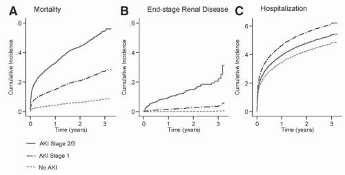
Past studies have also demonstrated a relationship between CIAKI and accelerated progression of CKD.103,108,113,114 In a study of 78 patients, Goldenberg et al.108 found that patients with a transient postprocedure rise in SCr ≥25% or ≥0.5 mg per deciliter following coronary angiography experienced a larger decrement in eGFR 2 years following the procedure than patients without these transient elevations in SCr (Δ eGFR of -20 ± 11 mL/min/1.73 m2 versus -6 ± 16 mL/min/1.73 m2, P= .02). Maioli et al.103 reported that patients with CIAKI had a 0.2 mg/dL higher mean SCr 1 month postangiography than did patients without CIAKI (P= .001). Finally, James et al.114 recently documented that patients who developed an increase in SCr ≥0.3 mg/dL or between 50% and 99% following coronary angiography experienced a more rapid loss of kidney function compared to patients without such changes in kidney function (loss of eGFR 0.8 mL/min/1.73m2 per year versus 0.2 mL/min/1.73m2 per year). For patients sustaining increases in SCr ≥100%, the long-term rate of loss of eGFR was even more marked (2.8 mL/min/1.73m2 per year).114 These investigations also demonstrated an increased risk of mortality and end-stage renal disease during 3 years of follow-up in patients who developed CIAKI with graded risks based on the severity of AKI (P< .001 for the trend in mortality and ESRD) (Fig. 33.2).113 As was noted with the associations of CIAKI with shorter term outcomes, although there is a strong association of CIAKI with longer term mortality and more rapid progression of CKD and ESRD, the causality of this association has not been determined.


THE PREVENTION OF CONTRAST-INDUCED ACUTE KIDNEY INJURY


Overview of Prevention

With a large body of evidence demonstrating associations of CIAKI with adverse short and longer term adverse outcomes, there has been a substantial emphasis placed on identifying interventions to prevent this form of renal injury. CIAKI is one of the few forms of AKI that is potentially preventable because patients at high risk are easily identifiable and most procedures that involve the intravascular administration of iodinated contrast are scheduled sufficiently in advance to allow implementation of prophylactic measures.

The initial step in the prevention of CIAKI is the identification of patients at an increased risk. Alternative imaging procedures that do not require the use of iodinated contrast media, including ultrasonography, nonenhanced CT scans, and MRIs and nuclear imaging, should be considered in patients at increased risk for CIAKI. This involves a careful assessment of both the risk of CIAKI and any potential loss of diagnostic yield with alternative imaging procedures. The use of gadolinium enhancement of MRI procedures is particularly problematic in patients with advanced CKD or ongoing AKI because gadolinium-containing agents are associated with the development of nephrogenic systemic fibrosis in patients with markedly decreased GFR—the subset of patients at highest risk of developing CIAKI.6,7


FIGURE 33.2 Cumulative incidence of (A) mortality, (B) end-stage renal disease (ESRD), and (C) hospitalization for all causes according to stage of acute kidney injury (AKI) defined according to the Acute Kidney Injury Network criteria (AKI stage 1:≥0.3 mg/dL absolute or 1.5-to twofold relative increase in serum creatinine; AKI stage 2: >two-to threefold increase in serum creatinine; AKI stage 3: >threefold increase in serum creatinine or serum creatinine ≥4.0 mg/dL with an acute rise of >0.5 mg/dL). Reprinted with permission from James MT, et al. Associations between acute kidney injury and cardiovascular and renal outcomes after coronary angiography. Circulation. 123(4):409-416.

Preventive measures for CIAKI can be categorized into four principal strategies: (1) the use of less nephrotoxic contrast agents; (2) the provision of preemptive renal replacement therapy to remove contrast from the circulation; (3) the use of pharmacologic agents to counteract the nephrotoxic effects of contrast media; and (4) the administration of IV fluids to expand the intravascular space and enhance diuresis.


The Choice of Contrast Agent

Various physicochemical properties of iodinated contrast media, including ionicity, osmolality, and viscosity, have been implicated in the development of CIAKI. All iodinated contrast agents contain either a single triiodobenzene ring or are dimeric structures with two triiodobenzene rings (Fig. 33.3).70

Only gold members can continue reading. Log In or Register to continue

Stay updated, free articles. Join our Telegram channel

May 29, 2016 | Posted by in NEPHROLOGY | Comments Off on Contrast-Induced Acute Kidney Injury

Full access? Get Clinical Tree

Get Clinical Tree app for offline access