Agent
Days preoperatively
Dabigatran, rivaroxaban
1–2a
NSAIDs
1–3
Aspirin
4–7
Warfarin
5
Clopidogrel
5–7
Despite the historic belief that aspirin needs to be discontinued 7 days before the procedure, recent data have demonstrated that 4 days is sufficient [12]. Warfarin requires a 5-day suspension to obtain an INR <1.5. If the patient is at moderate to high risk for thromboembolic accident, the recommendation is to use low-molecular-weight heparin (LMWH) to “bridge” the effect of the two anticoagulant therapies. LMWH should be started 3 days before surgery or 2 days after discontinuing warfarin and be stopped approximately 24 h before surgery [13]. All agents can be readministered the day after the procedure, once clinical evaluation has confirmed the absence of active bleeding. Moreover, cooperation with the anesthesiologist has a key role: the maintenance of adequate blood pressure and the use of appropriate drugs are key factors that can significantly affect the quality of the surgical field [13]. In particular, the patient’s position in a reverse Trendelenburg position can reduce bleeding by decreasing blood pressure and increasing venous drainage of nasal cavities.
There are several techniques that can help to decrease intraoperative bleeding. These include mucosal decongestion by topical vasoconstrictors and/or injection of vasoconstrictor before starting the procedure (i.e., placement of pledgets with 1:5,000 adrenalin or oxymetazoline 0.05 % in the nasal fossa, followed by injection of 1:200,000 adrenalin at level of the lateral nasal wall [14]). Copious bleeding can occur when a branch of the anterior ethmoidal artery (AEA) is inadvertently sectioned (Fig. 10.1), although it can be controlled by bipolar coagulation. However, in the rare event of an incorrect dissection leading to lesion of the main trunk of AEA, an intraorbital hematoma can occur requiring urgent orbital decompression to avoid visual loss due to stretching of the optic nerve and ischemia of the central retinal artery [1].


Fig. 10.1
In the specimen, the close relationship that may exist between the lateral branch of the AEA (black arrowhead) and the projection of the sac on the lateral nasal wall is outlined. (a) Macroscopic view; (b) endoscopic view; (c) the lacrimal sac (LS) and the agger nasi cell (ANC) have been exposed. MT middle turbinate, NS nasal septum
10.2.2 Orbital Wall Injury
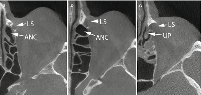
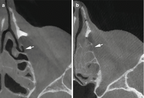
Orbital wall injury is usually consequent to incorrect identification of the lacrimal sac projection onto the lateral nasal wall, with the dissection that is carried posterolaterally to the lacrimal sac itself. When performing endo-DCR, the surgeon should always keep in mind that the lacrimal bone can be very thin and the lamina papyracea is a fragile structure. Moreover, the lamina papyracea can be dehiscent as a consequence of previous surgery, chronic inflammation, or even spontaneously. Thus, improper dissection can easily result in exposure of the periorbita. Preoperative CT is recommended in order to be aware of any anatomic variant (Figs. 10.2 and 10.3).


Fig. 10.2
The CT scans show the variable anatomic relationship between the agger nasi cell (ANC) and the lacrimal sac (LS), uncinate process (UP)

Fig. 10.3
The lamina papyracea in proximity of the lacrimal bone can be very thin (white arrow), either spontaneously (a) or as a consequence of chronic rhinosinusitis (b)
Lesion to the periorbita usually leads to fat exposure [15]. In case of fat herniation, a minimal cauterization with bipolar forceps usually helps retracting the fat into the orbit with no further risk for the patient. However, it is worth remembering that patients with orbital fat prolapse are reported to have a 5.3-fold higher risk of failure of the procedure [16]. Indeed, fat exposure is not a dangerous situation in itself unless the surgeon does not immediately recognize orbit penetration. In fact, further dissection into periorbital fat, especially with powered instrumentation, can easily result in damage to the medial rectus muscle and subsequent diplopia. In addition, the use of vasoconstrictors must be avoided when the periorbita is opened because of the risk of retinal ischemia with consequent irreversible blindness.
Other consequences potentially related to orbital wall injury are eyelid edema, ecchymosis, and emphysema. In the review of Leong et al. [1], periorbital edema, hematoma, and emphysema are reported at a frequency of about 0.1 %, 4.9 %, and 1.2 %, respectively. Similarly, in a series of 84 endo-DCR, Zuercher et al. [17] described only one case of subconjunctival hematoma (1.19 %) and three cases of edema or emphysema (3.57 %).
All these conditions have optimal outcomes with conservative management. When obstructing the opening of the eyelids, periorbital edema may be reduced by intravenous administration of steroid. Artificial teardrops and/or topical antibiotic drops may be useful to prevent conjunctivitis. Ecchymosis usually recovers spontaneously. Emphysema is due to the penetration of air in a breach of the medial orbital wall consequent to an increase in nasal cavity pressure. Asking the patient to avoid blowing the nose for some days is usually enough to resolve the problem.
10.2.3 CSF Leak
CSF leak is a very rare complication during endo-DCR. In children, inappropriate superior dissection can potentially cause injury to the skull base, with subsequent cerebrospinal fluid leak. In adults, the risk of such damage is very small. In the literature, only isolated cases are reported and the documented incidence is about 0.04 % [18].
Moreover, in most cases, the risk of CSF leak is increased by comorbidities. In fact, in the case report published by Friedel et al. [18], CSF leak was encountered while performing a DCR in a woman affected by ozena, and a fistula was created in an area of extensive bone resorption at the level of right ethmoidal cribra, near the course of AEA. Thus, preoperative identification at CT of anatomic alterations that increase the risk of violating the skull base is of utmost importance.
10.2.4 Injury to Canaliculi
Canaliculi injury can occur during insertion of a Bowman probe or lacrimal stent; therefore, these maneuvers need to be performed as gently as possible. A proper understanding of the anatomy of the lacrimal pathway is the prerequisite to properly insert the probe. The course of each canaliculus before their joining can be divided into two directions. From the puncta, the inferior and superior canaliculus head downward and upward, respectively, in a perpendicular manner for a tract of 2 mm before turning medially at an angle of about 90°. The second portion of each canaliculus is about 8 mm in length and converges on a plane passing through the medial canthus, joining in the common canaliculus.
While probing the lacrimal pathway, the surgeon should follow the anatomical bends without forcing at any time, since the risk of laceration or creation of false routes is high in view of the fragility of these structures.
Inappropriate probing can have serious functional consequences. In fact, the resulting stenosis/occlusion would determine a presaccal obstruction, causing the failure of the DCR and requiring conjunctivorhinostomy. Periorbital fluid collection along the inferior lid plus emphysema has also been described as a consequence of a false track created during canaliculi probing [19].
Notwithstanding, the incidence of canaliculi injuries is very low. In the review by Leong et al. [1], laceration of the puncta was described only in 2 of 4921 procedures (gathering external, endoscopic, laser-assisted DCR), for an incidence of 0.04 %. In the retrospective series of Zuercher et al. [17], injury to the inferior canaliculus occurred in 1 of 84 patients (1.19 %).
10.3 Postoperative Complications
10.3.1 Infections
As with any endoscopic sinonasal procedure, DCR is at risk of postoperative infection, and there is still some controversy in the literature about the need for systemic prophylactic antibiotic following the procedure. An interesting study [20] retrospectively reviewed a series of 82 external DCRs treated only by topical antibiotics (eye drops containing 0.1 % dexamethasone, 3.5 mg neomycin, and 10,000 units polymyxin B sulfate) three times a day for 1 week postoperatively without any systemic antibiotics. Only one (1.2 %) infection (wound infection) occurred. This complication was successfully treated by oral antibiotics without any sequelae. In 13 (15.9 %) cases with a clinical history of recurrent dacryocystitis or mucoceles, no postoperative infection was encountered. The authors concluded that systemic antibiotic prophylaxis in external DCR may be avoided. These results could be reasonably extrapolated and applied even to endo-DCR, especially considering the absence of external skin incision.
Acute postoperative rhinosinusitis (ARS) following endonasal DCR is an uncommon complication. In the retrospective study of Shams and Selva [21], a series of 196 patients (203 procedures) was reviewed to look for postoperative ARS. Prophylactic antibiotic therapy was not routinely administered. The incidence of ARS in the entire series was 1.5 %, but it increased to 15 % in patients preoperatively affected by chronic rhinosinusitis and asymptomatic at the time of surgery. All patients experiencing ARS recovered with antibiotics, and no further complication was reported, thus confirming that chronic rhinosinusitis may be considered a risk factor for postoperative ARS.
Chronic maxillary and frontal sinusitis are usually consequent to incorrect dissection at the level of osteomeatal complex, resulting in adhesions that may impair sinus drainage. The reported incidence is very low (0.1 %) [1] and can decrease to almost nil with the increasing experience of the surgeon. In the report by Fayet et al. [2], where the authors compared two homogenous series of 300 procedures performed by the same surgeons in subsequent periods, the incidence of maxillary and frontal sinusitis decreased from 2.4 % and 0.3 %, respectively, to zero.
If septoplasty is performed to gain access to the surgical field, a septal abscess may occur; in this case, it should be drained, and the patient should be treated with systemic antibiotics and topic irrigations.
As the lacrimal system represents a bridge between the orbit and the nasal cavity, infection may involve also the orbital content.
Incidence of canaliculitis is about 0.3 % [1, 5], even though in other series the incidence of inflammation and/or infection of lacrimal systems is reported to be as high as 7.14 % [17]. This condition requires prompt intervention with topical and, in selected cases, systemic antibiotics, to reduce the risk of extension of the infection to the orbit and sequelae as obstruction of canaliculi.
10.3.2 Epistaxis
Epistaxis after DCR, which presents as anterior bleeding, is rare, and is seen in 1.3–2.8 % of cases [2, 3, 11]. Moreover, when only copious epistaxis (defined as epistaxis requiring packing, cautery, surgical intervention, blood transfusion, or causing delayed discharge) is considered, the incidence decreases to 0.6 % [2, 11]. Management for postoperative epistaxis should follow a stepwise protocol.
Stay updated, free articles. Join our Telegram channel

Full access? Get Clinical Tree


