Adult or pediatric colonoscope with monitor (CO2 insufflation if available)
Indigo carmine diluted 50 % with injectable saline
Endoscopic injector needle
Endoscopic snare
Endoscopic Roth net® (US Endoscopy, Mentor, OH)
Suction trap
Bovie cautery
Laparoscopic monitors
High-definition flexible tip laparoscope
Trocars: 5 mm × 4, 10 mm × 1, 12 mm × 1
Laparoscopic bowel graspers and scissors
Laparoscopic needle driver
Laparoscopic energy device (surgeon preference)
Micro-laparoscopic (3 mm) instruments if available
Laparoscopic linear stapler with appropriate loads
Endo-catch bag (Covidien, Norwalk, CT)
Wound protector
Polysorb or Vicryl sutures
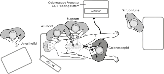
Laparoscopic monitors will be placed depending on the location of the lesion. For right colon polyps, monitors are placed on the patient’s right side and toward the head of the bed (Fig. 5.1). For left colon lesions, the monitors are placed at the patient’s left and toward the foot of the bed. For transverse colon or flexure lesions, the monitors are placed at the head of the bed as the endoscopist will stand between the patient’s legs.


Fig. 5.1
Suggested trocar and monitor placement for CELS technique for excision of a right colon polyp (Original artwork by Yuko Tonohira)
Endoscopic equipment may vary. Surgeons and endoscopists may prefer to use a pediatric versus an adult colonoscope. In addition, we feel it is a prerequisite to have CO2 colonoscopy available in the operating room. Simultaneous performance of laparoscopy and colonoscopy with room air can present technical challenges due to overdistension of the bowel. Luminal insufflation using room air can significantly obscure the laparoscopic view and compromise exposure. In institutions where CO2 is not available for endoscopy, a technique of laparoscopically clamping the terminal ileum to minimize bowel distension during laparoscopy has been described, but we have found that colonic distension alone is still a major impediment to this method [3, 4]. Since 2003, our group has been performing colonoscopy with the use of CO2 insufflation during laparoscopy. Because the bowel absorbs CO2 gas approximately 150 times faster than room air, there is minimal unwanted dilation of the colon and excellent simultaneous endoscopic and laparoscopic visualization. We have previously demonstrated that intraoperative CO2 colonoscopy is safe during simultaneous CO2 laparoscopy and can be used to avoid excessive bowel dilation during CELS procedures [9, 17]. Therefore, if available, it is preferred to have CO2 for insufflation during colonoscopy.
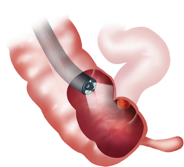
After the abdomen is prepped and draped in a sterile fashion, CO2 colonoscopy is performed to locate the lesion (Fig. 5.2). We feel it is important to perform colonoscopy first prior to port placement because intermittently the polyp, which may have previously been deemed unresectable by a referring gastroenterologist, may actually be amenable to traditional colonoscopic polypectomy alone. Dilute indigo carmine solution (50 % dilution of indigo carmine with injectable saline solution) is then used to mark the location of the polyp and to raise it up in the submucosal plane. If the polyp seems amenable to endoscopic removal alone, then this may be attempted. During this portion of the procedure, it is important to try and recognize the signs of a potential malignancy. Many times, polyps that have been biopsied or previously had attempts at snaring may be scarred and difficult to lift with submucosal injection. These findings must be contrasted with findings of a possible cancerous polyp. These findings include central umbilication, ulceration, vascular pattern on narrow band imaging, and firmness. If these findings are present, options are to continue with CELS and perform an intraoperative frozen section or to proceed to formal colectomy. We do not feel that it is necessary to perform frozen section on all polyps resected as this can add to the operative time and cost of the case. In our experience, the rate of cancer on polyps that were thought to be benign was only 2 % (1/48). Therefore, frozen section should only be done on patients with suspicion of malignancy. In our experience, 12 patients underwent colectomy instead of CELS for suspected malignancy, and only 4 (33 %) of these patients actually had cancer [7]. Although this is a low sensitivity, this may reflect our overly cautious attempts to avoid performing CELS for a case of malignancy.


Fig. 5.2
Endoscopic visualization of a right colon polyp (Original artwork by Yuko Tonohira)
If the polyp cannot be removed through a purely endoscopic approach, then laparoscopy is performed. This combined technique can be technically demanding, and the surgeon must be proficient in both laparoscopic and endoscopic techniques. For the first several cases, it is useful to have an assistant that is proficient in both of these techniques in order to be successful.
First, a periumbilical incision is made and the fascia is entered sharply. A blunt laparoscopic port is placed and pneumoperitoneum is established. A high-definition flexible tip laparoscope is preferred for better visualization. The abdomen is explored, and the site that was previously marked is located. Depending on the location of the lesion, typically two 5 mm trocars may be placed. For right colon lesions, trocars can be placed in the left lower quadrant and suprapubically. For left colon lesions, trocars can be placed in the right lower quadrant and suprapubically. For transverse colon lesions, trocars can be placed on both sides in both the lower and upper quadrants. If available, micro-laparoscopic (3 mm) instruments can also be used. A 5–12 mm port may be needed for a stapler if a colonoscopic-assisted laparoscopic wall excision is anticipated. Typically for CELS, a hand port is not necessary. However, if converting to a segmental or formal colectomy, then some may elect to place a hand port for hand-assisted laparoscopy.
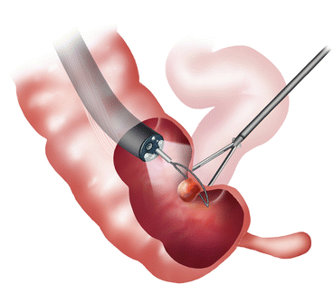
For laparoscopic-assisted colonoscopic polypectomy, the concept is simple; the laparoscopist manipulates the bowel wall to make polypectomy by the endoscopist more simple. The lesion is located by the endoscopist, and its position is confirmed by laparoscopic visualization with the use of transillumination and/or by endoscopic visualization during laparoscopic manipulation of the colon (Fig. 5.3). This maneuver can also expose areas that were not previously visualized because of mucosal folds or segmental kinks of the colon. The location of the polyp in relation to the peritoneum is important. Polyps that are located on the retroperitoneal side or mesenteric side require lateral mobilization of the colon for adequate exposure. If the polyp is in a difficult location (i.e., at a flexure or near the mesenteric border of the colon) and this area cannot be easily manipulated, the colon will need to be mobilized. This is done as in any laparoscopic procedure. We prefer to use an energy device along the line of Toldt and carried out in the native planes. Once the colon is mobilized adequately, the polyp can then be manipulated.


Fig. 5.3
Endoscopic visualization of a colon polyp with simultaneous laparoscopic manipulation of the colon wall (Original artwork by Yuko Tonohira)
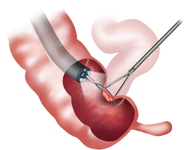
As stated previously, the polyp is lifted with dilute indigo carmine solution. This aids in visualizing the polyp in comparison to the normal surrounding mucosa and also aids in seeing the location of the polyp laparoscopically. It also provides a “buffer” zone to facilitate endoscopic resection without causing a full-thickness injury. If the polyp does not lift, we typically would err on the side of performing a colectomy even if it may be due to scarring from previous biopsy. More recently, we have added a viscous solution such as 25 % albumin to the injection solution to avoid dissipation of isotonic injection fluid and progressive bowel wall edema. Polypectomy is performed using an electrosurgical snare. This can be done using a single attempt or in a piecemeal fashion. For polyps that are either flat or situated in a difficult location, laparoscopic manipulation of the polyp during snare polypectomy will facilitate delivery of the polyp into the snare (Fig. 5.4).


