Laurie A. Tomlinson, David C. Wheeler
Clinical Evaluation and Management of Chronic Kidney Disease
Although many patients with chronic kidney disease (CKD) progress to end-stage renal disease (ESRD) and require renal replacement therapy (RRT), the majority die of nonrenal causes, particularly premature cardiovascular events.1 Early diagnosis of CKD is therefore important because it provides opportunities to delay progression of CKD (see Chapter 80) and to prevent cardiovascular complications (see Chapter 82).
Definitions
Chronic kidney disease is defined as abnormalities of kidney structure or function, present for at least 3 months, with implications for health (Table 81-1). The Kidney Disease: Improving Global Outcomes (KDIGO) guidelines recommend classification of CKD based on cause, category of glomerular filtration rate (GFR), and albuminuria (see Fig. 79-1).2 Because of the impracticalities of using radioisotopes and 24-hour urine collections, the KDIGO classification system recommends that kidney function be assessed by estimating the GFR (eGFR) from the serum creatinine level through use of an appropriate equation, except in circumstances in which eGFR estimations are known to be less accurate, such as when there is significant muscle wasting. The Modification of Diet in Renal Disease (MDRD) equation has been in common use in clinical laboratories until recently, but KDIGO has recommended that this be replaced by the Chronic Kidney Disease Epidemiology Collaboration (CKD-EPI) equation, which more accurately categorizes the risk of mortality and progression to ESRD (see Chapter 3).3 Although staging systems for CKD based on eGFR have limitations, they have proven to be useful in many clinical settings and are now deeply embedded into guidelines developed for CKD management.
Table 81-1
Criteria for definition of chronic kidney disease.
ACR, Albumin-creatinine ratio; AER, albumin excretion rate; GFR, glomerular filtration rate.
| Criteria for Definition of Chronic Kidney Disease (CKD) | |
| CKD is defined as abnormalities of kidney structure or function, present for more than 3 months, with implications for health. These may include the following. | |
| Markers of kidney damage | Albuminuria (AER ≥ 30 mg/24 h; ACR ≥ 30 mg/g [≥ 3 mg/mmol]) Urine sediment abnormalities Electrolyte and other abnormalities caused by tubular disorders Abnormalities detected through histology Structural abnormalities detected through imaging History of kidney transplantation |
| Decreased GFR | GFR <60 ml/min/1.73 m2 |
(From reference 2.)
The evidence base for the management of CKD is constantly evolving, and guidelines are updated in light of this. Although every effort has been made to ensure that this chapter reflects current recommendations, the reader is advised to check for any relevant guideline updates.
Clinical Presentation
Chronic kidney disease is usually asymptomatic until late stage 4 or stage 5 and is commonly detected by routine blood testing. There is some evidence that early diagnosis with appropriate management may slow the rate of decline of kidney function and reduce cardiovascular risk.4 Screening of the general population for CKD is not recommended, but in the United Kingdom the National Institute for Health and Care Excellence (NICE) proposes offering testing to people with conditions associated with an increased prevalence—those with diabetes, hypertension, cardiovascular disease (CVD), structural renal tract disease, renal calculi, prostatic hypertrophy, multisystem diseases with potential kidney involvement (e.g., systemic lupus), a family history of category G5 CKD, or hereditary kidney disease—and after opportunistic detection of hematuria or proteinuria.5
Evaluation of Chronic Kidney Disease
Establishing Chronicity
When an eGFR of less than 60 ml/min/1.73 m2 is detected in a patient, careful attention needs to be paid to previous blood and urine test results and the clinical history to determine if this is a result of acute kidney injury (AKI), an abrupt decrease in kidney function, or CKD that has been present but asymptomatic for some time.
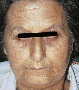
A detailed medical history covering issues, including other medical conditions, family history of kidney disease, prescribed medication, and illicit drug use, may suggest an underlying cause. There may be hints of a past history of kidney problems (e.g., hypertension, proteinuria, microhematuria) or symptoms suggestive of prostatic disease. The physical examination findings are not usually helpful, although skin pigmentation, scratch marks, left ventricular hypertrophy, and hypertensive fundal changes favor a chronic presentation (Fig. 81-1). Details of the social and personal circumstances are also crucial, particularly for patients with progressive kidney disease in whom RRT is likely to be required.
Blood tests for other conditions can be helpful if the findings indicate evidence of an acute illness that may be the cause of kidney failure, such as systemic vasculitis or multiple myeloma. A normochromic normocytic anemia is usual in CKD but may also be a feature of acute systemic illnesses and therefore is not discriminatory. Low serum calcium and raised phosphate levels also have little discriminatory value, but normal levels of parathyroid hormone (PTH) are more in keeping with AKI. Patients with grossly abnormal biochemical values—for example, blood urea nitrogen higher than 140 mg/dl, serum creatinine above 13.5 mg/dl (>1200 µmol/l), or blood urea greater than 300 mg/dl (>50 mmol/l)—who appear relatively well and are still passing normal volumes of urine are much more likely to have chronic than acute kidney disease.
Assessment of Glomerular Filtration Rate
For patients in whom the distinction between AKI and CKD is unclear, repeat testing of kidney function should be performed within 2 weeks of the initial finding of an eGFR below 60 ml/min/1.73 m2. However, if previous results confirm that this is a chronic finding, or if repeated blood test results over a 3-month period are consistent, CKD is confirmed. Other tests (such as cystatin C or an isotope-clearance measurement) may be required for confirmation of CKD in circumstances when eGFR based on serum creatinine is known to be less accurate.
Assessment of Proteinuria
Dipstick testing of the urine and urine culture are important.6 This may reveal microhematuria, which can be a useful pointer toward an underlying diagnosis. Workup of hematuria is discussed in Chapter 4. Whether or not proteinuria is detected by dipstick, further measurement of urinary protein excretion should be conducted. Proteinuria is an important diagnostic and prognostic marker, and its presence indicates a higher risk for both progression of kidney disease and cardiovascular complications.7 KDIGO recommends that the preferred method of assessing proteinuria is by measurement of the urinary albumin-creatinine ratio (ACR) using an early morning urine sample.2 The degree of albuminuria is graded by the A1 to A3 category system, replacing previous terms such as microalbuminuria (see Fig. 79-1). However, it is important to be aware that some patients will excrete proteins other than albumin, and a urine protein-creatinine ratio (PCR) may be more useful for certain conditions.8 Serial PCR measurements may be particularly useful in glomerular disease because of the higher variability of ACR and the greater cost of determining albumin in urine. Where appropriate, urine tests for Bence-Jones protein (immunoglobulin light chains) may be required because this is not detected by standard proteinuria or albuminuria testing.
Kidney Imaging
Imaging of the kidneys with ultrasound is useful for a number of reasons. Small kidneys with reduced cortical thickness, showing increased echogenicity, scarring, or multiple cysts, suggest a chronic process. Structural abnormalities such as autosomal dominant polycystic kidney disease (ADPKD), hydronephrosis caused by obstruction, or coarse renal scarring may be detected. NICE states that kidney ultrasound scanning is important only in certain circumstances and suggests counseling patients if ADPKD is suspected before imaging.5 In some situations, imaging with computed tomography, magnetic resonance, or angiography may be useful, taking into account the risks of administering contrast media (see Chapter 5).
Further Investigations
Establishing the cause of CKD is important when possible, and further specific testing, as indicated by the history and results of initial investigations, may be required. There may be an underlying treatable condition that requires appropriate management, or there may be a genetic cause such as ADPKD, for which counseling should be offered. Furthermore, some kidney diseases may recur after transplantation (see Chapter 108), and an accurate diagnosis may therefore influence later management. Despite thorough investigation, however, the cause of CKD is often unclear, with an unhelpful past medical history, minimal abnormalities on urinalysis, and small kidneys on ultrasound. In such patients, investigation should not be pursued relentlessly because the implications for treatment are often minimal. Attempting to obtain biopsy material from small kidneys is associated with risk, and even if a biopsy is performed, histologic assessment may simply show nonspecific chronic scarring rather than diagnostic features that explain the cause of kidney damage.
Predicting Prognosis
With the cause of CKD established if possible, the GFR and the level of proteinuria measured, and other comorbidities categorized, it may be possible to estimate the risk of CKD progression and likely future need for RRT. KDIGO recommends consideration of the GFR and the albuminuria categories according to a “heat map” of risk (see Fig. 79-1).2 Other factors associated with CKD progression will also help inform prognosis. These include cause of CKD, age, sex, ethnicity, dyslipidemia, smoking, obesity, history of CVD, ongoing exposure to nephrotoxic agents, and degree of control of hypertension and hyperglycemia. However, often the best guide to future change in kidney function is the previous pattern of decline, highlighting the importance of considering results of previous blood and urine testing during the initial assessment.
Monitoring and Defining Progression
Once a diagnosis of CKD has been established, arrangements need to be put in place to ensure regular monitoring of kidney function and proteinuria. In patients at low risk of decline, this could be done annually. However, assessment should be undertaken more regularly if the trajectory of the disease is not clear, and in patients at higher risk of progression.
Determining a true change in kidney function may be difficult because small fluctuations in eGFR are common and not necessarily indicative of progression. They may be caused by reversible factors, such as intravascular depletion or high meat intake, so repeat testing may be required. NICE defines progression as a decline in eGFR of more than 5 ml/min/1.73 m2 within 1 year or more than 10 ml/min/1.73 m2 within 5 years.5 KDIGO refers to a sustained decline in eGFR of more than 5 ml/min/1.73 m2 within 1 year as rapid progression and also specifies a drop in GFR category accompanied by a 25% or greater drop in eGFR from baseline as defining progression.2 In patients with CKD progression, current management should be reviewed to detect reversible causes, and specialist referral should be considered.
When to Refer to the Nephrologist
Although management of patients with early nonprogressive CKD is increasingly becoming the responsibility of primary care physicians, nephrologists need to assess those individuals likely to progress to ESRD and to require RRT. Substantially similar criteria for referral have been developed by NICE and KDIGO (Table 81-2). Such criteria are not absolute but should provide a guide to the primary care physician as to which patients are likely to benefit from specialist care. For example, many patients with stable category G4 CKD are successfully managed in the community, often after initial assessment by or with advice from secondary care colleagues.
Table 81-2
Suggested criteria for referral of patients with CKD to a nephrologist.
ACR, Albumin-creatinine ratio; eGFR, estimated glomerular filtration rate; KDIGO, Kidney Disease: Improving Global Outcomes; NICE, National Institute for Health and Care Excellence; PTH, parathyroid hormone; RBC, red blood cell.
| Suggested Criteria for Referral of Patients with Chronic Kidney Disease (CKD) to a Nephrologist | ||
| NICE 2008 | KDIGO 2012 | |
| Advanced CKD | Stage 4 and 5 CKD | Category G4 and G5 CKD |
| Proteinuria | High proteinuria: ACR ≥ 70 mg/mmol unless known to be caused by diabetes and appropriately treated | Consistent proteinuria: ACR ≥ 300 mg/g (≥30 mg/mmol) |
| Hematuria | Proteinuria (ACR ≥ 30 mg/mmol) together with hematuria | Urinary red cell casts, RBCs > 20 per high-power field sustained; not readily explained |
| Progression of CKD | Rapidly declining eGFR: >5 ml/min/1.73 m2 in 1 yr >10 ml/min/1.73 m2 in 5 yr | Progression of CKD: Sustained decline in eGFR > 5 ml/min/1.73 m2 in 1 yr Drop in GFR category with a ≥25% drop in eGFR from baseline |
| Uncontrolled hypertension | Hypertension that remains poorly controlled despite the use of at least four antihypertensive drugs at therapeutic doses | CKD and hypertension refractory to treatment with four or more antihypertensive agents |
| Hereditary kidney disease | Known or suspected rare or genetic causes of CKD | Hereditary kidney disease |
| Other conditions | Suspected renal artery stenosis | Recurrent or extensive nephrolithiasis Persistent abnormalities of serum potassium |
(Data from references 2
Related posts:


Stay updated, free articles. Join our Telegram channel

Full access? Get Clinical Tree


