Fig. 1
a Sonographic feature of a gastric carcinoid. A round-shaped hypoechoic tumor is demonstrated. Wall stratification is not demonstrated clearly in this figure. b Color Doppler ultrasound shows the rich vascularity of the tumor. c Contrast-enhanced ultrasound of the tumor also reveals the rich blood perfusion
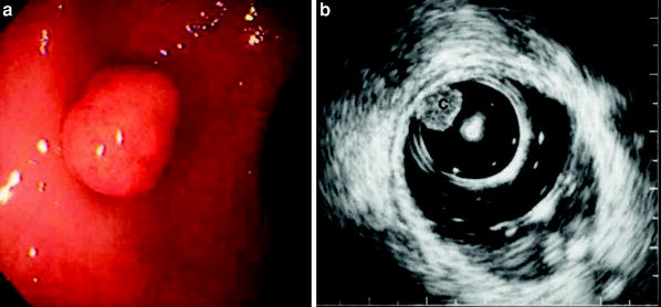
Fig. 2
a Endoscopic feature of rectal carcinoid. A small (5 mm in diameter) submucosal tumor is demonstrated. b Endosonography with a miniature ultrasound probe (12 MHz). A small round-shaped tumor (C) is demonstrated beneath the first layer and no changes in submucosal layer are seen
4 Submucosal Tumors
4.1 Cyst and Lymphangioma
Cysts and lymphangiomas are located in the submucosal layer (Figs. 3, 4). They are well demarcated by a thin wall and are anechoic. Posterior echo enhancement as an artifact is a characteristic figure. They show good compressibility and change their shapes with external compression. Color Doppler does not reveal any vascular structure inside. Often septal structures are seen inside the tumor.
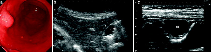
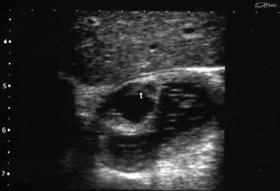
Fig. 3
a Endoscopy of a gastric cyst. The tumor is located in the anterior wall of the antrum and is covered with normal mucosa without indentation or erosion. b Sonography with a convex probe (3 MHz) of the gastric cyst (gc). A small anechoic tumor is demonstrated although the main layer where the tumor lies is not clear. c Scanning with a high-frequency linear probe (7 MHz) revealed that the lesion (gc) lies in the submucosal layer (sml). S stomach
Fig. 4
Lymphangioma at the duodenal bulb. A cystic tumor (t) with septa is seen in the submucosal layer
4.2 Lipoma
Lipomas also lie in submucosal layer (Fig. 5). They are very soft, their shape can be changed easily by external compression or peristalsis. Their internal echoes are generally equivalent or slightly lower than that of the adjacent submucosal layer. Vascular signals are seldom detected with color Doppler. Usually no changes are seen in the mucosal and proper muscle layers.
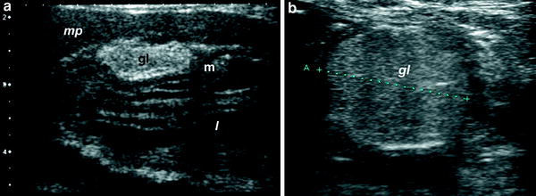
Fig. 5
a Sonographic features of gastric lipomas (gl). a Hyperechoic tumor located in the submucosal layer of the gastric wall is demonstrated. b Sonography of duodenal lipoma, which resembles that of gastric lipoma
4.3 Ectopic Pancreas
An ectopic pancreas is characterized as a hypoechoic tumor mainly located in the submucosal layer with thickening of the adjacent proper muscle layer (Fig. 6). A tiny cystic area representing the ductal structure may be demonstrated.
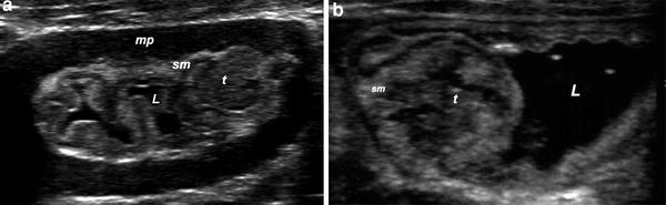
Fig. 6
a Aberrant pancreas at the greater curvature of the stomach. A hypoechoic tumor (t) with a small cystic area inside mainly located in the submucosal layer (sm) is observed. b Aberrant pancreas at the ileum. A hypoechoic tumor (t) involving cystic areas in the submucosal layer is demonstrated. mp Muscularis propria. L Lumen
4.4 Hemangioma
A hemangioma is demonstrated as a hyperechoic tumor in the submucosal layer. Calcification is occasionally seen inside the tumor (Fig. 7). Since the blood flow of the capillaries making up the tumor is extremely slow, it is difficult to detect blood flow signals using the conventional color Doppler method.
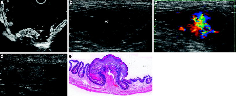
Fig. 7


a Endoscopic ultrasound of a gastric hemangioma (gh). The tumor lies in the submucosal layer and calcifications inside the tumor are seen (asterisks). b Sonography of a pyogenic granuloma (pg) in the ileum. Hypoechoic tumor with irregular contour is demonstrated. c Color Doppler image of the tumor. Extremely rich vascularity is shown. d Contrast ultrasound also shows rich perfusion of the tumor. e Hematoxylin and Eosin stain of the resected specimen
Related posts:


Stay updated, free articles. Join our Telegram channel
Full access? Get Clinical Tree
