Urological Surgery and Equipment


Urological Surgery and Equipment


Preparation of the patient for urological surgery

The degree of preparation is related to the complexity of the procedure. Certain aspects of examination (pulse rate, BP) and certain tests (haemoglobin, electrolytes, creatinine) are important, not only to assess fitness for surgery, but also as a baseline against which changes in the post-operative period may be measured.


  • Assess cardiac status (angina, arrhythmias, previous MI, BP, ECG, CXR). We assess respiratory function by pulmonary function tests (FVC, FEV1) for all major surgery and any surgery where the patient has symptoms of respiratory problems or a history of chronic airways disease (e.g. asthma).


  • Arrange an anaesthetic review where there is, for example, cardiac or respiratory comorbidity.


  • Culture urine, treat active (symptomatic) infection with an appropriate antibiotic, starting a week before surgery, and give prophylactic antibiotics at the induction of anaesthesia.


  • Consider stopping aspirin and NSAIDs 10 days prior to surgery.


  • Obtain consent.


  • Measure haemoglobin and serum creatinine and investigate and correct anaemia, electrolyte disturbance, and abnormal renal function. If blood loss is anticipated, group and save a sample of serum or crossmatch several units of blood, the precise number depending on the speed with which your blood bank can deliver blood, if needed. In our own unit, our policy is (other units may have a different policy) (Box 17.1):


    • The patient may choose to store their own blood prior to the procedure.


Should aspirin and other antiplatelet drugs be stopped prior to minor urological procedures and urological surgery?


Aspirin and TRUS biopsy

In the UK, 65% of urologists routinely stop aspirin prior to TRUS biopsy; 35% do not.1 Four of 297 urologists (1.3%) reported cerebrovascular side effects from stopping aspirin. There remains no consensus guidance on whether to stop or continue aspirin.


Aspirin and TURP

There is wide variation in the management of aspirin in men undergoing TURP. In a recent audit of UK urologists, 38% said they did not stop aspirin prior to TURP, but of those that said they did stop it, a substantial number still proceeded with TURP if the aspirin had inadvertently not been stopped.2 Overall, 75% either did not bother stopping aspirin or proceeded with TURP if patients were inadvertently still taking it, presumably because of a perceived increased risk of serious cardiovascular events. Some studies suggest an increased risk of bleeding and the need for blood transfusion in those on aspirin while others report no increased risk. There is only one RCT and this showed that aspirin did increase blood loss after TURP, but not enough to increase the requirement for blood transfusion.3 The risks of short-term withdrawal of aspirin prior to TURP have not been established, although there are anecdotal reports of serious adverse cardiovascular events. So should aspirin be stopped or continued prior to TURP? The short answer is that there is no substantial body of evidence to support stopping it or continuing it and as the majority continue to do TURP with patients on aspirin, but a substantial minority stop it, either behaviour is reasonable. Since bleeding times return to normal within 48h of stopping aspirin (the time taken for new platelets to reach sufficient numbers to compensate for impaired function of circulating platelets), it seems reasonable to stop it 2 days before surgery and to restart it within a few days of surgery when it is obvious that post-operative bleeding has stopped (usually when it is deemed safe to remove the catheter).


Drug-eluting cardiac stents and antiplatelet agents

Be careful in patients receiving the newer antiplatelet drugs such as clopidogrel or ticlopidine (with or without aspirin) since bleeding times can increase 3-fold.4 Severe intractable bleeding can occur following ‘minor’ procedures such as prostate biopsy or bladder biopsy. Patients with coronary artery stents are treated with dual anticoagulation with aspirin and clopidogrel for several months after stent insertion to reduce the risk of stent thrombosis. The precise duration of antiplatelet therapy has not been established, but 9-12 months is currently recommended by the American Heart Association. Seek advice from a cardiologist about the safety of stopping these drugs. Consider delaying invasive procedures (e.g. prostate or bladder biopsy) if the risk of bleeding is deemed to be unacceptable in the presence of the continued need for anticoagulation.


Bowel preparation

Indicated if large bowel is to be used (bowel prep is not required if small bowel alone is to be used, e.g. ileal conduit, ileal neo-bladder reconstruction). Use a simple mechanical prep (Citramag® or Picolax®—magnesium salts), two doses starting the morning before surgery with clear fluid-only diet.


1 Masood J, Hafeez, Calleary J, Barua JM (2007) Aspirin use and transrectal ultrasonographyguided prostate biopsy: a national survey. Br J Urol Int 99:965-6.

2 Enver MK, Hoh I, Chinegwundoh FI (2006) The management of aspirin in transurethral prostatectomy: current practice in the UK. Ann R Coll Surg Engl 88:280-3.

3 Nielsen JD, Holm-Nielsen A, Jespersen J, et al. (2000) The effect of low-dose acetylsalicylic acid on bleeding after transurethral prostatectomy—a prospective, randomized, double-blind, placebocontrolled study. Scan J Urol Nephrol 34:194-8.

4 Stephen Jones J (2007) Urologists: be aware of significant risks of stopping anticoagulants in patients with drug eluting coronary stents. BJU Int 99:1330-1.


Antibiotic prophylaxis in urological surgery

The precise antibiotic prophylaxis policy that you use will depend on your local microbiological flora. Your local microbiology department will provide regular advice and updates on which antibiotics should be used, both for prophylaxis and treatment. The policy shown here and in Table 17.1 is our own local policy.

Since the last edition of this book, there has been a move away from cefuroxime in an attempt to reduce the risk of antibiotic-induced C. difficile colitis. There has also been a similar move away from the use of fluoroquinolones (ciprofloxacin, norfloxacin, ofloxacin, levofloxacin) because they are a risk factor for the development of C. difficile-associated diarrhoea and pseudomembranous colitis (and also for MRSA since they are secreted onto the skin and many staphylococci are resistant to them).* Trimethoprim, gentamicin, penicillin, and co-amoxiclav are less likely to cause C. difficile-associated disease.

Culture urine before any procedure and use specific prophylaxis (based on sensitivities) if culture positive.

We avoid ciprofloxacin in inpatients because it is secreted onto the skin and causes MRSA colonization. For most purposes, nitrofurantoin provides equivalent cover without being secreted onto the skin. We do use ciprofloxacin if there is known Proteus infection (all Proteus species are resistant to nitrofurantoin).


Patients with artificial heart valves

Patients with heart murmurs and those with prosthetic heart valves: 1g of IV amoxicillin with 120mg of gentamicin should be given at induction of anaesthesia, with an additional dose of oral amoxicillin, 500mg 6h later (substituting vancomycin 1g for those who are penicillin-allergic).


Patients with joint replacements

The advice is conflicting.


AAOS/AUA advice

Joint advice of the American Academy of Orthopaedic Surgeons (AAOS) and the American Urological Association (AUA)—antibiotic prophylaxis is not indicated for urological patients with pins, plates, or screws or for most patients with total joint replacements. It is recommended for all patients undergoing urological procedures, including TURP within 2 years of a prosthetic joint replacement, for those who are immunocompromised (e.g. rheumatoid patients, those with SLE, drug-induced immunosuppression, including steroids), and for those with a history of previous joint infection, haemophilia, HIV infection, diabetes, and malignancy.

Antibiotic regime: single dose of a quinolone, such as 500mg of ciprofloxacin, 1—2h preoperatively + ampicillin 2g IV + gentamicin 1.5mg/kg 30-60min preoperatively (substituting vancomycin 1g IV for penicillinallergic patients).


UK advice

In the UK, a Working Party of the British Society for Antimicrobial Chemotherapy has stated that patients with prosthetic joint implants (including total hip replacements) do not require antibiotic prophylaxis and consider that it is unacceptable to expose patients to the adverse effects of antibiotics when there is no evidence that such prophylaxis is of any benefit. This advice is based on the rationale that joint infections are caused by skin organisms that get onto the prosthesis at the time of the operation and that the role of bacteraemia as a cause of seeding outside the immediate post-operative period has never been established.

We use the same antibiotic prophylaxis as for patients without joint prostheses.


Table 17.1 Oxford Urology procedure: specific antibiotic prophylaxis protocol for urological surgery


Procedure


Antibiotic prophylaxis


Catheter removal


Nitrofurantoin 100mg PO 30min before catheter removal


Change of male long-term catheter


Gentamicin 1.5mg/kg IM or IV 20min before*


Flexible cystoscopy or GA cystoscopy


Nitrofurantoin 100mg PO 30-60min before procedure


Transrectal prostatic biopsy


Ciprofloxacin 500mg PO and metronidazole 400mg 20min prebiopsy and for 48h post-biopsy (ciprofloxacin 500mg bd , metronidazole 400mg tds )


ESWL


500mg PO ciprofloxacin 30min before treatment (nitrofurantoin does not cover Proteus, a common ‘stone’ bacterium)


PCNL


Co-amoxiclav 1.2g IV tds starting the day before, hours before operation, and 3 doses post-operatively; gentamicin (3-5mg/kg ) commencing the day prior to surgery, with a further dose 24 hours later


Ureteroscopy


Gentamicin 1.5mg/kg IV at induction


Urogynaecological procedures (e.g. colposuspension)


Co-amoxiclav 1.2g IV and metronidazole 500mg IV at induction of anaesthesia


TURPs and TURBTs — both for non-catheterized patients (i.e. elective TURP for LUTS ) and patients with catheters (undergoing TURP for retention)


Nitrofurantoin 100mg + IV gentamicin at induction (1.5mg/kg ); nitrofurantoin 100mg PO 30min before catheter removal


Radical prostatectomy


Co-amoxiclav 1.2g IV + 240mg IV gentamicin + 500mg IV metronidazole at induction


240mg of gentamicin


24h post-op; 48h IV co-amoxiclav 1.2g tds ; ciprofloxacin PO 5 days


Cystectomy or other procedures involving the use of bowel (e.g. augmentation cystoplasty)


Co-amoxiclav 1.2g IV + 500mg IV metronidazole at induction; further 2 doses of co-amoxiclav 1.2g and metronidazole 500mg post-operatively


Artificial urinary sphincter insertion


Vancomycin 1g 1.5h before leaving the ward (infuse over 100min)** + co-amoxiclav 1.2g IV + 3mg/kg IV gentamicin at induction; continue IV cefuroxime, gentamicin, and vancomycin (1g bd ) for 48h


* Sepsis rate (necessitating admission to hospital) may be as high as 1% without antibiotic cover.
** OR teicoplanin if vancomycin allergic—400mg at induction and bd thereafter for a total of 48h; meropenem may be substituted for vancomycin in ‘vancomycin-free’ hospitals. About 9% of patients undergoing flexible cystoscopy develop bacteruria (>105 CFU /mL of urine). A randomized, placebo controlled trial of ciprofloxacin 500mg or trimethoprim 200mg in 2083 patients undergoing flexible cystoscopy showed a significant reduction of bacteriuria to 3 and 5%, respectively. While both antibiotics reduce the risk of bacteriuria, ciprofloxacin is more effective—after adjustment for baseline bacteriuria (˜74% had bacteriuria before cystoscopy), the odds of bacteriuria for those taking trimethoprim were 4 times greater than those on ciprofloxacin (Johnson MI , Merrilees D, Robson WA et al. (2007) Oral ciprofloxacin or trimethoprim reduces bacteriuria after flexible cystoscopy. Br J Urol Int 100:826-9).


* C. difficile is a Gram-positive, anaerobic, spore-forming bacillus. Most common cause of nosocomial diarrhoea and antibiotic-associated colitis. Disease arises as a consequence of faeco-oral transmission of C. difficile spores (Ribotype 027 seems to be particularly pathogenic). Once colonization has occurred, progression to diarrhoea or colitis depends on coexisting conditions and host immune response. C. difficile toxins A and B are responsible for pathogenicity. They bind to intestinal epithelial receptors. Inflammatory cytokines cause fluid secretion, mucosal destruction, and tissue necrosis. Other risk factors for C. difficile-associated disease: age >65y. Use of proton pump inhibitors, laxatives, nasogastric tubes, prolonged hospital stay. Treatment for diarrhoea and colitis: stop causative antibiotics, isolate and barrier nurse (wash hands with soap and water as alcohol hand rubs are ineffective against spores), oral metronidazole (oral vancomycin reserved for serious or recurrent infection).


Complications of surgery in general: DVT and PE

Venous thromboembolism (VTE) is uncommon after urological surgery, but it is considered the most important non-surgical complication of major urological procedures. Following TURP, 0.1-0.2% of patients experience a pulmonary embolus (PE)1 and 1-5% of patients undergoing major urological surgery experience symptomatic VTE.2 The mortality of PE is in the order of 1%.3


Risk factors for DVT and PE

Increased risk: open (vs endoscopic) procedures, malignancy, increasing age, duration of procedure.


Categorization of VTE risk

American College of Chest Physicians (ACCP) Guidelines on the prevention of VTE2 and British Thromboembolic Risk Factors (THRIFT) Consensus Group4 categorize the risk of VTE:


  • Low-risk patients: those <40 undergoing minor surgery (surgery lasting <30min) and no additional risk factors. No specific measures to prevent DVT are required in such patients other than early mobilization. Increasing age and duration of surgery increases the risk of VTE.


  • High-risk patients: include those undergoing non-major surgery (surgery lasting >30min) who are aged >60.


Additional risk factors (that indicate the requirement for additional prophylactic measures, e.g. the addition of SC heparin and/or intermittent pneumatic calf compression (IPC)


  • Active heart or respiratory failure.


  • Active cancer or cancer treatment.


  • Acute medical illness.


  • Age >40y.


  • Antiphospholipid syndrome.


  • Behcet’s disease.


  • Central venous catheter in situ.


  • Continuous travel >3h up to 4 weeks before surgery.


  • Immobility (paralysis or limb in plaster).


  • Inflammatory bowel disease (Crohn’s disease/ulcerative colitis).


  • Myeloproliferative diseases.


  • Nephrotic syndrome.


  • Obesity (BMI >30kg/m2).


  • Paraproteinaemia.


  • Paroxysmal nocturnal haemoglobinuria.


  • Personal or family history of VTE.


  • Recent myocardial infarction or stroke.


  • Severe infection.


  • Use of oral contraceptive or hormone replacement therapy.


  • Varicose veins with associated phlebitis.


  • Inherited thrombophilia.


  • Factor V Leiden.


    • Prothrombin 2021A gene mutation.


    • Antithrombin deficiency.


    • Protein C or S deficiency.


    • Hyperhomocysteinaemia.


    • Elevated coagulation factors (e.g. Factor VIII).


Prevention of DVT and PE


Diagnosis of DVT

Signs of DVT are non-specific (i.e. cellulitis and DVT share common signs—low-grade fever, calf swelling, and tenderness). If you suspect a DVT, arrange a Doppler USS. If the ultrasound probe can compress the popliteal and femoral veins, there is no DVT; if it cannot, there is a DVT.


Diagnosis of PE

Small PEs may be asymptomatic. Symptoms: include breathlessness, pleuritic chest pain, haemoptysis. Signs: tachycardia, tachypnoea, raised JVP, hypotension, pleural rub, pleural effusion.


Tests


  • CXR: may be normal or show linear atelectasis, dilated pulmonary artery, oligaemia of affected segment, small pleural effusion.


  • ECG: may be normal or show tachycardia, right bundle branch block, inverted T waves in V1-V4 (evidence of right ventricular strain). The ‘classic’ SI, QIII, TIII pattern is rare.


  • Arterial blood gases: low PO2 and low PCO2.


  • Imaging: CT pulmonary angiogram (CTPA)—superior specificity and sensitivity when compared with ventilation perfusion (VQ) radioisotope scan.


  • Spiral CT: a negative CTPA rules out a PE with similar accuracy to a normal isotope lung scan or a negative pulmonary angiogram.


Treatment of established DVT


  • Below-knee DVT: above-knee thromboembolic stockings (AK-TEDs), if no peripheral arterial disease (enquire for claudication and check pulses) + unfractionated heparin 5000U SC 12-hourly.


  • Above-knee DVT: start a low molecular weight heparin (LMWH) and warfarin and stop heparin when INR is between 2 and 3. Continue treatment for 6 weeks for post-surgical patient; lifelong if underlying cause (e.g. malignancy).


Treatment of established PE

Fixed dose of SC LMWH seems to be as effective as adjusted dose IV unfractionated heparin for the treatment of PE found in conjunction with a symptomatic DVT.3 Rates of haemorrhage are similar with both forms of heparin treatment. Start warfarin at the same time and stop heparin when INR is 2-3. Continue warfarin for 3 months.


Options for prevention of VTE


  • Early mobilization.


  • AK-TEDs—provide graduated, static compression of the calves, thereby reducing venous stasis. More effective than below-knee TEDS for DVT prevention.5


  • SC heparin (low-dose unfractionated heparin (LDUH) or LMWH).

    In unfractionated preparations, heparin molecules are polymerized— molecular weights from 5000-30 000Da. LMWH is depolymerized— molecular weight 4000-5000Da.


  • IPC boots, which are placed around the calves, are intermittently inflated and deflated, thereby increasing the flow of blood in calf veins.6


  • For patients undergoing major urological surgery (radical prostatectomy, cystectomy, nephrectomy), AK-TEDS with IPC intraoperatively, followed by SC heparin (LDUH or LMWH) should be used. For TURP, many urologists use a combination of AK-TEDS and IPCs; relatively few use SC heparin.7


Contraindications to AK-TEDS


  • Any local leg conditions with which stockings would interfere, such as dermatitis, vein ligation, gangrene, recent skin grafts.


  • Peripheral artery occlusive disease (PAOD).


  • Massive oedema of legs or pulmonary oedema from congestive cardiac failure.


  • Extreme deformity of the legs.


Contraindications to heparin


  • Allergy to heparin.


  • History of haemorrhagic stroke.


  • Active bleeding.


  • Significant liver impairment—check clotting first.


  • Thrombocytopenia (platelet count <100 × 109/L).


Management of anticoagulation in the perioperative period

Liaise with whoever is responsible for the patient’s anticoagulation (e.g. anticoagulant clinic). Warfarin should be stopped either 4 days (if the target INR is 2.5) or 5 days (if the target INR is higher) before surgery. Determine the INR the day before surgery to reduce the risk of cancellation. Administer oral vitamin K (2.5mg) if the INR is ≥2.0. Check the INR on the day of surgery.

The main decision is whether to give bridging therapy with treatment dose heparin (unfractionated heparin or LMWH) and if not, whether preoperative prophylactic LMWH is advised when the INR is <2.0. For pragmatic purposes, to save monitoring the INR as an outpatient, this could be instituted 2-3 days after warfarin is stopped, i.e. on the morning after two doses have been omitted.

A controversial group of patients are those with a prosthetic (noncaged) aortic valve and no other risk factor. It is acceptable not to use bridging therapy with treatment dose heparin in these patients particularly if the bleeding risk is high.8, 9


Table 17.2 Pre- and post-operative risks


Pre-operative


Post-operative*


High risk, e.g. VTE within 1 month. Prosthetic mitral valve, AF, and history of stroke


Treatment dose heparin (either IV UFH or SC LMWH )**


Treatment dose heparin (either IV UFH or SC LMWH )


Non-high Risk, e.g. AF without previous stroke


Nil/prophylactic LMWH ***


Prophylactic LMWH


* Continue until INR >2.0 for two consecutive days.
** Stop full dose IV UFH 6h preoperatively and check APTT, omit full dose SC LMWH on day of surgery.
*** For patients with VTE within 1-3 months or cancer, we would suggest prophylactic LMWH preoperatively.


1 Donat R, Mancey-Jones B (2002) Incidence of thromboembolism after transurethral resection of the prostate (TURP). Scan J Urol Nephrol 36:119-23.

2 Geerts WH, Heit JA, Clagett PG, et al. (2001) Prevention of venous thromboembolism. (American College of Chest Physicians (ACCP) Guidelines on prevention of venous thromboembolism) Chest 119:132S-175S.

3 Quinlan DJ, McQuillan A, Eikelboom JW (2004) Low molecular weight heparin compared with intravenous unfractionated heparin for treatment of pulmonary embolism. Ann Intern Med 140:175-83.

4 Lowe GDO, Greer IA, Cooke TG, et al. (1992) Risk of and prophylaxis for venous thromboembolism in hospital patients. Thromboembolic Risk Factors (THRIFT) Consensus Group. BMJ 305:567-74.

5 Howard A, Zaccagnini D, Ellis M, Williams A, Davies AH, Greenhalgh RM (2004) Randomized clinical trial of low molecular weight heparin with thigh-length or knee-length antiembolism stockings for patients undergoing surgery. Br J Surg 91:842-7.

6 Soderdahl DW, Henderson SR, Hansberry KL (1997) A comparison of intermittent pneumatic compression of the calf and whole leg in preventing deep venous thrombosis in urological surgery. J Urol 157:1774-6.

7 Golash A, Collins PW, Kynaston HG, Jenkins BJ (2002) Venous thromboembolic prophylaxis for transurethral prostatectomy: practice among British urologists. J R Soc Med 95:130-1.

8 Dunn AS, Turpie AG (2003) Perioperative management of patients receiving oral anticoagulants: a systematic review. Arch Intern Med 163:901-8.

9 Kearon C (2003) Managment of anticoagulation before and after elective surgery. Am Soc Hematol Educat Program Book, pp. 528-34.


Fluid balance and the management of shock in the surgical patient


Daily fluid requirement

Can be calculated according to patient’s weight:


  • For the first 10kg: 100mL/kg per 24h (= 1000mL).


  • For the next 10kg (i.e. from 10-20kg): 50mL/kg per 24h (= 500mL).


  • For every kg above 20kg: 20mL/kg per 24h (= 1000mL for a patient weighing 70kg).

Thus, for every 24h, a 70kg adult will require 1000mL for their first 10kg of weight, plus 500mL for their next 10kg of weight, and 1000mL for their last 50kg of weight = total 24h fluid requirement, 2500mL.

Daily sodium requirement is 100mmol and for potassium, 70mmol. Thus, a standard 24h fluid regimen is 2L of 5% dextrose + 1L of normal saline (equivalent to about 150mmol Na+), with 20mmol K+ for every litre of infused fluid.

Fluid losses from drains or nasogastric aspirate are similar in composition to plasma and should be replaced principally with normal saline.


Shock due to blood loss

Inadequate organ perfusion and tissue oxygenation. The causes are hypovolaemia, cardiogenic, septic, anaphylactic, and neurogenic. The most common cause in the surgical patient is hypovolaemia due to blood and other fluid loss. Haemorrhage is an acute loss of circulating blood volume. Haemorrhagic shock may be classified as:


  • Class I: up to 750mL of blood loss (15% of blood volume); normal pulse rate (PR), respiratory rate (RR), BP, urine output, and mental status.


  • Class II: 750-1500mL (15-30% of blood volume); PR >100; decreased pulse pressure due to increased diastolic pressure; RR 20-30; urinary output 20-30mL/h.


  • Class III: 1500-2000mL (30-40% of blood volume); PR >120; decreased BP and pulse pressure due to decreased systolic pressure; RR 30-40; urine output 5-15mL/h; confusion.


  • Class IV: >2000mL (>40% of blood volume); PR >140; decreased pulse pressure and BP; RR >35; urine output <5mL/h; cold, clammy skin.


Management


  • Remember ‘ABC’: 100% oxygen to improve tissue oxygenation.


  • ECG, cardiac monitor, pulse oximetry.


  • Insert two short and wide IV cannulae in the antecubital fossa (e.g. 16 G). A central venous line may be required.


  • Infuse 1L of warm Hartmann’s solution or if severe haemorrhage, then start a colloid instead (e.g. Gelofusin®). Aim for a urinary output of 0.5mL/kg/h and maintenance of BP.


  • Check FBC, coagulation screen, U & E, and cardiac enzymes.


  • Cross-match 6U of blood.


  • Arterial blood gases to assess oxygenation and pH.


  • Obvious and excessive blood loss may be seen from drains, but drains can block so assume there is covert bleeding if there is a tachycardia (and low BP). If this regimen fails to stabilize pulse and BP, return the patient to the operating room for exploratory surgery.


Patient safety in the urology theatre

It is a fundamental part of safe surgical practice to cross-check that the following have been done prior to starting an operation or procedure. The process of cross-checking should be done with another member of staff, using several sources of information (e.g. the notes, consent form, X-ray images) to confirm the following.


  • Patient identification: confirm you are operating on the right patient by a process of ‘active’ identification (i.e. ask the patient their name, date of birth, and their address to confirm that you are talking to the correct patient).


  • Ensure you are doing the correct procedure and on the correct side by cross-checking with the notes and X-rays: for lateralized procedures (e.g. nephrectomy, PCNL), the correct side of the operation should be confirmed by cross-checking with the X-rays and with the X-ray report as well as referring to the notes. Where it is possible for the sides of an IVU to be incorrectly labelled, this cannot happen with a CT scan where the location of the liver (right side) and the spleen (left side) provides confirmation of what side is what.


  • Appropriate antibiotic prophylaxis has been given.


  • DVT prophylaxis has been administered (e.g. heparin, AK-TEDS, IPC boots).


  • Blood is available, if appropriate.


  • The patient is safely and securely positioned on the operating table: pressure points padded, not touching metal (to avoid diathermy burns), body straps securely in place.

Develop an approach to operating that involves members of your team. Listen to the opinions of staff who are junior to you. They may sometimes be able to identify errors that are not obvious to you. Cultivate the respect of the recovery room staff. They may express concern about a patient under their care—listen to their concerns, take them seriously, and if all is well, reassure them. It does no harm for your patients or for your reputation to develop the habit of visiting every patient in the recovery room to check that all is well. You may be able to identify a problem before it has developed into a crisis and at the very least, you will gain a reputation for being a caring surgeon.


Transurethral resection (TUR) syndrome

Arises from the infusion of a large volume of hypotonic irrigating solution into the circulation during endoscopic procedures (e.g. TURP, TURBT, PCNL). Occurs in 0.5% of TURPs.


Diagnosis: symptoms, signs, and tests

Confusion, nausea, vomiting, hypertension, bradycardia, visual disturbances, seizures. If the patient is awake (spinal anaesthesia), they may report visual disturbances (e.g. flashing lights).


1 Madsen PO, Naber KG (1973) The importance of the pressure in the prostatic fossa and absorption of irrigating fluid during transurethral resection of the prostate. J Urol 109:446-52.

2 Coppinger SW, Lewis CA, Milroy EJG (1995) A method of measuring fluid balance during transurethral resection of the prostate. Br J Urol 76:66-72.

3 Hahn RG (1993) Ethanol monitoring of extravascular absorption of irrigating fluid. Br J Urol 72:766-9.


Catheters and drains in urological surgery


Catheters

Made from latex or silastic (for patients with latex allergy or for long-term use—better tolerated by the urethral mucosa).


Types


  • Self-retaining (also known as a Foley, balloon, or 2-way catheter) (Fig. 17.1). An inflation channel can be used to inflate and deflate a balloon at the end of the catheter, which prevents the catheter from falling out.


  • A 3-way catheter (also known as an irrigating catheter). Has a third channel (in addition to the balloon inflation and drainage channels) which allows fluid to be run into the bladder at the same time as it is drained from the bladder (Fig. 17.2).


Size

The size of a catheter is denoted by its circumference in mm. This is known as the ‘French’ or ‘Charriere’ (hence Ch) gauge. Thus a 12 Ch catheter has a circumference of 12mm.


Uses


  • Relief of obstruction (e.g. BOO due to BPE causing urinary retention— use the smallest catheter that you can pass; usually a 12 Ch or 14 Ch is sufficient in an adult).


  • Irrigation of the bladder for clot retention (use a 20 Ch or 22 Ch 3-way catheter).


  • Drainage of urine to allow the bladder to heal if it has been opened (trauma or deliberately, as part of a surgical operation).


  • Prevention of ureteric reflux, maintenance of a low bladder pressure, where the ureter has been stented (post-pyeloplasty for PUJO).


  • To empty the bladder before an operation on the abdomen or pelvis (deflating the bladder gets it out of harm’s way).


  • Monitoring of urine output post-operatively or in the unwell patient.


  • For delivery of bladder instillations (e.g. intravesical chemotherapy or immunotherapy).


  • To allow identification of the bladder neck during surgery (e.g. radical prostatectomy, operations on or around the bladder neck).


Drains

Principally indicated for the prevention of accumulation of urine, blood, lymph, or other fluids. Particularly used after the urinary tract has been opened and closed by suture repair. A suture line takes some days to become completely watertight and during this time, urine leaks from the closure site. A drain prevents accumulation of urine (a urinoma), the very presence of which can cause an ileus and if it becomes infected, an abscess can develop.


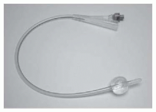
Fig. 17.1 A Foley catheter with the balloon inflated.


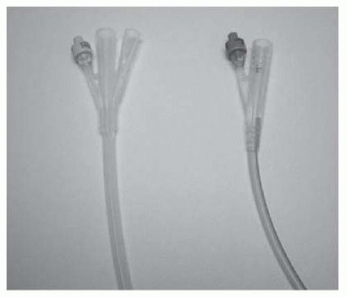
Fig. 17.2 2- and 3-way catheters.


  • Tube drains (e.g. a Robinson’s drain; Figs. 17.3 and 17.4): provide passive drainage (i.e. no applied pressure). Used to drain suture lines at a site of repair or anastomosis of the urinary tract. Avoid placing the drain tip on the suture line as this may prevent healing of the repair. Suture it to adjacent tissues to prevent it from being dislodged.


  • Suction drains (e.g. Hemovac®; Figs. 17.5 and 17.6): provide active drainage (i.e. air in the drainage bottle is evacuated, producing a negative pressure when connected to the drain tube to encourage evacuation of fluid). Used for the prevention of accumulation of blood (a haematoma) in superficial wounds. Avoid in proximity to a suture line in the urinary tract—the suctioning effect may encourage continued flow of urine out of the hole, discouraging healing.

As a general principle, drains should be brought out through a separate stab wound, rather than through the main wound, since the latter may result in bacterial contamination of the main wound with subsequent risk of infection. Secure the drain with a thick suture to prevent it from inadvertently ‘falling out’.


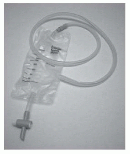
Fig. 17.3 A Robinson’s (passive) drainage system.


Fig. 17.4 Note the eyeholes of the Robinson’s catheter.


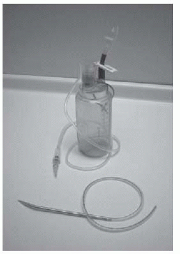
Fig. 17.5 A Redivac suction drain showing the drain tubing attached to the needle used for insertion and the suction bottle.


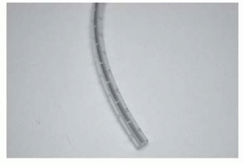
Fig. 17.6 The eyeholes at the tip of the suction drain.


Guidewires

An essential tool for endourological procedures.


Uses

As a track over which catheters or instruments can be passed into the ureter, collecting system of the kidney (retrograde or antegrade), or the bladder.


Types

Many different types of guidewire are available. They are classified according to their size, tip design, rigidity, and surface coating. These specific properties determine their use. All are radio-opaque so X-ray screening can be used to determine their position. They come prepackaged in a coiled sheath to allow ease of handling and storage (Fig. 17.7).


Size

‘Size’ refers to diameter measured in inches (length is usually around 150cm). Most common size are 0.035 inches (2.7 Ch) and 0.038 inches (2.9 Ch). Also available as 0.032 inches (2.5 Ch).


Tip design

Shape of tip—straight or angle (Fig. 17.8); a straight tip is usually adequate for most uses. Occasionally, an angled tip is useful for negotiating an impacted stone or for placing the guidewire in a specific position. Similarly, a J-shaped tip can negotiate an impacted stone—the curved leading edge of this guidewire type can sometimes suddenly flick past the stone (in this situation, a straight guidewire can inadvertently perforate the ureter, thereby creating a false passage).


Surface coating

Most standard guidewires are coated with polytetrafluoroethylene (PTFE) which has a low coefficient of friction, thus allowing easy passage of the guidewire through the ureter and of instruments over them. Some guidewires are coated with a polymer which, when wet, is very slippery (hydrophilic coating). In some cases, the entire length of the guidewire is so coated (e.g. Terumo Glidewire) and in others, just the tip (e.g. Sensor guidewire). The virtually friction-free surface of Glidewires makes them liable to slip out of the ureter and they, therefore, make unreliable safety wires (they can be exchanged for a wire with greater friction via a ureteric catheter). If allowed to become dry, these wires have a high coefficient of friction, which makes them difficult to manipulate.


Tip rigidity

The tip of all guidewires, over at least 3cm, is soft and, therefore, flexible. This reduces—although does not completely remove—the risk of ureteric perforation.


Fig. 17.7 Guidewires come prepackaged in a sheath for ease of handling.


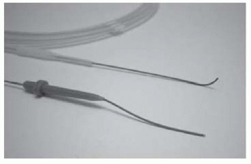
Fig. 17.8 Examples of straight-tip and angled-tip guidewires.

Only gold members can continue reading. Log In or Register to continue

Stay updated, free articles. Join our Telegram channel

Jul 22, 2016 | Posted by in UROLOGY | Comments Off on Urological Surgery and Equipment

Full access? Get Clinical Tree

Get Clinical Tree app for offline access