Chapter 84 URETEROVAGINAL FISTULA
DEFINITION AND INCIDENCE
Ureteral injury has occurred in about 0.5% to 1% of all pelvic surgeries.1 Total abdominal hysterectomy is the most common procedure leading to injury to the ureter and subsequent ureterovaginal fistula, accounting for 75% of all cases in one study.2 Enlarged uteri, pelvic adhesions, and significant intraoperative bleeding also appear to increase the risk of ureteral injury at the time of gynecologic surgery.3 Other operations that may lead to ureterovaginal fistula include anterior colporrhaphy, colorectal surgery, and oophorectomy. Obstetric causes rarely lead to ureterovaginal fistula formation and are primarily attributed to cesarean section (especially after prior cesarean section),3 although traumatic vaginal delivery has been reported as a cause of ureterovaginal fistula.4 Other unusual causes of ureterovaginal fistula include vaginal foreign body5 and residual stone fragments after shockwave lithotripsy.6 Radiation therapy by itself or in conjunction with surgery can place a patient at risk for ureteral injury and subsequent formation of ureterovaginal fistula.7 Persistent urinary leakage resulting from an injury to the ureter during any of these procedures places the patient at risk for developing an ureterovaginal fistula. Later series looking at laparoscopic pelvic surgery found a rate of ureteral injury of less than 1% to 2%, with a much lower incidence of injury associated with diagnostic laparoscopy compared with laparoscopic intervention.8,9
The injury to the ureter usually occurs in the distal third. Types of injury include ischemia, transaction, excision, and ligation. Ischemia is most commonly associated with radical (Wertheim’s) hysterectomy, which requires the ureter to be stripped from its fascial encasement in the broad ligament. More commonly, the ureter is injured by a suture, with resultant extravasation leading to fistula formation. The left ureter is much more at risk, and most prior series reveal ureterovaginal fistula formation on the left three to five times more often than on the right side. This increased risk to the left ureter is related to its course, which places it much closer to the cervix than the right ureter. Another common site of injury is at the level of the pelvic brim, where the ureter crosses the iliac vessels. Injury at this site is thought to occur during the manipulation and division of the infundibulopelvic ligament.3 Patients with pelvic inflammatory disease, prior pelvic surgery, and other conditions that can distort the normal pelvic anatomy may be at greater risk for ureteral injury and subsequent fistula formation.
EVALUATION
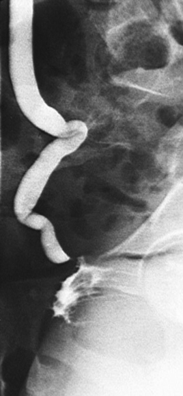
Renal ultrasound may be helpful as a screening tool, but the result may be normal if distal obstruction is absent. The most commonly used modality to evaluate for ureteral injury is intravenous urography (IVU). It can identify n ureteral injury in most cases, and it enables evaluation of renal function and potential obstruction on the affected side (Fig. 84-1). IVU may not identify a ureterovaginal fistula in every patient. Lask and colleagues10 evaluated iatrogenic ureteral injuries in 44 patients, 10 of whom had a ureterovaginal fistula. The fistula was identified in only 3 of 10 patients undergoing IVU.10 These findings highlight the importance of retrograde pyelography, which may be required to identify the fistula in some patients.11 This is especially true if the IVU result is abnormal but does not reveal ureterovaginal fistula and the patient complains of urinary incontinence after recent pelvic surgery.
Any obstruction at the level of the injury should be identified because an untreated distal blockage makes spontaneous healing of the fistula extremely unlikely. If conservative attempts at therapy are to be attempted, such as placement of a ureteral stent as discussed in the next section, the retrograde pyelogram can be done simultaneously. Computed tomography should be considered for patients who are systemically ill to evaluate for possible urinoma or abscess formation, which should be percutaneously drained.
In addition to radiographic evaluation, the double-dye test can be used. This was initially described using intravenous indigo carmine,12 but it has been modified to eliminate the need for intravenous access to perform the test. With the modified test, a patient is given Pyridium orally until the urine turns orange. A tampon is placed vaginally, and the bladder is emptied with a catheter. Through that catheter, the bladder is filled with a mixture of 300 mL of normal saline and 5 mL of methylene blue dye. After 5 minutes, the bladder is emptied, and the tampon is removed and inspected. An orange stain at the top of the tampon indicates a ureterovaginal fistula, a blue stain in the middle of the tampon indicates a vesicovaginal fistula, and a blue stain at the distal tip of the vagina indicates leakage of urine through the urethra.13
ENDOSCOPIC THERAPY
A variety of minimally invasive therapies have been described for the management of ureterovaginal, and they have a wide range of results. The least invasive option is observation. This has been reported with periodic success in the past and was primarily used before the use of fluoroscopy and modern endoscopic equipment made ureteral stent placement commonplace.11,14,
Related posts:
 DEVELOPMENTAL ANATOMY AND UROGENITAL ABNORMALITIES
 USE OF CADAVERIC FASCIA FOR PUBOVAGINAL SLINGS
 ANTERIOR COLPORRHAPHY FOR CYSTOCELE REPAIR
 TRANSVAGINAL CLOSURE OF THE BLADDER NECK IN THE TREATMENT OF URINARY INCONTINENCE
 FEMALE SEXUAL FUNCTION AND DYSFUNCTION
 FUNCTIONAL ANATOMY AND PATHOPHYSIOLOGY OF PELVIC ORGAN PROLAPSE
Stay updated, free articles. Join our Telegram channel

Full access? Get Clinical Tree


