Ulcerative Colitis


Ulcerative Colitis


Victor W. Fazio


Five things are proper to the duty of a Chirurgian; To take away that which is superfluous; To restore to their places such things as are displaced; To separate those things which are joined together; To join those that are separated; and To supply the defects of nature.

—AMBROISE PARÉ: Works, Book I, Chapter 2

The expression nonspecific inflammatory bowel disease (IBD) is used to describe two conditions of unknown etiology: ulcerative colitis (UC) and Crohn’s disease (CD). With respect to the differential diagnosis, the two diseases often have similar characteristics. The symptoms are frequently quite alike, radiologic investigation may pose confusion in differentiation, and even pathologic evaluation may reveal overlapping features, with an indeterminate colitis reported in as many as 15% of patients. Some have even reported that both conditions can coexist in the same individual.773 Because of the often diverse approaches to management and the fact that a vulnerable age group is frequently affected (young persons in their late teens and early 20s), these diseases are among the most challenging confronting the physician today. Ulcerative colitis is discussed in this chapter; Crohn’s disease and indeterminate colitis are presented in Chapter 30.


▶ HISTORICAL PERSPECTIVE

It is difficult to know whether ulcerative colitis was truly recognized as a disease prior to the 19th century. Infectious and noninfectious diarrheas have existed since antiquity, but most of the descriptions are of a clinical syndrome— diarrhea and rectal bleeding, the so-called bloody flux. Samuel Wilks is generally credited with coining the term ulcerative colitis.775 In a letter to the editor of the Medical Times and Gazette published in 1859, he described the postmortem appearance of the intestine. Subsequently, the surgeon general of the Union Army after the Civil War referred to the disease, ulcerative colitis, and included photomicrographs of the condition.216 Other detailed descriptions followed,776 and by the early 20th century, more than 300 case reports of ulcerative colitis had been collected for presentation to the Royal Society of Medicine.216


▶ EPIDEMIOLOGY (INCIDENCE, PREVALENCE) AND ETIOLOGY

As suggested, the nonspecific IBDs, especially Crohn’s disease, have become pervasive worldwide, and the two conditions have thus emerged as one of the most important biomedical problems of our time.352 Unfortunately, our understanding of their pathogenesis still remains obscure.

Evaluation of the epidemiology of the two conditions is made difficult by the plethora of diarrheal states found throughout the world that may be infectious or parasitic in nature and that present with symptoms not unlike those of nonspecific IBD.352 Furthermore, because of international failure to classify the two diseases as distinct from the numerous specific inflammatory bowel problems, our
ability to obtain meaningful data is compromised. Most of the information, therefore, has been accumulated from Western countries, where the diseases are relatively prevalent. In the United States, however, IBD is not a reportable condition.


Available evidence suggests considerable variation in the incidence rates—common in developed countries and unusual in Asia, Africa, and South America. Because a true surveillance of prevalence requires an exhaustive clinical, endoscopic, and radiologic evaluation of all members of a sample population, the true prevalence is based on inference rather than analysis of well-established data.487 There even appears to be a seasonal variation, not only of onset, but also of relapse.624 A statistically significant increase has been observed in the months of August to January.

The incidence of ulcerative colitis and Crohn’s disease in England, United States, and Scandinavia is reported to be at least 4 to 6 cases per 100,000 white adults per year, with prevalence rates of between 40 and 100 cases per 100,000.353 Most studies have demonstrated an increased incidence of Crohn’s disease over the past 25 years.56,67,147,378,472,501,551

The condition is more common in whites, among Jewish people, and among those of Western origin (especially northern Europe and the northern part of eastern Europe).353 Other countries such as South American nations, the former Soviet Union, and Japan have a much lower incidence and prevalence. More recent reports suggest that UC affects approximately 500,000 individuals in the United States, with an incidence of approximately 12 per 100,000 population per year.324,325,437,438,536 Moreover, UC posts a considerable health care burden. The disease accounts for more than a quarter million physician visits annually with 30,000 hospitalizations and is also associated with loss of more than a million workdays per year.694 The direct health care costs are astoundingly high, exceeding $4 billion annually, which includes estimated hospital costs of more than $960 million and drug costs of $680 million.

As with carcinoma of the colon, the prevalence of IBD in many industrialized countries and the development of the conditions among those from low-risk populations who emigrate to higher risk areas suggest an environmental cause.

Investigative efforts to identify the etiologic agent responsible for the nonspecific IBDs have thus far been unsuccessful. IBD that appears nonspecific in nature has been found in hamsters, horses, swine, and the canine population, but an experimental animal model for induction or transmission of the disease still eludes investigators. The three primary areas of investigation that continue to be pursued actively are genetics, immunology, and infection.


Genetics

Several studies have shown the existence of family aggregations with the disease, implying that genetic factors also must play a role.5,119,148,351,354,384,769 For example, there is a high degree of concordance in monozygotic twins.599 Although ulcerative colitis and Crohn’s disease are not classic genetic disorders, the occurrence of IBD in family members born in widely separated areas or living apart for long periods, along with the increased incidence among Jews and the tendency toward familial aggregation of cases with ankylosing spondylitis in Crohn’s disease, suggest a genetically mediated mechanism in the causation of the conditions.148,353,566 Roth and colleagues, in a study of Ashkenazi Jews with IBD, calculated the true lifetime risk for the development of IBD in relatives to be 8.9% for offspring, 8.8% for siblings, and 3.5% for parents.634 Although the possibility of a common environment may contribute to the increased risk of IBD, the shared genetic pool is much more likely to be the primary factor. A familial occurrence has been noted in 17.5% of more than 600 patients with IBD.688 In a report from the Cleveland Clinic, Farmer and colleagues evaluated the family histories of more than 800 patients with an onset of IBD before the age of 21.148 Twenty-nine percent of those with ulcerative
colitis had a positive history, and 35% of those with Crohn’s disease had a positive family history for IBD.

The first IBD susceptibility locus was found on chromosome 16 and identified as IBD1. This is apparently a purely Crohn’s locus, however. Most of the current studies seem to demonstrate a consistent association with various genetic mutations and Crohn’s disease, but not that of ulcerative colitis.

Satsangi and colleagues propose that the ethnic, familial, twin, disease association, and genetic marker studies of IBD are best explained by the concept that the conditions represent multifactorial diseases.660 Environmental and genetic factors contribute to disease susceptibility. They conclude that it is possible for different susceptibility genes to underlie phenotypic differences between Crohn’s disease and ulcerative colitis, leading to diverse manifestations of the two conditions as well as varied degrees of severity. Mendeloff believes that there is a deficiency in the investigation of these diseases because the mortality is low and the genetic determinants are multiple.487 He suggests that in the future it will be necessary to develop a better fundamental means of acquiring and recording data, and that at least for the immediate present a concerted effort should be made to identify those families in which multiple cases of IBD exist and to investigate these people thoroughly. This would include genetic, psychological, and metabolic studies.


Autoimmunity

The idea of circulating antiepithelial antibodies combining with antigens on the intestinal cell surface and damaging the cells seems a reasonable theory to explain the etiology of IBD. This is the concept of autoimmunity. Snook clearly defines the criteria for the classification of a disease as autoimmune.692 According to him, such a condition “… requires the demonstration of autoreactive lymphocytes or autoantibodies, or both, which are specific for the disease concerned, present in all cases, and most important, capable of reproducing the disease on syngeneic transfer.”692 However, despite the demonstration of anticolon antibodies in both blood and tissue of patients with IBD, current evidence seems to militate against the likelihood that these play a primary pathogenetic role in the two conditions.642


Immune complex mediation of IBD has been thought by some to be a responsible factor, but studies have failed to corroborate a significantly increased frequency or concentration of these complexes regardless of disease activity.642 Other immunologic mechanisms that have been investigated include abnormality and variability of circulating lymphocytes, lymphocyte cytotoxicity, defective cell-mediated immunity, immediate hypersensitivity, leukocyte chemotaxic impairment, and immunoregulatory cellular imbalance.353,642 At the level of the immune response, a genetic influence is suggested by the association of ulcerative colitis with HLADR2 and by the occurrence of various autoantibodies in the unaffected relatives of patients with ulcerative colitis.677 Furthermore, studies of monozygotic twins have indicated that the altered mucosal production of immunoglobulin G1 (IgG1) and IgG2 in ulcerative colitis may be genetically determined.677 James and colleagues suggest that the presence of circulating antigen-nonspecific suppressor T cells in patients with Crohn’s disease in its early stages is the result of an immunoregulatory abnormality of antigen-specific helper and suppressor T cells.305 A useful concept of the pathogenesis for IBD may involve an interaction between host responses, immunologic genetic influences, and external agents, but no definitive proof has yet been forthcoming.


Infection

With respect to infectious agents, ulcerative colitis in particular has been attributed to bacterial causes for more than 60 years. In 1928, Bargen reported that the condition was caused by a transmissible diplococcus.30 He prepared a vaccine from this organism, an approach that was believed to be a valuable part of the treatment of the condition. In the article that introduced the technique of skin-grafted ileostomy for the surgical management of ulcerative colitis (see Chapter 31), Dragstedt and colleagues were persuaded that “bacterium necrophorum, together with other factors … plays an etiologic role in the disease.”140 Crohn’s disease specifically was often confused with tuberculosis. This observation led Crohn and coworkers to suggest that the disease might be caused by a mycobacterial agent.114 Subsequent studies have failed to demonstrate conclusively an association with an infective agent, and, in fact, the incidence of IBD correlates inversely
with that of the infectious dysenteries.353 An exception is that of cytomegalovirus infection, which has been shown to complicate ulcerative colitis in immunocompromised patients, such as individuals with AIDS.761

The concept of a microbial infection as the offending agent has been resurrected with the recognition of newer bacterial causes of enteritis and colitis (especially Campylobacter jejuni and Clostridium difficile).63,380,534,739 Although several studies suggest the possibility of these two organisms contributing to relapse of IBD, Gurian and colleagues, in examination of stool specimens from 32 patients who had exacerbation of IBD, revealed no Clostridium difficile cytotoxin and negative cultures for Campylobacter jejuni.235 Other bacteriologic agents (Shigella, Salmonella, Streptococcus faecalis, Pseudomonas variant, Chlamydia, Mycobacterium, and many others) have been proposed, but their role has not been confirmed.89,203,242,730 More recently, it has been suggested that probiotics play an important role in preventing overgrowth of potentially pathogenic bacteria and in maintaining the integrity of the gut mucosal barrier.756 Therefore, there may be a role for such agents in the treatment of IBD (see Medical Management).

There has been considerable interest in a possible viral etiology of ulcerative colitis and Crohn’s disease. Transmission of granulomatous lesions has been successfully carried out in experimental animals.98,135,722 Tissue culture and electron microscopic investigation have also suggested that a viral agent is present in tissue from patients with IBD.14,204,774 However, considerable controversy continues concerning the specificity of these findings. Whether the evidence is sufficiently compelling to permit accurate identification of viral particles on electron microscopic examination of affected tissue remains unresolved.642,787


Diet

Dietary factors, especially cow’s milk, have been implicated as possible causative agents for the development of IBD. Early studies seemed to demonstrate an elevated milk protein antibody level in patients with ulcerative colitis in comparison with a control population. Subsequent studies in which milk was excluded from the diet failed to demonstrate an improvement in the clinical response, and later studies with milk and milk products failed to demonstrate any correlation. Other factors that have been under investigation include chemical food additives, mercury ingestion, inadequate fiber, excess intake of refined sugar, and even the increased consumption of corn flakes.471,642 Generally, articles dealing with diet and IBD are conflicting, confusing, and frequently unreliable. One can safely state that there is no clear consensus to suggest that dietary factors play a role in the etiology of either ulcerative colitis or Crohn’s disease.


Oxidative Metabolism

Recent evidence suggests that abnormal oxidative metabolism may be of significance in the activity of IBD. Increased attention has been placed on the role of free radicals in both normal metabolism and defense against disease.687 A free radical is defined as any species capable of independent existence that contains one or more unpaired electrons, an unpaired electron being defined as one that is alone in an orbital.687 It appears that reactive oxygen metabolites are produced in excess in active IBD. The effects of specific anti-inflammatory antioxidants, such as aminosalicylates, are compatible with the proposition that free radicals play a major role in the pathogenesis of IBD.687


Stool

Studies of the role of the fecal stream in causing an exacerbation of symptoms in Crohn’s colitis have yielded confusing results. For example, Harper and colleagues evaluated the effects of introducing small bowel effluent and a sterile ultrafiltrate of the effluent into the defunctionalized colon after a loop ileostomy had been created.254 There was little response to the ultrafiltrate challenge, but there was a definite clinical exacerbation following introduction of the effluent. Conversely, Korelitz and colleagues reported that diverting the fecal stream had an adverse effect on the clinical course in four patients.368 Following reestablishment of intestinal continuity, the bowel returned to normal.


Smoking

Two distinct patterns of cigarette smoking seem to be relevant in patients with IBD; those with ulcerative colitis are much less likely to smoke than those with Crohn’s disease. Additionally, cigarette smoking has been found to have a negative correlation with ulcerative colitis.77,122,255,310,737,759 In some cases, complete remission of symptoms was obtained through the use of nicotine-laced chewing gum. In other cases, exacerbation of the disease was noted when patients ceased smoking. Conversely, Crohn’s disease is more common in smokers than in those who have never smoked.330,737,759 The increased risk seems to be more apparent in women and may also be associated with a greater likelihood of recurrence.714


Oral Contraceptives

Both ulcerative colitis and Crohn’s disease have been found to be more common among women using oral contraceptives than in those who do not.759 A possible vascular (ischemic) basis for this observation has been suggested. There is no information to date on the effect of stopping the contraceptive pill on the activity of IBD.


Psychological Factors

The psychological aspects and the possible psychosomatic factors contributing to the onset and exacerbation of IBD, and of ulcerative colitis in particular, have been a subject of considerable debate since publication of the original article by Murray in 1930.522 Karush and colleagues, in a book on Psychotherapy in Chronic Ulcerative Colitis, state that susceptibility to the problem develops in colitic patients through “disruption and distortion of the relationship to the parents and to other significant persons as early as the second year of life.”328 They further contend that this results in exaggerated emotional manifestations, egocentricity, dependency conflicts, and poor mechanisms for coping with the stresses of life.328 Frequent anxiety or depression, in the opinion of the authors, is also characteristic, and this predisposition, they believe, explains the relatively high incidence of schizophrenia in patients with ulcerative colitis. In their study of precipitating emotional factors, the authors reported that a well-defined event of powerful emotional impact preceded the onset of the disease by a few days or weeks. Subsequent recurrences were also heralded by such events.


Although many articles have been published to support this description of the susceptible personality, opponents of the psychosomatic theory point out that the concept is based on either anecdotal or uncontrolled studies.642 Several publications have compared patients with ulcerative colitis with normal subjects and those with other illnesses and have found no evidence of an increased frequency of psychiatric illness.49,141,267,488 Furthermore, those with ulcerative colitis who had a psychiatric illness did not appear to have more serious gastrointestinal involvement, nor did severity of the ulcerative colitis predict a more frequent or more severe psychiatric disorder.267 Bercovitz noted that there are no long-term, prospective psychological studies available of any large group of people.51 North and colleagues reviewed all known English-language articles at the time (138 studies) on the association between psychiatric factors and ulcerative colitis and found that most contained serious flaws in research design (e.g., absence of control subjects and lack of diagnostic criteria).547 In the seven publications that truly represented meaningful, systematic, investigative efforts, no association was identified. Cunnien concluded that patients with ulcerative colitis have no universal unique personality characteristics and no documented increase in psychiatric illness when compared with medically ill and population controls.117 He further observed that no demonstrable emotional precipitating factors have been uniformly identified, no symbolic emotional conflicts have been documented in controlled studies, and psychotherapy has not been effective in altering the disease course.


Comment

Serious illness during an extremely vulnerable period of emotional development, the need for hospitalization and psychotropic medications (e.g., corticosteroids), or the fear of surgery (especially the “mutilation” of an ileostomy) may induce considerable stress. In fact, it would be the remarkable patient indeed who was not emotionally troubled by the consequences of such illness. The importance of providing emotional support to the patient cannot be overestimated. Psychotherapy has been demonstrated to be of value, but an internist or a surgeon who understands the role of emotional conflicts and anxieties either as a cause or a result of the patient’s illness can provide such counsel.


Appendectomy

As has been suggested, the current concept of the etiology of IBD in most patients is that the condition may be triggered by genetic predisposition to a variety of environmental factors. The association of one of these factors, appendectomy, has been the subject of numerous investigations. Koutroubakis and colleagues undertook a meta-analysis of 17 case- controlled studies and found that appendectomy was inversely associated with the development of ulcerative colitis (P < .0001).372 The role of the appendix in the development of mucosal immunity is currently the subject of intensive study.


Age, Sex, and Race

Ulcerative colitis and Crohn’s disease occur at any age but are most commonly seen in persons younger than the age of 30 years. The incidence is highest among teenagers, but a small secondary peak in the incidence of the two conditions occurs late in the sixth decade.353 In most series, both sexes are equally affected. Farmer and colleagues noted, in a study covering 20 years ending in 1974, that 838 patients were 20 years old or younger at the time of diagnosis at the Cleveland Clinic.148 Thirteen percent with ulcerative colitis and 5% with Crohn’s disease were younger than 11 years of age. Approximately one-third of the patients in each group were between the ages of 11 and 15. There were 316 patients with ulcerative colitis, with a male-to-female ratio of almost 1:1, and 522 patients with Crohn’s disease, 57% of whom were male. In Goligher’s experience, approximately one-half of the patients were between 20 and 39 years of age when the disease was diagnosed, with a 4:3 predominance of female over male patients.212 With Crohn’s disease, Goligher reported that in his own personal series, female patients predominated in a 3:2 ratio.214 There seemed to be a tendency for the disease to develop later than ulcerative colitis does, 70% of patients being between the ages of 20 and 49 years.

In the Lahey Clinic experience of 151 patients who underwent proctocolectomy, the mean age at surgery for both ulcerative colitis and Crohn’s disease was 36 years.108 Fifty-six percent of patients with ulcerative colitis were men, whereas 57% who had Crohn’s colitis were women.

Goldman and colleagues suggest that Crohn’s disease in black patients may be more common than is generally appreciated.207 This group represented 11% of the patients in their experience during a 10-year period. The authors noted that extraintestinal manifestations developed in all patients. Furthermore, the disease generally appeared to be associated with more severe complications than had been observed in whites.


▶ DIFFERENTIAL DIAGNOSIS: ULCERATIVE COLITIS VERSUS CROHN’S DISEASE

Ulcerative colitis and Crohn’s disease can usually be distinguished based on the clinical course, symptomatology, manifestations, and endoscopic findings. Ulcerative colitis is a disease characterized by exacerbations and remissions. In contrast, the individual with Crohn’s disease has less clear-cut periods of flare-up and remission; the disease often tends to run a more smoldering course. Frequently, the patient is really not well and yet is not sufficiently ill to warrant hospitalization. Table 29-1 summarizes a number of the characteristic features that may help to differentiate between the two conditions.

Rectal bleeding is virtually a sine qua non for the diagnosis of ulcerative colitis. A physician might seriously question the accuracy of the diagnosis in the absence of this symptom. Bleeding is much less frequently seen in Crohn’s colitis; in fact, 25% of patients with Crohn’s disease never manifest bleeding. This should not be surprising because ulcerative colitis is an inflammatory disease of the mucosa, whereas with Crohn’s colitis, ulceration may be minimal. However, in rare instances, massive lower gastrointestinal bleeding may be associated with Crohn’s disease.477

Ulcerative colitis is confined to the colon and rectum; Crohn’s disease can occur anywhere in the digestive tract, from the mouth to the anus. Anorectal disease in particular (fissures, abscesses, and fistulas) is more commonly noted in patients with Crohn’s disease than in those with ulcerative colitis (see Chapters 12, 13, and 14). The diagnosis is often suspected on examination of the perianal skin (see Figure 14-51).


TABLE 29-1 Features of Nonspecific Inflammatory Bowel Disease


ULCERATIVE COLITIS


CROHN’S DISEASE


Course


Exacerbations and remissions


Smoldering


Bleeding


Virtually always


Uncommon


Abdominal pain


Uncommon


Common


Perianal disease


Rare


Up to 40%


Fistulas


Never


Occasional


Abdominal mass


Never


Occasional


Carcinoma


Increased association


Increased, but less than in ulcerative colitis


Extraintestinal manifestations


Not unusual


Not unusual


Radiologic and Endoscopic


Distribution


In continuity with rectum


Skip areas often observed


Uniform distribution


Often eccentric


Rectal involvement always


Often rectal sparing


Small bowel


Spared (backwash only)


Often involved


Stricture


Rare, virtually always malignant


Frequent, virtually always benign


Mucosa


Contact bleeding, granularity, superficial ulcers, pseudopolyps


Longitudinal ulcers, fissuring, cobblestone appearance


Microscopic


Extent


Mucosa and submucosa


Transmural


Granulomas


Never


Common


Dysplasia


Yes


Yes


Lymph nodes


Reactive


With granulomas


Crypt abscesses


Present


Present


Mucus production


Decreased


Increased


Proctosigmoidoscopic examination may be of value in differentiating between the two conditions (see later discussion). The rectum is always diseased during attacks of ulcerative colitis. Characteristic changes include contact bleeding, granularity, and ulceration. In Crohn’s colitis, 40% of the patients have sparing of the rectum, irrespective of anal or perianal involvement. However, when the rectum is involved by Crohn’s disease, differentiation between the two may be quite difficult.

Extracolonic manifestations had been presumed to be found only with ulcerative colitis, but it is now recognized that these can be observed in both conditions. They are discussed in Chapter 30.

It may not be possible to differentiate between the two diseases either by radiologic or clinical means. In approximately 10% to 15% of cases, distinction cannot be made even pathologically; these patients are thus placed into the so-called indeterminate category (see Chapter 30). The appropriateness of using the ileal pouch-anal operation as an alternative in this group of patients is also discussed in Chapter 30.


Physical Examination

Physical examination is usually unrewarding in patients with ulcerative colitis who do not have fulminant disease. Abdominal tenderness is usually absent, and there is no abdominal distension. No masses are palpable. However, in the acutely ill person, abdominal distension may be associated with toxic megacolon. Diffuse tenderness may be apparent, and if perforation has ensued, all the usual signs and symptoms of an intra-abdominal catastrophe may be noted.


Endoscopic Examination

Proctosigmoidoscopy, flexible sigmoidoscopy, and colonoscopy are important tools for evaluating the bowel and for confirming the presence or absence of IBD. Proctosigmoidoscopic examination is particularly useful in differentiating Crohn’s disease from ulcerative colitis; the rectum is always diseased during attacks of ulcerative colitis. The earliest manifestation of inflammation is the loss of a normal vessel pattern, the result of edema of the bowel wall. Contact bleeding, granularity, and ulceration are more obvious signs of inflammatory disease. The rectum is spared in 40% of patients with Crohn’s colitis, irrespective of anal or perianal involvement. However, when the rectum is involved by this condition, differentiation between the two diseases may be quite difficult.

In individuals with distal disease (i.e., ulcerative proctitis or proctosigmoiditis), complete endoscopic examination by means of the colonoscope is unnecessary at the time of presentation. The extent of disease can usually be determined with the rigid instrument or the flexible sigmoidoscope. The presence of diffuse, confluent, symmetric disease from the
dentate line cephalad to the limit of the inflammatory reaction is consistent with ulcerative proctitis or proctosigmoiditis, depending on the extent of involvement (Figure 29-1). In individuals with treated ulcerative colitis, the finding of rectal sparing or patchiness should not necessarily alter the diagnosis to Crohn’s disease.54 If the patient’s symptoms are appropriate to the endoscopic findings, treatment can be initiated without further contrast study or colonoscopy. Conversely, if the patient’s disease extends beyond the limit of the endoscopic procedure performed, it will be necessary at some point to undertake total colonic evaluation.


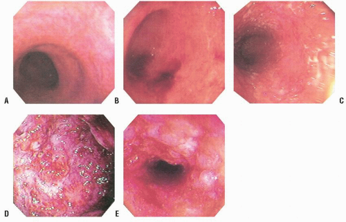
FIGURE 29-1. Colonoscopic changes in ulcerative colitis. A: Loss of the normal vessel pattern is the earliest endoscopic change. B: Contact bleeding. The friability of the mucosa is demonstrated by contact with the instrument. C: Granularity appears in ulcerative colitis of longer duration. D: Florid changes in an ulcerated mucosa. E: A colonic stricture in a patient with ulcerative colitis proved on biopsy to be malignant.

Colonoscopy has replaced barium enema examination for the evaluation of IBD. Generally, endoscopic examination will identify more proximal inflammatory changes than will the radiologic study. Furthermore, histologic examination of random biopsy specimens will often reveal more proximal disease than was suspected by endoscopic examination.146,783 Das and colleagues reported 31 patients with idiopathic proctitis who underwent colonoscopy while they were asymptomatic.118 Multiple biopsy samples were taken from throughout the colon and rectum. Although the obvious disease appeared limited to the distal 20 cm, microscopic abnormalities were seen commonly in more proximal locations. The authors postulated that the clinical course would be more consistent with distal disease when this indeed could be confirmed by biopsy, whereas more proximal involvement usually implied that the disease would be refractory to conventional management (e.g., topical steroids). A corollary to this observation is to perform biopsies distal to obvious inflammatory changes if the rectum appears to be spared because one may discover that the rectum is not truly normal. This may cause the physician to reassess the accuracy of a diagnosis of Crohn’s disease based on what was initially thought to be a lack of rectal involvement.

The place of colonoscopy in the evaluation and followup of IBD has been extensively reviewed by many authors. Teague and Waye recommend colonoscopy for five indications: differential diagnosis, resolution of radiographic abnormalities (e.g., filling defects and strictures), preoperative and postoperative evaluation in Crohn’s disease, examination of stomas, and screening for premalignant and malignant changes.727


Preparation

Often, colonoscopy can be performed without prior bowel cleansing in patients with mild-to-active IBD, with essentially the same comprehensive evaluation achieved as when a full preparation is used.34 In the patient who has a history of IBD, the preparation for colonoscopy includes a modified diet. Clear liquids are suggested for 24 hours. Vigorous cleansing enemas such as those that may be used in the evaluation of a noninflamed colon are contraindicated. For someone with a relatively active colitis, no laxative is suggested. In more severe cases, a clear liquid diet as the sole modality for bowel preparation is probably the safer alternative. If the colitis is minimal or relatively inactive, a reduced dose of a laxative is suggested, although it is probably wiser to use a balanced electrolyte solution (e.g., CoLyte, GoLytely, MoviPrep).


Appearance

Ulcerative colitis and Crohn’s disease are usually recognized endoscopically by excluding IBD due to specific cause, such as amebic colitis, ischemic colitis, pseudomembranous colitis, and so forth. The most common differential diagnostic problems are related to the numerous and varied infectious colitides (see Chapter 33). Other sources of confusion include radiation changes, the so-called solitary ulcer
syndrome, and, of course, the differentiation between the two nonspecific IBD conditions, themselves—ulcerative colitis and Crohn’s disease.727 Biopsy may be helpful because histologic changes suggestive of Crohn’s disease in particular may be apparent. Up to 20% of such patients may exhibit granulomas (see Figure 30-2). In patients with ulcerative colitis, rectal biopsy is extremely important for recognizing dysplasia, especially in those with long-standing disease (see Relationship to Carcinoma).

Waye has described a number of colonoscopic features in the differential diagnosis of IBD.765 He suggests that patients with ulcerative colitis always have rectal involvement from the anal verge cephalad in continuity with whatever proximal involvement is present. Erythema of the colonic wall is one of the early manifestations. More obvious changes include granularity, friability, bleeding, edema with interhaustral septal thickening and blunting, ulceration, mucosal bridging, the presence of pseudopolyps, and the superimposition of carcinoma.

With granulomatous colitis, Waye observed the following major colonoscopic findings: a normal rectum (obviously this is not always the case), asymmetry or eccentricity of involvement, cobblestone appearance, normal vasculature (because friability is not usually encountered except in advanced disease), edema of the bowel wall (as seen in ulcerative colitis), normal mucosa intervening between areas of ulceration, serpiginous or rake ulcers (these may course for several centimeters), pseudopolyps (as in ulcerative colitis), and skip areas (lack of continuity of involvement).765 He adds another observation, the presence of amyloidosis in the biopsy specimen.

Pera and colleagues undertook a prospective study by means of colonoscopy in 357 patients in order to obtain the best predictive information that would enable the authors to differentiate between the two conditions.584 An “endoscopic score” was calculated by means of “likelihood ratios.” Errors were more frequently noted when severe inflammation was encountered. The most useful endoscopic features in the differential diagnosis were discontinuous involvement, anal lesions, and cobblestone appearance of the mucosa for Crohn’s disease, and erosions or microulcers and granularity for ulcerative colitis.584

Myren and colleagues performed routine and random histologic evaluation by means of colonoscopic biopsy in patients with and without IBD.524 In 110 individuals, 278 biopsy specimens were obtained at different levels of the colon. Clinical information, including colonoscopic diagnosis, was available to the pathologist at the initial routine examination. Later, the sections were examined blindly and independently by two pathologists. Agreement was obtained in only two-thirds of the patients. The authors concluded that the limiting factor in making reliable diagnoses was the small size of the tissue specimens available. Also, discrepancies may have been caused by the fact that biopsy specimens were not always representative of the process in the colonic mucosa.

Farmer and colleagues reviewed 100 patients who underwent colonoscopy for distal ulcerative colitis.148 They classified their patients into two groups: those whose disease was confined to the distal 25 cm and those whose mucosal changes extended above that level but not beyond the splenic flexure. Because flexible sigmoidoscopy or colonoscopy is not as accurate in defining disease in the rectum, whether it is inflammatory or neoplastic, there was a 5% disagreement about the presence of inflammation in that area of the bowel. Certainly, the rigid instrument is far more useful in evaluating the rectum than the colonoscope. Biopsy specimens were taken at multiple levels, and the retrospective review was performed during several years. It was determined that patients whose disease was initially limited to the rectum and sigmoid colon had a good prognosis—only 10% progressed to more extensive involvement. However, 25% of patients experienced recurrence. Prognosis was similar whether the disease was confined to the lower 25 cm or was distal to the splenic flexure.


Comment

Additional biopsy specimens for the evaluation of a patient with IBD should be obtained from an area that appears macroscopically, at least, to be relatively uninvolved. The true extent of the inflammation as well as the significance of the presence of granulomas (granulomas may be seen in patients with ulcerative colitis underlying an ulcer) can then be interpreted properly.


Colonoscopy versus Barium Enema

In general, colonoscopy permits identification of segmental involvement and microulceration better than does barium enema. However, radiographic studies yield more information about haustra, especially in the right colon.193 One must remember, however, that the procedure is contraindicated in patients with acute exacerbation of the colitis and certainly in those who have a toxic megacolon. Myren and colleagues performed colonoscopic evaluation in 40 patients and compared the results with that of conventional barium enema.523 Colonoscopic diagnosis and biopsy results were corroborating in 80% of the patients, whereas colonoscopic evaluation and radiologic survey revealed agreement in only 55%. The area in which radiology seemed to have an advantage was the decreased haustration that was more apparent by x-ray techniques. Erosions, mucosal edema, and vascular injection were not detected by barium enema. Others have confirmed the merits of colonoscopy in the evaluation of patients with nonspecific IBD.185,723,766,777


Radiographic Features


Plain Films

In any evaluation of a patient with IBD, the importance of a plain x-ray film of the abdomen (KUB) should not be underestimated. Without submitting patients to the rigors of a contrast study or colonoscopy, particularly when they may be acutely ill, the physician can obtain valuable information about the extent of disease (Figure 29-2).

The importance of a plain film of the abdomen is further increased in those who have toxic dilatation, a condition usually seen in the transverse colon (Figure 29-3). When the radiographic appearance of toxic dilatation is noted, a barium enema examination or colonoscopy is contraindicated due to the risk of iatrogenic perforation, but serial abdominal films are clinically extremely valuable. The effectiveness of medical therapy can be evaluated by determining the increase or decrease in the degree of dilatation. The abdominal examination should not be relied on exclusively because the patient may frequently be confused or even obtunded. However, the degree of dilatation may not necessarily be predictive of
the need for surgery. Furthermore, the high dose of steroids often used in the treatment of toxic megacolon may mask abdominal signs.


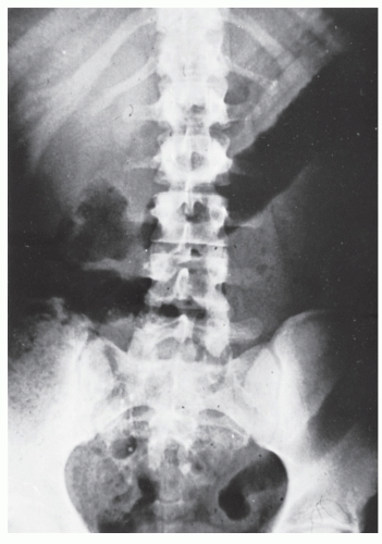
FIGURE 29-2. Ulcerative colitis. This plain abdominal film of an acutely ill patient reveals some thickening of the colonic wall, especially in the region of the sigmoid where it overlies the iliac crest, and loss of haustral markings throughout the left colon and transverse colon to the region of the hepatic flexure. Subsequent barium enema study demonstrated disease extending exactly to the point seen on the plain film. (From Corman ML, Veidenheimer MC, Nugent FW, et al. Diseases of the Anus, Rectum and Colon. Part II: Nonspecific Inflammatory Bowel Disease. New York, NY: Medcom; 1976.)


Barium Enema

The radiologic findings during the acute phase of ulcerative colitis include edema, ulceration, and changes in colonic motility. Edema may be apparent even on the plain film of the abdomen, such as is seen in Figure 29-3. Initially, ulceration may be minimal and difficult to identify. As the disease becomes fulminant, the ulceration becomes more obvious and may take on a “collar button” appearance (Figure 29-4). Edema and inflammation of the mucosa may result in the radiologic appearance that has been called “thumbprinting,” a phenomenon characteristically observed in patients with ischemic colitis (Figure 29-5).

When the disease enters a more chronic phase, other features are characteristic on the barium enema examination. These include fibrosis, which results in shortening of the bowel, depression of the flexures, pseudopolyposis, and stricture formation. The bowel wall is less distensible, and the motility pattern is disturbed. Diffuse, confluent, symmetric disease, beginning with the anorectal junction, are the hallmarks of the radiologic manifestations of chronic ulcerative colitis (Figures 29-6 and 29-7). The presence of polypoid lesions throughout the entire colon may confuse the uninitiated with the radiologic picture seen in familial polyposis (Figure 29-8). Both diseases commonly present with rectal bleeding and diarrhea. In ulcerative colitis, however, foreshortening of the bowel may be evident, particularly at the flexures, and if the outline of the colon is carefully examined, numerous discrete ulcerations can usually be appreciated (Figure 29-9).


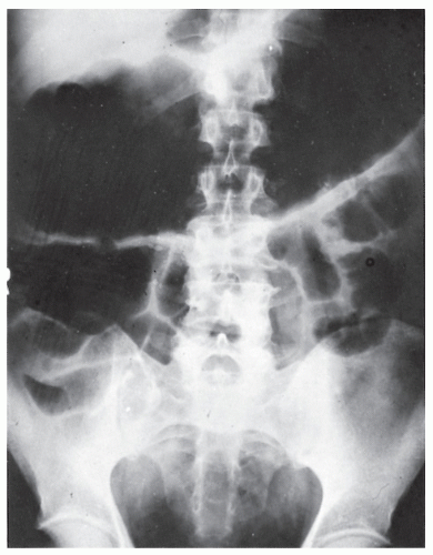
FIGURE 29-3. Ulcerative colitis. This plain abdominal x-ray film reveals marked dilatation of the transverse colon (toxic megacolon). The extent of dilatation may not necessarily be predictive of the need for urgent surgery. However, pneumoperitoneum, sometimes missed by the presence of free gas trapped by the omentum at the inferior border of the transverse colon, is an indication for urgent surgery. (From Corman ML, Veidenheimer MC, Nugent FW, et al. Diseases of the Anus, Rectum and Colon. Part II: Nonspecific Inflammatory Bowel Disease. New York, NY: Medcom; 1976.)

Benign strictures are extremely uncommon in ulcerative colitis. In fact, a stricture in this condition should be considered malignant until proved otherwise (see Relationship to Carcinoma).385 Radiologic examination will usually reveal a smoothly outlined, concentric lumen with tapering margins (Figure 29-10). Areas of spasm are frequently seen in ulcerative colitis and may be difficult to differentiate from stricture. The administration of propantheline (10 mg of Pro-Banthine intravenously) or glucagon (2 mg intramuscularly) may eliminate the stricture caused by such spasm. If the lumen is concentric and the margins are smooth, the lesion may be benign. Conversely, if the lumen is eccentric and the margins are irregular, a carcinoma must be suspected.462


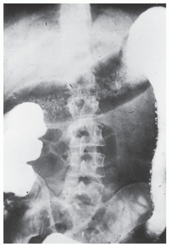
FIGURE 29-4. Acute ulcerative colitis. Note the loss of haustral markings up to and including the mid-ascending colon and numerous discrete ulcerations deep in the submucosa along the descending colon. (From Corman ML, Veidenheimer MC, Nugent FW, et al. Diseases of the Anus, Rectum and Colon. Part II: Nonspecific Inflammatory Bowel Disease. New York, NY: Medcom; 1976.)

Another radiologic finding sometimes observed in patients with ulcerative colitis is “backwash ileitis.” This is a very poor term because it implies that the ulcerative colitis has somehow regurgitated through the ileocecal valve to cause the disease in the distal ileum. Marshak and Lindner were able to demonstrate the presence of this phenomenon in approximately 10% of patients with ulcerative colitis whom they had studied.462 The changes may vary from lack of distensibility, as the head of pressure is increased when the barium is inserted, to ileal dilatation, narrowing, rigidity, and changes that may mimic those of regional enteritis (Figure 29-11). The clinical and therapeutic implications of backwash ileitis are uncertain. There is, however, evidence to suggest higher rates of sclerosing cholangitis in those with this manifestation. Moreover, this observation does not affect the outcome of restorative proctocolectomy in a negative fashion.

Portal venous gas has been reported to be a benign, albeit unusual, consequence of air-contrast barium enema in patients with IBD. Although the implication of such an observation when made in other patients is that antibiotic treatment is required, it may not be necessary if the patient is without bacteremic symptoms.331


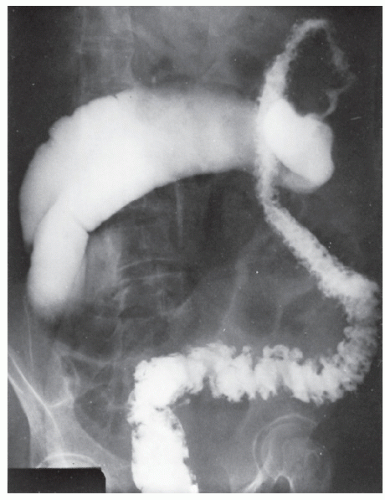
FIGURE 29-5. Edema of the bowel wall in acute ulcerative colitis; flocculation of barium caused by mucus (fuzzy appearance) and thumbprinting in the region of the splenic flexure.


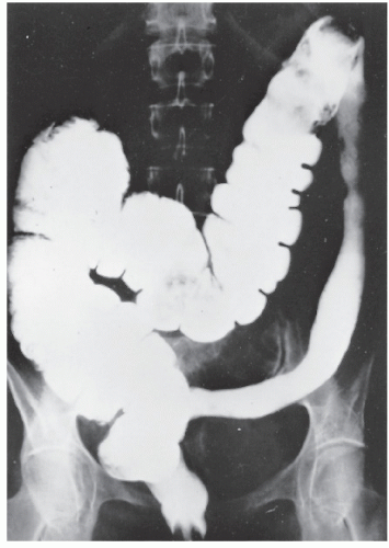
FIGURE 29-6. Chronic ulcerative colitis. Typical changes of left-sided disease include loss of haustral pattern, shortening of the sigmoid, and narrowing of the entire descending and sigmoid colon. (From Corman ML, Veidenheimer MC, Nugent FW, et al. Diseases of the Anus, Rectum and Colon. Part II: Nonspecific Inflammatory Bowel Disease. New York, NY: Medcom; 1976.)


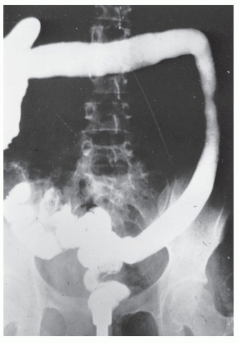
FIGURE 29-7. Chronic ulcerative colitis: a classic example of diffuse, symmetric, confluent disease. Characteristically, the left side is more involved than the right. Note that there is more foreshortening of the splenic flexure than of the hepatic flexure, and there is a suggestion of haustra on the right but not on the left. (From Corman ML, Veidenheimer MC, Nugent FW, et al. Diseases of the Anus, Rectum and Colon. Part II: Nonspecific Inflammatory Bowel Disease. New York, NY: Medcom; 1976.)


Computed Tomography

The advent of computed tomography (CT) has permitted direct visualization of the entire thickness of the bowel wall and mesentery, and determination of the presence or absence of fluid, fistula, or abscess.440 With the exception of its unique application to abscess drainage, the role of this study in the diagnosis and management of patients with IBD is controversial. One thing is certain, however; CT is of no value in assessing the extent of mucosal disease. Its advantages over contrast enema are primarily in delineating the presence and severity of pericolonic inflammation and in evaluating other organ disease. Although the place of CT in IBD is still a matter of conjecture, it is unlikely to prove to be of benefit in patients with ulcerative colitis. Because Crohn’s disease is a transmural process, one may anticipate a greater application with this condition.


Ultrasonography

Hata and colleagues performed ultrasonographic examinations in individuals with ulcerative colitis and Crohn’s disease and in 50 patients with no bowel disease.259 Crohn’s disease and ulcerative colitis could be detected by ultrasonography with a sensitivity of 86% and 89%, respectively. The primary benefit appeared to be in the demonstration of thickening of the bowel wall. However, because the study is less invasive than other alternatives and can be done without preparation, it may be used
to reduce the frequency of repeated colonoscopic or barium enema studies in patients already known to harbor IBD.259


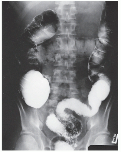
FIGURE 29-8. Ulcerative colitis. This barium enema study reveals extensive pseudopolyposis, especially in the region of the sigmoid colon. Close examination also reveals ulceration, so there should be no confusion with familial polyposis.


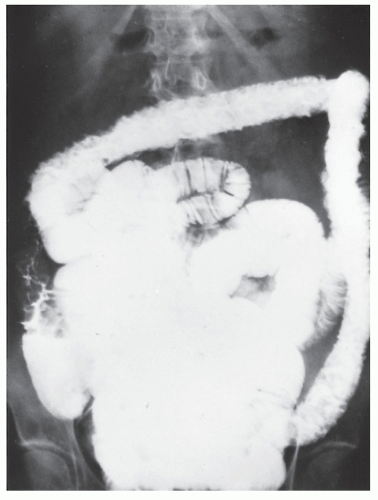
FIGURE 29-9. Ulcerative colitis. Extensive pseudopolyposis with foreshortening of the bowel. Note that the colon is outlined by ulcerations. (From Corman ML, Veidenheimer MC, Nugent FW, et al. Diseases of the Anus, Rectum and Colon. Part II: Nonspecific Inflammatory Bowel Disease. New York, NY: Medcom; 1976.)


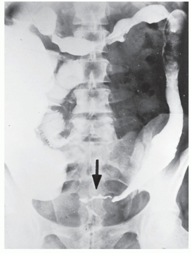
FIGURE 29-10. Ulcerative colitis with stricture. Inflammatory changes are noted to the hepatic flexure, with stricture in the distal sigmoid (arrow). The patient subsequently underwent resection, and the lesion was found to be benign. (From Corman ML, Veidenheimer MC, Nugent FW, et al. Diseases of the Anus, Rectum and Colon. Part II: Nonspecific Inflammatory Bowel Disease. New York, NY: Medcom; 1976.)


Pathology


Macroscopic Appearance

Ulcerative colitis is a disease confined to the mucosa and submucosa of the bowel. The only exception to this occurs when transmural involvement produces so-called toxic megacolon. The bowel wall is not thickened, no granulomas are present (except a foreign body giant cell reaction may occasionally be seen in an area of acute inflammation), and there are no skip areas. The rectum is always involved, and the disease extends proximally for varying distances, but
always with continuity of involvement to the proximal extent of the disease process (Figure 29-12). Characteristically, ulcerative colitis tends to involve the bowel more severely in a distal location than proximally. Despite extensive inflammatory reaction, the bowel wall retains its normal thickness (Figure 29-12). The results of the confluence of numerous ulcers are the longitudinal furrows of denuded mucosa that alternate with islands of heaped-up mucosa, the so-called pseudopolyps (Figures 29-13,29-14 and 29-15). Pseudopolyps are inflammatory polyps, not neoplastic lesions. These are seen during a quiescent phase of ulcerative colitis and are a later manifestation of this condition. They may be confused with familial polyposis (see Chapter 22), but the absence of normal mucosa between these polyps suggests the correct diagnosis.


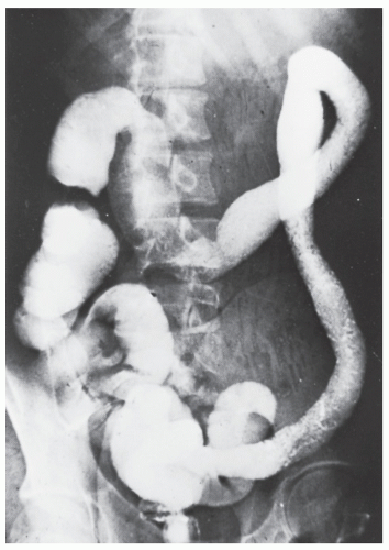
FIGURE 29-12. Ulcerative colitis. A resected specimen (rectum removed separately) shows continuity of involvement from the sigmoid to the mid-ascending colon. Loss of mucosa, deep ulceration, and extensive granularity are evident. (From Corman ML, Veidenheimer MC, Nugent FW, et al. Diseases of the Anus, Rectum and Colon. Part II: Nonspecific Inflammatory Bowel Disease. New York, NY: Medcom; 1976.)


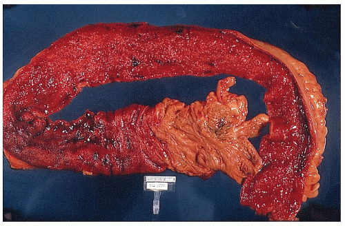
FIGURE 29-11. Extensive ulcerative colitis with loss of haustral markings and destruction of the normal mucosa throughout. Dilatation of the terminal ileum may be evidence of “backwash ileitis.” However, if signs of ileal inflammation extend beyond 10 cm, the diagnosis of Crohn’s disease is more likely. (From Corman ML, Veidenheimer MC, Nugent FW, et al. Diseases of the Anus, Rectum and Colon. Part II: Nonspecific Inflammatory Bowel Disease. New York, NY: Medcom; 1976.)


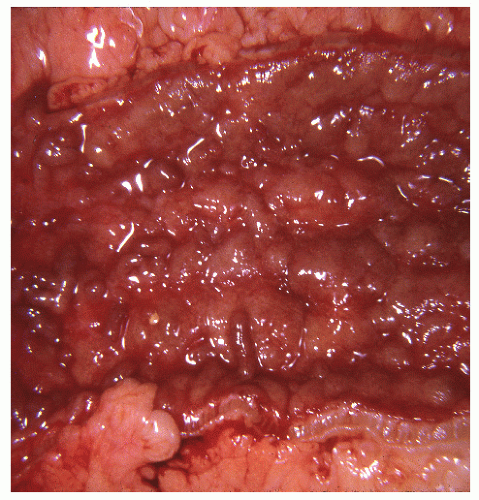
FIGURE 29-13. Ulcerative colitis. Longitudinal furrows of denuded mucosa alternate with islands of heaped-up mucosa, demonstrating how loss of mucosal integrity contributes to fluid and electrolyte depletion. (From Corman ML, Veidenheimer MC, Nugent FW, et al. Diseases of the Anus, Rectum and Colon. Part II: Nonspecific Inflammatory Bowel Disease. New York, NY: Medcom; 1976.)


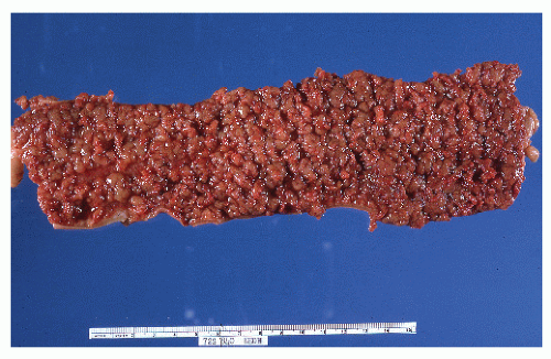
FIGURE 29-14. Extensive pseudopolyps in active ulcerative colitis. Note the relative uniformity of the polyps in comparison with the varied sizes seen in familial polyposis (see Chapter 22). (From Corman ML, Veidenheimer MC, Nugent FW, et al. Diseases of the Anus, Rectum and Colon. Part II: Nonspecific Inflammatory Bowel Disease. New York, NY: Medcom; 1976.)


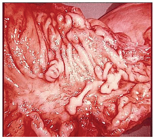
FIGURE 29-15. Ulcerative colitis. Islands of heaped-up mucosa and inflammatory polyps (pseudopolyps). (From Corman ML, Veidenheimer MC, Nugent FW, et al. Diseases of the Anus, Rectum and Colon. Part II: Nonspecific Inflammatory Bowel Disease. New York, NY: Medcom; 1976.)

The entire colon, including the cecum and appendix, may be involved (Figure 29-16). Characteristically, however, the disease does not affect the ileum. In fact, if the small bowel is involved for more than a few centimeters, the diagnosis is not ulcerative colitis. One exception to this is the so-called backwash ileitis seen occasionally when the entire colon is affected. This reversible condition, which may be demonstrated radiographically as edematous, thickened mucosal folds, is a nonspecific inflammatory reaction resulting from proximity of the ileum to the diseased colon.

The entire mucosa may be denuded in patients with longstanding, chronic ulcerative colitis (Figure 29-17). Under
these circumstances, the physician may be lulled into a false sense of security because the patient’s symptoms are often minimal. It is unlikely that someone will experience discharge of mucus, diarrhea, or bleeding if no inflamed mucosa is present. It is this individual who is particularly susceptible to the development of carcinoma (see Relationship to Carcinoma).


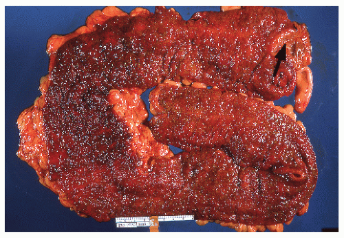
FIGURE 29-16. Ulcerative colitis. The entire colon is involved by inflammatory change, with sparing of the ileum (arrow). (From Corman ML, Veidenheimer MC, Nugent FW, et al. Diseases of the Anus, Rectum and Colon. Part II: Nonspecific Inflammatory Bowel Disease. New York, NY: Medcom; 1976.)


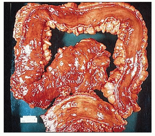
FIGURE 29-17. Ulcerative colitis. Complete desquamation of colonic mucosa. Note the normal bowel wall thickness. (From Corman ML, Veidenheimer MC, Nugent FW, et al. Diseases of the Anus, Rectum and Colon. Part II: Nonspecific Inflammatory Bowel Disease. New York, NY: Medcom; 1976.)

Toxic megacolon is a condition in which an acute inflammatory reaction extends throughout the entire thickness of the bowel wall to the serosa. Gangrene and perforation can result (Figure 29-18). This is the only manifestation of ulcerative colitis that is not limited to the mucosa and submucosa. The term is a poor one because it is not the colon that is toxic; it is obviously the patient.


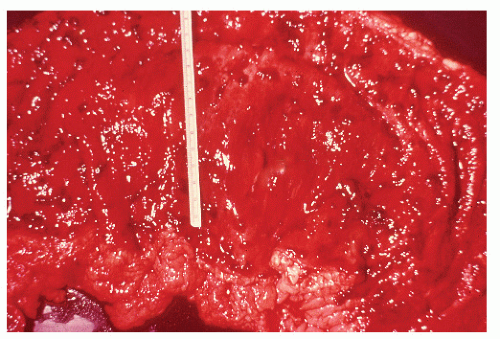
FIGURE 29-18. Ulcerative colitis. Portion of a resected transverse colon showing increased circumference of the bowel. There is practically no mucosa remaining, and circular muscle is exposed in some areas. (From Corman ML, Veidenheimer MC, Nugent FW, et al. Diseases of the Anus, Rectum and Colon. Part II: Nonspecific Inflammatory Bowel Disease. New York, NY: Medcom; 1976.)


Histologic Appearance

Ulcerative colitis is characterized histologically by an intense inflammation of the mucosa and submucosa, in addition to the presence of multiple crypt abscesses. Too much emphasis, however, should not be placed on the significance of crypt abscesses. Acute, self-limited colitis (in which cultures are negative) as well as infectious colitides (see Chapter 33) often have overlapping histopathologic features and must be distinguished from ulcerative colitis.268,282 Marked vascular engorgement accounts for the propensity to rectal bleeding (Figure 29-19). There is an obvious decrease in production of mucus by the crypt epithelial cells (Figure 29-20). The decrease may be explained by injury to these cells.352 Conversely, increased secretion of mucus is seen in patients with Crohn’s disease.

If the bowel is cut longitudinally, it becomes apparent that the deeper parts of the colonic wall are spared. Confinement of the disease to the mucosa and submucosa is the most characteristic finding in ulcerative colitis (Figure 29-21). Abscesses may enlarge to undermine the mucosa, which may then be shed into the bowel lumen, leaving an ulcer behind. When multiple ulcers form, the remaining nonulcerated mucosa extends above the muscularis as polypoid projections, resulting in the well-known pseudopolyps of ulcerative colitis (Figures 29-22 and 29-23). If ulceration continues, the entire mucosa may become denuded, and broad areas of the submucosa may be exposed to the fecal stream.

In toxic megacolon, there is full-thickness involvement of the bowel, necrosis, and friability, the histologic manifestation of which is shown in Figure 29-24.

Lymphoid hyperplasia involving the mucosa and submucosa occurs in up to 25% of patients with ulcerative colitis. This may be present beneath an area of relative inactivity (Figure 29-25).

Seldenrijk and colleagues prospectively performed blind evaluations of multiple colonic mucosal biopsy specimens in individuals with ulcerative colitis and Crohn’s disease to identify reproducible histologic features that could be used to distinguish between the two conditions.672 Three features— an excess of histiocytes in combination with a villous or irregular aspect of the mucosal surface and granulomas—had a high predictive value.


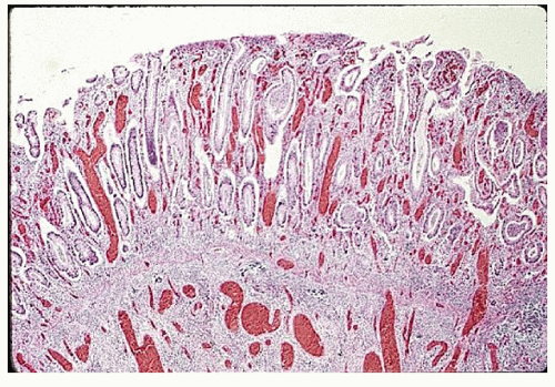
FIGURE 29-19. Ulcerative colitis. Intense inflammation of the mucosa with multiple crypt abscesses. (Original magnification × 80; from Corman ML, Veidenheimer MC, Nugent FW, et al. Diseases of the Anus, Rectum and Colon. Part II: Nonspecific Inflammatory Bowel Disease. New York, NY: Medcom; 1976.)


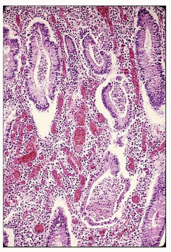
FIGURE 29-20. Ulcerative colitis. Crypt abscesses with degeneration of crypt epithelium and communication between the crypt lumina and lamina propria. Note vascular engorgement and decrease in mucus production by crypt epithelial cells. (Original magnification × 280; from Corman ML, Veidenheimer MC, Nugent FW, et al. Diseases of the Anus, Rectum and Colon. Part II: Nonspecific Inflammatory Bowel Disease. New York, NY: Medcom; 1976.)


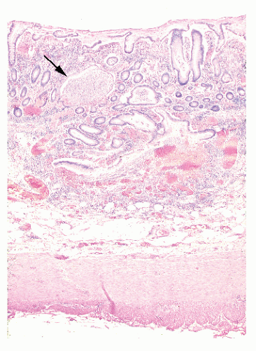
FIGURE 29-21. Ulcerative colitis. Marked inflammation of the mucosa and submucosa. Note the large crypt abscess (arrow) that has penetrated into the submucosa and has been lined partially by epithelial cells growing down into the abscess cavity. (Original magnification × 80; from Corman ML, Veidenheimer MC, Nugent FW, et al. Diseases of the Anus, Rectum and Colon. Part II: Nonspecific Inflammatory Bowel Disease. New York, NY: Medcom; 1976.)


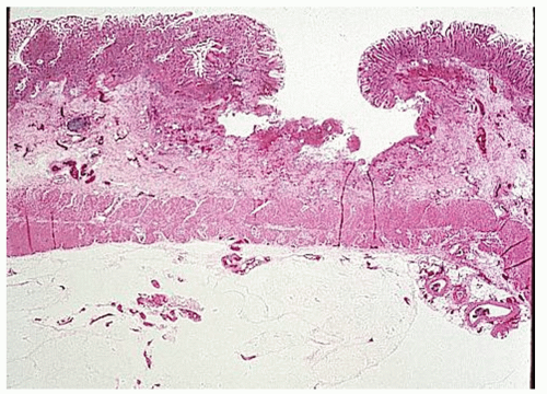
FIGURE 29-22. Ulcerative colitis. An enlarged abscess undermines the mucosa, leaving an ulcer. (Original magnification × 80; from Corman ML, Veidenheimer MC, Nugent FW, et al. Diseases of the Anus, Rectum and Colon. Part II: Nonspecific Inflammatory Bowel Disease. New York, NY: Medcom; 1976.)

Watanabe and coworkers studied rectal biopsy specimens from patients with ulcerative colitis undergoing colonoscopic examinations for the presence of substance P-containing nerve fibers.763 It is well known that the bowel is rich in peptidergic innervation, which contributes to the mucosal immune responses. Because substance P has stimulatory effects on various immunocytes in inflammatory diseases, its increased presence paralleling increased disease activity suggests that alterations play an important role in the pathogenesis of ulcerative colitis.763


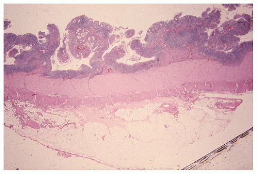
FIGURE 29-23. Ulcerative colitis. Confluence of ulcers results in pseudopolyps. (Original magnification × 80; from Corman ML, Veidenheimer MC, Nugent FW, et al. Diseases of the Anus, Rectum and Colon. Part II: Nonspecific Inflammatory Bowel Disease. New York, NY: Medcom; 1976.)


Antineutrophil Cytoplasmic Antibody Determination

Various studies have shown that antineutrophil cytoplasmic antibodies (ANCAs) with a perinuclear staining pattern
(pANCA) are present in up to 86% of patients with ulcerative colitis.655 Theoretically, this autoimmunity may represent a possible pathogenetic mechanism for the development of ulcerative colitis. A set of marker antibodies is available for the screening and differential diagnosis of ulcerative colitis and Crohn’s disease (Prometheus Laboratories, Inc., San Diego, CA). Proven applications of this technology include the following:


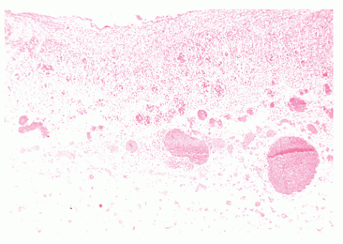
FIGURE 29-24. Ulcerative colitis. Toxic megacolon. Note the loss of epithelium, transmural necrosis, and hemorrhage. (Original magnification × 80; from Corman ML, Veidenheimer MC, Nugent FW, et al. Diseases of the Anus, Rectum and Colon. Part II: Nonspecific Inflammatory Bowel Disease. New York, NY: Medcom; 1976.)


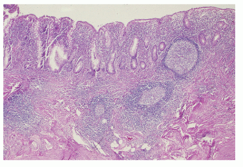
FIGURE 29-25. Lymphoid hyperplasia. Although mucosal disease is relatively inactive, lymphoid hyperplasia is pronounced. (Original magnification × 80; from Corman ML, Veidenheimer MC, Nugent FW, et al. Diseases of the Anus, Rectum and Colon. Part II: Nonspecific Inflammatory Bowel Disease. New York, NY: Medcom; 1976.)


  • as an adjunct to clinical and tissue pathology in the differential diagnosis of IBD


  • confirmation of the correct diagnosis before surgery


  • identification of those patients with left-sided ulcerative colitis that may be resistant to treatment


  • identification of those patients prone to the development of pouchitis following ileal pouch-anal anastomosis

Although the data provided by the Los Angeles group is persuasive, the reality is that pANCA testing is not commonly used in our practice, except where inflammation is additive to other proven features. Such knowledge might suggest an alternative operative approach.


Breath Pentane Analysis

As previously mentioned, neutrophils, macrophages, and other cells are capable of producing free oxygen radicals that can stimulate lipid peroxidation, especially during periods of active inflammation.366 To assess the degree of inflammation in IBD, Kokoszka and colleagues quantitatively determined breath pentane and alkane generated by peroxidation of cellular fatty acids.366 Individuals underwent indium-labeled granulocyte nuclear imaging to assess the presence and location of inflammation. The production of pentane, the product of the peroxidation of polyunsaturated fatty acids, can be quantified by measuring the content of exhaled breath. The investigators concluded that pentane analysis may be correlated with IBD activity.366


Leukocyte Scan

Abdominal scintigraphy by means of autologous- labeled leukocytes has been used to assess activity in IBD. Indium 111 (111In) and technetium 99m (99mTc) have been the most helpful in this regard. Stählberg and colleagues undertook an evaluation using scintigraphy with 99mTclabeled leukocytes to assess disease extent and activity in acute colitis.573 With colonoscopy as the reference method, the maximum extent of colitis was correctly assessed by the scan in two-thirds of patients, but rectal involvement was not perceived in 19%. The intensity of inflammatory activity correlated significantly with the colonoscopic assessment. The authors concluded that the noninvasive nature of this particular approach makes it a reasonable alternative to other investigations of the extent and activity of IBD.


▶ SIGNS, SYMPTOMS, AND PRESENTATIONS

Patients with ulcerative colitis and Crohn’s disease may present with very minimal symptoms and moderate complaints, or they may have fulminant manifestations. There is a considerable overlap in the symptomatology of the two conditions, but there are some differences in the presentation between the two. Rectal bleeding is always seen in patients with ulcerative colitis at some time during the course of the illness. It can be safely said that if the patient does not bleed, the diagnosis is not ulcerative colitis. Individuals with Crohn’s disease also may bleed, but this is not as frequent a manifestation and may not be as severe. Abdominal pain may be mild or absent in patients with ulcerative colitis, but it is rarely severe except possibly when toxic megacolon supervenes. However, patients with Crohn’s disease frequently have abdominal pain.229 An abdominal mass is occasionally found on physical examination in a patient with Crohn’s disease, but it is never seen in a patient with ulcerative colitis, except rarely if carcinoma supervenes.

The presence of diarrhea and the passage of mucus are frequently observed in both conditions and do not serve as distinguishing characteristics. Diarrhea may be manifested as two or three loose stools a day or may be as severe as 20 or more bowel movements within a 24-hour period. Often, patients with ulcerative colitis are more troubled by the frequency of the bowel movements than are those with Crohn’s disease. This is, perhaps, because distal disease tends to be associated with more urgency and, in some cases, tenesmus. Patients with Crohn’s disease may have rectal sparing and are less likely to experience urgency.

Anal disease is much more commonly seen in Crohn’s colitis than in ulcerative colitis. The presence of anal pain, swelling, and discharge may be a presenting feature of the former condition and may be the only abnormality observed on examination and subsequent investigation (see Chapters 14 and 30).

Fever is usually not a concern in patients with ulcerative colitis unless the patient is severely ill (toxic megacolon or toxic colitis). However, in patients with Crohn’s disease, a pyrexia is not uncommonly noted and is usually caused by an intra-abdominal abscess or undrained septic focus. Nausea and vomiting are not frequently seen in either condition unless there is evidence of intestinal obstruction. Anorexia, weight loss, anemia, and general debility are associated with relatively long-standing or fulminant disease.


Disease in the Older Adult

The development of IBD in the older adult population has been a source of some confusion. Many older patients who have signs and symptoms suggestive of IBD are thought to have ischemic colitis. Conversely, patients thought to have IBD subsequently have been proved to have ischemia as the cause of their symptoms. Brandt and colleagues reviewed 81 patients with colitis whose symptoms began after the age of 50 years.68 In this retrospective review, one-half of patients classified as having nonspecific IBD were really thought to have had ischemic colitis. In older persons, ulcerative colitis may have a sudden and fulminating onset progressing to a fatal outcome.


Disease in Children and Adolescents

Data from Edinburgh suggest that the rising incidence of IBD in young people is entirely a consequence of Crohn’s disease, a condition now more common than ulcerative colitis in this age group.670,671 When the condition occurs in children, there may be a more rapid onset and progression than when the disease occurs in young adults. Symptoms are the same as in adults, but toxic megacolon, bowel perforation, and massive hemorrhage are not uncommon sequelae. These youngsters often become chronically ill, have impaired growth and decreased mental acuity, and are less developed physically than their healthy peers (Figure 29-26).120,121,499,728,747 Growth failure, especially, is the result of prolonged inadequate caloric intake.343 It is because of these concerns that implementation of an elemental diet and parenteral nutrition are often part of the management of patients in this age group (see Medical Management).343,658 However, to be maximally effective, therapy must be initiated before puberty.343 Furthermore, unless medical treatment can achieve a sustained remission, operative intervention may be the only appropriate method for addressing the problem of retarded development (see Surgical Management).137 Particular emphasis should be placed on the assessment of growth and development as well as psychological support for both patient and family.65,633,658 Close cooperation between the physician and the surgeon is perhaps even more than usually appropriate in the management of these vulnerable individuals.626


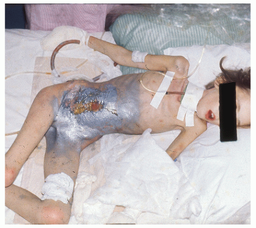
FIGURE 29-26. Crohn’s disease. Severe wasting in a 17-year-old girl, who looks much younger. Note the external abdominal wall fistula. (From Corman ML, Veidenheimer MC, Nugent FW, et al. Diseases of the Anus, Rectum and Colon. Part II: Nonspecific Inflammatory Bowel Disease. New York, NY: Medcom; 1976.)


Disease in Pregnancy

Because IBD is common in patients of childbearing age, the possibility of becoming pregnant is often an issue in medical and surgical care. However, pregnancy is not that frequent an event in patients with IBD. The reason probably is related to the fact that these patients may suffer any number of hormonal imbalances as a result of acute and chronic illness, often severely impairing their ability to become pregnant.

However, ulcerative colitis and Crohn’s disease do not adversely affect fertility, nor do they necessarily impede the progress of a pregnancy or the delivery of a normal, term infant. According to Zetzel, pregnancy in association with a preexisting colitis or complicated by the development of IBD is attended by the same prospect of a full-term delivery of a healthy child as is pregnancy in a healthy woman.792 However, Schade and colleagues found a significantly greater incidence of low birth weight (less than 2,500 g) in infants of mothers with ulcerative colitis than in a control population.662 Baird and colleagues found no evidence of an increased risk for pregnancy loss, but the likelihood of preterm birth was significantly greater.21

Levy and colleagues reviewed the clinical course of ulcerative colitis with respect to 60 pregnancies in 31 patients.411 Twenty percent were improved, 18% deteriorated, and 62% demonstrated no change during the course of pregnancy. Fourteen percent of the pregnancies were ended by spontaneous abortion and two by artificial abortions. One premature birth was noted in 50 full-term deliveries. All the births produced healthy children. The authors concluded that pregnancy does not seem to exacerbate preexisting ulcerative colitis, nor does colitis interfere with the outcome of the pregnancy.

Crohn and colleagues reported 74 pregnancies in 47 women whose colitis was inactive at the time of conception.115 All subsequent therapeutic and spontaneous abortions (including one stillbirth) occurred in patients in whom the colitis became activated. There was no difference in the incidence of abortion in patients who conceived during an active phase of colitis in comparison with those who conceived during an inactive phase. These observations have been confirmed by others.124

What happens to the colitis in patients who are pregnant? Zetzel reviewed a number of reported series and found it helpful to group the patients into several categories.792 Only 30% of patients who became pregnant during a quiescent phase of the colitis had an exacerbation of their disease, but a recrudescence developed in 60% when the pregnancy occurred during an active phase of the illness. In patients whose colitis developed initially during pregnancy or in the postpartum period, a particularly severe result was noted, with more than 60% having worsening of their symptoms. Khosla and colleagues noted that the infertility rate of patients with Crohn’s disease was similar to that seen in the
general population (12%).346 Those whose condition was in remission at the time of conception had a normal pregnancy, with the disease remaining quiescent in most individuals. However, active disease at the time of conception tended to inhibit remission despite therapy.346 In studies specifically in women with Crohn’s disease, pregnancy entailed no increased risk for exacerbation of the bowel inflammation.540,784 However, an increased risk for premature delivery and spontaneous abortion has been observed in those with active disease or in whom resection is required.

In the counseling of a colitis patient who is contemplating pregnancy, there is no justification for suggesting that attempts at conception be avoided, except when the possibility of teratogenic effects of a medication exists (e.g., the use of metronidazole [Flagyl]). Certainly, immunosuppressive treatment should be avoided in a patient wishing to conceive.

Concerns are also expressed about the safety of drug therapy in men, which could damage sperm and theoretically be associated with teratogenicity. Infertility in men is commonly associated with sulfasalazine administration. Sperm analysis may be helpful in determining whether a problem in conception can be attributed to this cause.

As mentioned, women who have a quiescent form of the disease are unlikely to experience problems with pregnancy and delivery. Conversely, if the patient is experiencing an exacerbation of the colitis, the illness itself may preclude the possibility of pregnancy. If the disease is more than moderately active, Zetzel counsels a temporary waiting period and introduction of appropriate medical therapy to secure a remission.792 However, even in this situation, the chances of a normal pregnancy and delivery approximate 50%.792 Contraception need be considered only in those women whose disease is so severe that surgery is imminent.791 Certainly, if the prospective parents wish to have a child, no benefit may be expected from a therapeutic abortion. Even in the severely ill pregnant woman, there is no evidence to suggest that the pregnancy cannot be brought to a successful conclusion with the birth of a healthy child.

If surgery becomes necessary during pregnancy, the method of treatment should be identical to that of a patient who is not pregnant. In other words, drug management (steroids, sulfasalazine) is not contraindicated. It has also been determined that azathioprine is safe and that termination of the pregnancy is not mandatory for those who conceive while taking the drug.6 Similarly, if an operation becomes necessary, the procedure should be performed as if the patient were not pregnant, although one should probably defer the implementation of a major reconstructive procedure (see Surgical Management). Closer to term, the enlarged uterus may preclude the possibility of performing even a conventional proctectomy; a staged operation, sparing the rectum, is therefore appropriate.

A high fetal and maternal mortality has been reported if surgical intervention becomes necessary for fulminant colitis.476,575 Bohe and colleagues noted two cases of fulminant disease during pregnancy that required subtotal colectomy and ileostomy in one, and proctocolectomy in the other.61 These operations were undertaken in the 32nd and 33rd week of pregnancy, respectively. I have performed surgery in two women during pregnancy: one patient underwent a proctocolectomy at 3 months, and the other underwent a total abdominal colectomy and ileostomy at 5 months. Both proceeded to uneventful conclusions of their pregnancy and delivered normal, healthy infants. In my opinion, it is not in the interest of the mother or the fetus to delay surgery until the pregnancy can be terminated with a viable child. In other words, I do not advocate waiting until the 32nd to perform a cesarean section while the mother is forced to postpone needed surgery.

Lindhagen and colleagues assessed the fertility and outcome of pregnancy in 78 women who had previously undergone resection for Crohn’s disease.426 Neither the number of live births nor the frequency of abortions differed from that which would be expected in the general population. The major factor, again, appeared to be that the disease was in remission or, as in the aforementioned reference, removed.

Successful childbirth has been reported following restorative proctocolectomy with pelvic ileal reservoir (see later discussion in that section).490,532,590 Twenty-eight patients carried 37 pregnancies to term after continent (Kock) ileostomy, according to a report from Göteborg, Sweden.562 Problems encountered were an increased urge to empty the reservoir, especially in the last trimester, and some difficulties with intubation. In most patients, a vaginal delivery was successful, with cesarean section reserved for obstetric indications.

If pregnancy develops following proctocolectomy and ileostomy, the question arises whether the prospective mother should undergo a cesarean section or deliver vaginally. My own feeling is that if the pregnant woman has an adequate pelvis for a normal vaginal delivery, this should be attempted. A cesarean section is not mandatory simply because the patient has an ileostomy, but if an episiotomy is performed, there may be a delay in healing of the perineal wound. However, it has been my experience that the obstetrician almost invariably will opt for a cesarean section. A national registry of ostomates is being maintained; a 1985 United States published survey indicated that about 1,000 had become pregnant.217 Parenthetically, prolapse of a loop or end colostomy is sometimes seen, especially in the last trimester. This usually will resolve spontaneously following delivery.


Extraintestinal Manifestations

Extraintestinal manifestations were at one time thought to be primarily associated with Crohn’s disease, but with several exceptions, they can be found in both conditions. These are discussed in Chapter 30.


▶ COURSE AND PROGNOSIS

As with many diseases, the prognosis for ulcerative colitis today is very different from that of half a century ago. Generally, this is attributed to improved medications, advances in surgical technique, and associated support during major abdominal surgery. Nordenholtz and colleagues examined the causes of death in patients with Crohn’s disease and ulcerative colitis through an analysis of death certificates in Rochester, New York.544 Of the total of 1,358 patients with IBD followed from 1973 to 1989, 130 (59 with ulcerative colitis and 71 with Crohn’s disease) were found to have recorded death certificates. Sixty-eight percent of patients with Crohn’s disease and 78% of those with ulcerative colitis died of causes unrelated to their IBD.544 Deaths caused by Crohn’s disease decreased from 44% in the first 8-year period to 6%
in the second. Colorectal cancer caused 14% of the deaths in patients with ulcerative colitis, three times more often than in persons with Crohn’s disease. Excluding cancer, only two deaths were directly attributable to ulcerative colitis, both occurring within the first 2 years after diagnosis.544

With respect to course and prognosis, Langholz and associates at the University of Copenhagen examined 1,161 patients during a 25-year period.381 The distribution of disease activity was remarkably constant each year, with about 50% of individuals in clinical remission. After 10 years, the colectomy rate was 24%. With 25 years of follow-up, the cumulative probability of a relapsing course was 90%.381 The probability of maintaining working capacity up to 10 years was approximately 93%. The authors concluded that although ulcerative colitis is a troublesome condition, most patients can manage their lives with little interference.

Maunder conducted a MEDLINE search for articles relating to ulcerative colitis and Crohn’s disease published since 1981 to determine the quality of life.468 Health-related quality of life is a quantitative measurement of the subjective perception of the state of one’s health, including emotional and social aspects. The investigators determined that the articles published indicated a trend toward a higher quality of life during the period from 1984 to 1987 in comparison with the period from 1981 to 1984. Although one can certainly question the validity of such an investigative effort, it appears that the previous comment concerning the general improvement in our ability to treat these two conditions is supported.


▶ RELATIONSHIP TO CARCINOMA

Carcinoma of the colon arising in a patient with ulcerative colitis was initially described by Crohn and Rosenberg in 1925. Since that time, numerous cases have been reported, so that there is uniform agreement with respect to the association between chronic ulcerative colitis and the subsequent development of adenocarcinoma. Primary malignant lymphoma complicating ulcerative colitis, although extremely rare, is nevertheless also thought to be associated with ulcerative colitis.1 Unfortunately, the true incidence of carcinoma is often a matter of conjecture, depending on the referral nature of the institution from which the report emanates. For example, some population-based studies have shown a lower incidence of colorectal cancer than that reported from major medical centers.200,601


Predisposing Factors and Incidence

Various factors predispose a colitic patient to colon cancer. These include total colonic or pancolonic disease; prolonged duration of the illness (the earliest reported case is in a patient with the disease of 7 years’ duration); continuous active disease, as opposed to intermittent symptoms; and possibly the severity of disease. An early age of onset probably poses no increased cancer risk save for the fact that cancer risk often parallels duration. The cumulative risk for cancer increases with the duration of colitis, reaching 25% to 30% at 25 years, 35% at 30 years, 45% at 35 years, and 65% at 40 years.560 The data also indicates a greater risk in patients with a family history of colon cancer and in those with primary sclerosing cholangitis. Sugita and colleagues observed a strong correlation between the age of onset of ulcerative colitis and the age of onset of cancer, a correlation found both in patients with extensive disease and in those with illness affecting the left side.712 However, colitis and cancers developed in patients with left-sided colitis about a decade later than in those with extensive disease, although the mean duration of the colitis before the development of cancer was virtually the same in both groups (approximately 21 years), irrespective of the age at onset of the disease.712 The incidence of cancer in patients with ulcerative colitis has been variously reported to be between 2% and 5%. Öhman reported the overall incidence to be 2.7%.560 Johnson and coworkers noted long-term findings in more than 1,400 patients with ulcerative colitis and found that colorectal cancer developed in 63 (4.4%).313 No statistically significant difference was observed in the probability of carcinoma of the colon and rectum developing following proctitis in comparison with total colitis. Patients with left-sided disease seemed to fare considerably better with respect to risk for the development of colorectal cancer. Greenstein reported that neoplasms developed in 11.2% of 267 patients with ulcerative colitis.226 Colorectal cancers developed in 13% of patients with universal colitis, and malignant change developed in 5% with left-sided colitis. Cancer tended to develop in patients with left-sided disease at least a decade later than in those with universal colitis. The median duration from onset of colitis to diagnosis of cancer was 20 years for those with universal colitis and 32 years for individuals with left-sided colitis.226 Cancer did not develop in any patient with left-sided colitis before the 23rd year of disease. In a review of more than 1,200 patients seen at the Cleveland Clinic, the cumulative risk for colorectal cancer was significantly higher in those with extensive colitis t han with left-sided disease.504 Ekbom and colleagues reviewed more than 3,000 patients with ulcerative colitis and noted that the absolute risk for the development of colorectal cancer 35 years after diagnosis was 30%.145

In patients who undergo resective surgery for ulcerative colitis, the incidence of associated cancer is considerably higher. Van Heerden and Beart reported the Mayo Clinic experience with 726 patients who underwent surgical exploration for chronic ulcerative colitis between the years 1961 and 1975.754 Seventy patients (9.6%) were found to have a carcinoma of the colon. These individuals represented 1.4% of all patients in whom chronic ulcerative colitis was diagnosed during the period of study.

Generally, the incidence of carcinoma is the same in both sexes. This is not surprising in light of the fact that ulcerative colitis affects each in approximately equal numbers. Welch and Hedberg reported a bimodal distribution of the incidence—one peak in the fourth decade and the other in the seventh.768

An increased risk of the development of colon carcinoma also appears to be evident in Crohn’s disease, especially of the small bowel.53 This is discussed in Chapter 30.


Characteristics

The distribution of tumors in patients having ulcerative colitis and carcinoma was reported by Öhman to demonstrate multicentricity much more commonly than in those having colorectal cancer without IBD.560 He also noted that 28% of patients had lesions of the transverse colon. Similarly, only 28% of patients had cancers involving the rectum and rectosigmoid, whereas patients with lesions of the sigmoid colon comprised 17% of the series. Conversely, Riddell and
colleagues reported that the rectum was the most common site of involvement and that this was more noticeable in men than in women.623 In their study, more than 40% of the cancers were found in the rectum, and approximately 25% of patients had multiple tumors. It is certainly clear that multicentricity of the cancers is a frequently reported phenomenon (Figure 29-27).


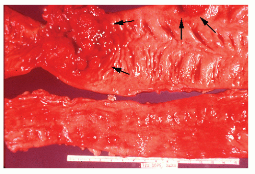
FIGURE 29-27. Carcinomas in ulcerative colitis (arrows). (From Corman ML, Veidenheimer MC, Nugent FW, et al. Diseases of the Anus, Rectum and Colon. Part II: Nonspecific Inflammatory Bowel Disease. New York, NY: Medcom; 1976.)

Another characteristic of colorectal cancer with ulcerative colitis is that very often the cancer tends to be infiltrative and scirrhous. Visible tumor involving the mucosa may not be observed even by careful endoscopic examination (Figure 29-28). Although such is the most common clinical presentation of cancer in this disease, it can also appear as a typical ulcerating or polypoid appearance (Figure 29-29).

Another pathologic feature of carcinoma arising in ulcerative colitis is the tendency of the lesion to be highly aggressive and poorly differentiated. More than half of young patients with ulcerative colitis and colorectal cancers have colloid carcinomas with histologically apparent mucus-secreting tumors of the signet-ring cell type696 (Figure 29-30; see also Figure 23-22). The fact that there may be few or no symptoms tends to lull both patient and physician into a false sense of security. Witness the situation illustrated in Figure 29-17, in which the mucosa has been completely denuded. When no mucosa is present, bleeding does not occur and mucous discharge is no longer evident. This may cause the physician and the patient to believe the medical measures that have been implemented are effectively controlling the disease. It is in just this kind of circumstance, a patient with long-standing ulcerative colitis, that a carcinoma can supervene. As suggested, physical examination, barium enema, and even endoscopic evaluation may fail to identify the lesion. But when stricture occurs, the patient
must be presumed to have a carcinoma until it can be proved otherwise (Figures 29-31,29-32 and 29-33). The presence of a stricture in a patient with ulcerative colitis is an indication for operative intervention. Lashner and colleagues demonstrated that of 15 patients with strictures, 11 had dysplasia and 2 were found to have cancer on colonoscopy/biopsy.385 An additional 4 patients were found to have carcinomas at the stricture site at the time of colectomy.


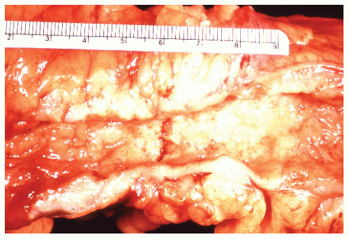
FIGURE 29-28. Carcinoma of the sigmoid in ulcerative colitis. Note the characteristic infiltration of the bowel wall, with an appearance resembling that of the linitis plastica type of carcinoma seen in the stomach. (From Corman ML, Veidenheimer MC, Nugent FW, et al. Diseases of the Anus, Rectum and Colon. Part II: Nonspecific Inflammatory Bowel Disease. New York, NY: Medcom; 1976.)


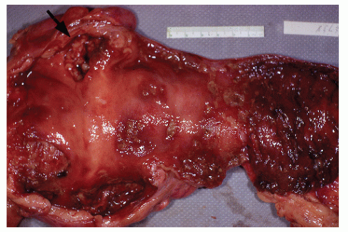
FIGURE 29-29. Ulcerative colitis with ulcerating carcinoma (arrow) in the hepatic flexure. (From Corman ML, Veidenheimer MC, Nugent FW, et al. Diseases of the Anus, Rectum and Colon. Part II: Nonspecific Inflammatory Bowel Disease. New York, NY: Medcom; 1976.)


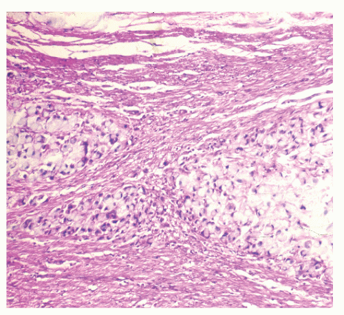
FIGURE 29-30. Signet-ring cell carcinoma infiltration of the muscularis propria in a patient with ulcerative colitis. (Original magnification × 600.)


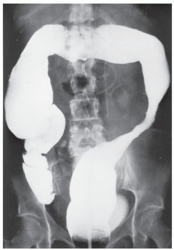
FIGURE 29-31. Stricture in ulcerative colitis. Foreshortening of the lower descending and sigmoid colon with stricture. Laparotomy revealed extensive carcinoma, hepatic metastases, and two additional unsuspected primary cancers in the resected specimen. (From Corman ML, Veidenheimer MC, Nugent FW, et al. Diseases of the Anus, Rectum and Colon. Part II: Nonspecific Inflammatory Bowel Disease. New York, NY: Medcom; 1976.)

Carpeting with “pseudopolyps” makes interpretation of biopsies very difficult. One can make a good case for definitive proctocolectomy and ileo-pouch anal surgery under these circumstances.


Results of Surgery for Cancer Complicating Ulcerative Colitis

Ritchie and colleagues reviewed the St. Mark’s Hospital (Harrow, United Kingdom) experience of carcinoma complicating ulcerative colitis between the years 1947 and 1980; 67 patients with carcinoma were identified.627 In comparison with those who underwent surgery for carcinoma of the colon and rectum in the same time period, it was felt that the colitic group had a higher proportion of inoperable and high-grade tumors, but the prognosis was found to be very similar in patients with and without colitis for the same stage of lesion. In the Mayo Clinic series, 40% of patients with carcinoma in chronic ulcerative colitis had a Dukes’ A or B growth, in comparison with a 63% incidence if carcinoma arose in the absence of the disease.754 Conversely, 60% with carcinoma and ulcerative colitis had Dukes’ C and D lesions, in comparison with 37% who had carcinoma alone. The mean age of their patients at the onset of the colitis was 26 years, with a duration of disease of 17 years before the development of malignancy; 23% exhibited multicentric tumors. Those whose carcinoma was identified incidentally during prophylactic colectomy had a 5-year survival of 72%, whereas those with clinical or radiographic evidence suggestive of cancer had a much poorer survival rate (35%).


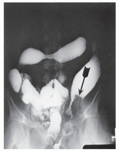
FIGURE 29-32. Carcinoma in ulcerative colitis. Note the loss of haustrations, marked shortening, and sigmoid stricture. The tumor extends cephalad from the stricture to appear as a polypoid filling defect (arrow).

The advanced nature of the cancerous change is attested to by the report of Johnson and colleagues.313 Only 57% of their patients underwent curative resection, and the overall survival rate in this group was 61%. In Öhman’s experience, two-thirds of those operated on for cure survived 5 years, a percentage virtually identical to the 69% of noncolitic patients.560 All with Dukes’ A lesions survived 5 years. Lavery and colleagues reviewed the Cleveland Clinic experience with 79 patients found to have carcinoma arising in ulcerative colitis.388 In comparing their survival s tatistics with those of patients with noncolitic cancer, they too noted no statistically significant difference in survival rates for the same stage of invasion. The poorer results were a consequence of the fact that a higher percentage of patients presented with more advanced or incurable disease at the time of surgery. All reports confirm that the prognosis for colitis-associated colorectal cancers, as for noncolitic cancers, is directly related to the degree of invasion (i.e., Dukes’ stage).102,711


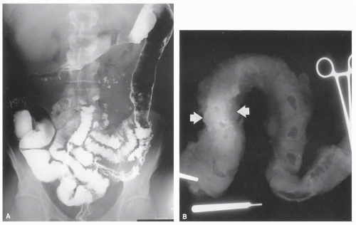
FIGURE 29-33. Carcinoma in ulcerative colitis involving the entire colon. A: Barium enema study shows a long stricture in the proximal transverse colon, which proved to be malignant. The short stricture in the distal transverse colon was benign. Note also the dilated terminal ileum, a characteristic of backwash ileitis. B: On x-ray film, resected specimen shows calcification (arrows) in the wall at the site of the stricture. This is seen with signet-ring malignant tumors and mucinous adenocarcinomas and is believed to be caused by inspissated mucin.


Dysplasia

In 1967, Morson and Pang described a phenomenon they called dysplasia, a frequent and widespread histologic change in patients with carcinoma complicating ulcerative colitis.512 They suggested that biopsy of the rectum could detect this premalignant situation and possibly dictate the requirement for surgical intervention.

This appeared to be a singular advance in the management of patients with long-standing disease. Before the introduction of the concept of dysplasia, “prophylactic” proctocolectomy was advocated to protect patients from the development of malignancy. This was justified on the basis that although the patient might be asymptomatic, the cancer could be far advanced when discovered. Many individuals with few or no complaints related to the gastrointestinal tract were submitted to surgery because of this understandable concern. With the promulgation of this concept, one ideally can seek to identify those patients who on biopsy and histologic examination are found to harbor this dysplastic phenomenon.


Definition and Interpretation

Dysplasia may be interpreted to be mild, moderate, or severe, but the significance of these distinctions has become less important. Clearly, however, the correct interpretation of the biopsy results rests on the talent and experience of the pathologist. It is imperative, therefore, to have available someone who is not only competent but also interested in this particular aspect of colon pathology.

The criteria for diagnosing dysplasia are problematic and vary from institution to institution. Dysplasia includes adenomatous and villous changes in the mucosa, irregular budding tubules beneath the muscularis mucosae, and cellular alterations consisting of a reduced number of goblet cells and the presence of hyperchromatic nuclei, stratified nucleoli, and coarse chromatin352,638 (Figures 29-34,29-35 and 29-36). The following criteria have been proposed by Nugent and colleagues549:

Mild dysplasia


  • Preservation of crypt architecture


  • Nuclear stratification, but not reaching the luminal surface


  • Nuclear crowding and hyperchromasia


  • Mitoses in upper portion of crypt


  • Usually, moderate diminution of goblet cell mucin

Moderate dysplasia


  • Distortion of crypt architecture with branching and lateral buds


  • Nuclear abnormalities as in mild dysplasia, but stratification reaching luminal surface


  • Usually, depletion of goblet cell mucin

Marked dysplasia


  • More marked distortion of crypt architecture, frequently with villous configuration of surface epithelium


  • Nuclear abnormalities as in moderate dysplasia, but with loss of polarity frequently present


  • Frequently, presence of “back-to-back” glands


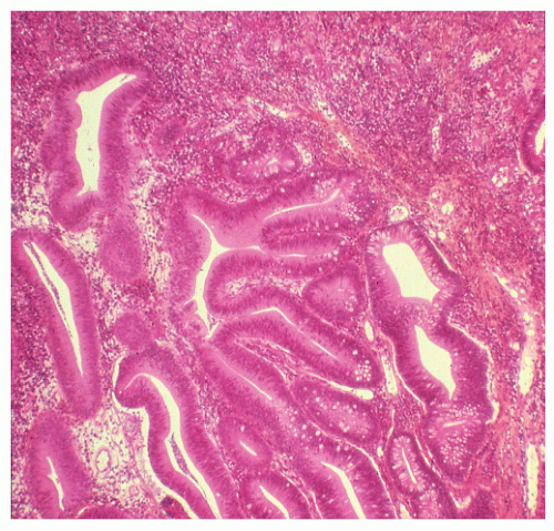
FIGURE 29-34. Moderate dysplasia in a patient with ulcerative colitis. Note the loss of polarity and decreased mucus production. (Original magnification × 80; courtesy of Rudolf Garret, MD.)

The last category includes all abnormalities short of invasive carcinoma and encompasses what some might designate as carcinoma in situ.


Results of Evaluation for Dysplasia

Nugent and colleagues initially reviewed the clinical and histologic data in a retrospective fashion of 23 patients with known colon carcinoma and chronic ulcerative colitis.549 All but one were found to have dysplasia at a remote site from the cancer. Based on this experience, the authors enrolled 151 patients with more than 7 years of ulcerative colitis in an annual colonoscopy/biopsy surveillance program.548 The number of specimens taken ranged from 3 to 10. Initial biopsies were positive for high-grade dysplasia in 4 patients, 3 of whom were found to harbor a carcinoma at the time of colectomy. Of 12 with low-grade dysplasia or high-grade dysplasia on initial biopsy, 11 had undergone colectomy; five cancers were found. Of 10 patients in whom dysplasia developed on follow-up, 9 underwent colectomy, and only 1 cancer was found. Carcinoma did not develop in any patient with left-sided disease. A later experience from the same group revealed that carcinoma subsequently developed in none of the 148 patients whose biopsy findings remained negative for dysplasia throughout the study.550 In a still later review from the same institution, carcinoma associated with ulcerative colitis developed in 41 patients, 19 of whom were under colonoscopic surveillance and 22 of whom were not.90 It seems then that carcinoma was detected at a significantly earlier Dukes’ stage in the surveillance group. The 5-year survival rate was 77.2% for those surveyed, but only 36.3% for the no-surveillance group. The authors concluded that colonoscopic surveillance reduces colorectal carcinoma-related mortality by permitting the detection of carcinoma at an earlier Dukes’ stage.90


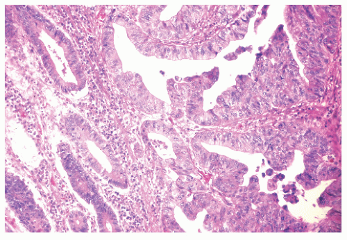
FIGURE 29-35. Moderate dysplasia in ulcerative colitis. Loss of polarity and proliferation of epithelial cells. (Original magnification × 260; courtesy of Rudolf Garret, MD.)


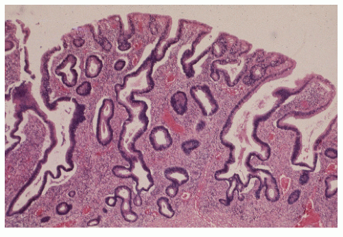
FIGURE 29-36. Severe dysplasia. Atypical hyperplasia with irregularly shaped crypts lined by crowded cells with hyperchromatic nuclei. (Original magnification × 80; from Corman ML, Veidenheimer MC, Nugent FW, et al. Diseases of the Anus, Rectum and Colon. Part II: Nonspecific Inflammatory Bowel Disease. New York, NY: Medcom; 1976.)

Dickinson and colleagues surveyed 43 patients with longstanding ulcerative colitis extending proximal to the splenic flexure.133 Dysplasia was found in nine patients in one or more biopsy specimens (severe in two, moderate in one, and mild in six). The two patients with severe dysplasia were subsequently found to harbor carcinomas.

Blackstone and colleagues performed colonoscopy on 112 patients with long-standing ulcerative colitis during a 4-year period.58 In 12 patients, the procedure revealed a polypoid mass that on biopsy exhibited dysplasia. Seven of these patients were subsequently found to have dysplasia in the absence of a polypoid excrescence (i.e., a flat mucosa), and only one carcinoma was found later. The authors thought the identification of a single polypoid mass to be highly significant for the presence of concurrent invasive cancer. As such, this was strong evidence to support the need for colectomy.

Löfberg and colleagues studied 72 patients with total ulcerative colitis in a 15-year surveillance program.431 The cumulative risk for the development of at least low-grade dysplasia was found to be 14% after 25 years of disease. Others also recommend the use of surveillance colonoscopy in following patients with high-risk ulcerative colitis.73,395,454,632,785 Furthermore, Lindberg and associates
demonstrated in their 20-year surveillance program of 143 patients with ulcerative colitis that primary sclerosing cholangitis (see Chapter 30) is an independent risk factor for the development of dysplasia or cancer, especially in the proximal colon.424


Dysplasia Surveillance Limitations and Concerns

Although the increased risk for the development of colorectal cancer in individuals with long-standing ulcerative colitis is well-known and accepted, in recent years in particular there has been increased concern about the utility of surveillance colonoscopy.116 For example, Jonsson and colleagues undertook a prospective study of 131 patients with ulcerative colitis and concluded that the surveillance program was resource-consuming, questioning the cost-to-benefit ratio.318 Of greater concern, however, is the validity of the concept. Taylor and coworkers, reporting from the Mayo Clinic, evaluated the reliability of this premise by means of multiple random biopsies on resected specimens of patients with chronic ulcerative colitis, with and without cancer.724 Using a standard technique of multiple random biopsies (see later discussion), they used ordinary colonoscopic biopsy forceps to obtain four biopsy specimens from mucosa that was not macroscopically suggestive of dysplasia or cancer in eight defined regions of each of 100 colon specimens obtained. Although an overall association between the presence of cancer and high-grade dysplasia was detected, the sensitivity and specificity to detect concomitant carcinoma were both 0.74. Their findings prompt concern that reliance on random biopsies obtained during colonoscopic surveillance may be inappropriate.724

Gorfine and associates reviewed 590 specimens for the presence of dysplasia and found that 77 (13.1%) contained at least one focus.219 Cancers were significantly more common in those specimens with dysplastic changes than in those without such changes (33/77 vs. 5/513; P < .001). Colonoscopically diagnosed dysplasia as a marker for synchronous cancer had a sensitivity of 81% and a specificity of 79%. The authors concluded that the concept of dysplasia is an unreliable marker for the detection of synchronous carcinoma, but when any degree is discovered colectomy is indicated.

Bernstein and coworkers summarized 10 prospective studies to ascertain whether colonoscopic surveillance is the appropriate alternative to prophylactic colectomy.55 First of all, the risk for progression to dysplasia was found to be only 2.4% for individuals whose initial evaluation was negative. Therefore, surveillance might perhaps be less frequently applied for those patients. Of a greater concern, however, is the fact that 32% of the patients with high-grade dysplasia were found to have invasive cancer. For this group, at least, the surveillance program failed to prevent the development of malignancy. However, when an unsuspected cancer is found at the time of surgery performed for dysplasia, it tends to be at a lower TNM stage than the cancer in patients in whom the diagnosis is made prior to surgery. In order for reasoned decisions to be made, therefore, it has been advised that patients be informed about the limitations of colonoscopic surveillance so that they can rationally take part in their management.51 Unfortunately, a highly malignant carcinoma was reported to have developed in a patient without prior dysplasia or DNA aneuploidy who was enrolled in a colonoscopic surveillance program.436

An opposing viewpoint concerning the value of surveillance for dysplasia has been expressed by Collins and colleagues.100 It is their contention that no compelling evidence proves that such follow-up evaluation is beneficial. Others have also expressed reservations.383,400,401 and 402,602,622 This is especially true with respect to the significance of lowgrade dysplasia. In the experience of Befrits and coworkers (Stockholm, Sweden), no progression to high-grade dysplasia was observed during 10 years of follow-up in 60 patients, leading to the conclusion that colectomy in cases with single or even repeated low-grade dysplasia is not justified.46 Others opine that clinicians have to take not one but two giant leaps of faith to reject the null hypothesis and recommend surgery for low-grade dysplasia.423 But today, most gastroenterologists are of the opinion that, because dysplasia implies concomitant neoplasia, individuals with even low-grade dysplasia should be counseled to undergo colectomy.750

The cost-effectiveness of surveillance colonoscopy has become a hotly debated issue, and as of this writing, accord with respect to the appropriate frequency of such examinations is still an unattained ideal (see the following section).181,238 There is no controversy, however, with respect to those individuals who harbor a dysplasia-associated lesion or mass (DALM). These patients require colectomy.46,423 This includes any benign neoplasm (tubular adenoma, villous adenoma) that arises in a segment of inflammatory disease.


Surveillance Program

In a surveillance program, evaluation is advised for those who have had a minimum of 7 years of total or subtotal colonic disease. These persons are then submitted to total colonoscopy with biopsy of any demonstrable lesion. As suggested, the biopsy of a specific, elevated lesion will yield a much higher incidence of dysplasia. Multiple random biopsy samples should be taken throughout the colon. Although traditionally 10 such biopsies have been advised, in recent years some have suggested sampling 30 to 40 specimens. The examination should be performed every other year or more often if dysplasia is identified. Whether one wishes to wait a year and repeat the study once dysplasia, irrespective of degree, has been identified is a subject of controversy (see earlier). It must be remembered, however, that colonoscopy in ulcerative colitis is not necessarily a benign procedure, and performing multiple biopsies may invite complications (Figure 29-37). Therefore, an experienced endoscopist should be selected to examine these patients. Preferably, biopsy specimens should be obtained in areas free from obvious inflammation.548


Comment

It is certainly true that cost-benefit analysis has not been determined, that incurable cancer may still supervene, that patient compliance is problematic, and that willingness of patients to commit themselves to a resection if the biopsy reveals dysplasia is doubtful. However, the alternative course is even less agreeable. Barium enema examination is useful only to demonstrate the macroscopic anatomy of the colon: loss of haustrations, shortening, and possible stricture. It is unlikely that this study will reveal a carcinoma earlier than will endoscopic examination with biopsy. Prophylactic colectomy after 8 to 10 years is one option; denial
is another. However, until a better alternative is available, one should continue to recommend the protocol as outlined, with the performance of flexible sigmoidoscopy in alternate years.


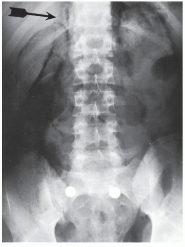
FIGURE 29-37. Retroperitoneal gas from a colon perforated during colonoscopic biopsy for dysplasia in a 19-year-old patient with ulcerative colitis. The renal outlines are clearly evident, as is the right adrenal gland (arrow).


Aneuploidy

Another method for identifying precancerous changes is flow cytometry. Several studies have demonstrated that DNA aneuploidy correlates with the presence of dysplasia and, therefore, a high risk for developing cancer.286,425,432,485,637,715 Suzuki and colleagues showed that 77% of dysplastic tissue demonstrated aneuploidy or polyploidy, whereas 94% of specimens of nondysplastic tissue exhibited diploidy.715 Löfberg and colleagues found that 20% of 59 patients with long-standing total ulcerative colitis harbored an aneuploid DNA pattern on colonoscopy/biopsy, and that this correlated with the presence of dysplasia.431 The frequency of aneuploidy is higher in patients with disease longer than 10 years and with a greater extent of involvement.286

As mentioned, one of the problems with the concept of dysplasia alone is interobserver and intraobserver variability.432 Sampling error as well as total reliance on histologic information has its own inherent limitations. In the experience of Rubin and coworkers, a significant correlation between aneuploidy and severity of histologic abnormality was found in patients at high risk for cancer (negative, indefinite, dysplasia, or cancer).637 In a prospective study from their institution of 25 high-risk individuals without dysplasia, 20% were found to have aneuploidy, and all of these patients progressed to dysplasia within 2.5 years. Conversely, all 19 individuals who failed to demonstrate aneuploidy did not progress to either aneuploidy or dysplasia within the limits of the study (up to 9 years). The authors concluded that patients who demonstrate aneuploidy should be submitted to more extensive and frequent colonoscopic surveillance, whereas those who do not require less frequent investigations.637 Löfberg and colleagues confirmed that nuclear DNA content appears to be an earlier phenomenon than dysplasia in the malignant transformation of the colorectal mucosa, and that the use of flow cytometry in surveillance programs might be of particular value for selecting individuals at an increased risk for the development of cancer.432

In summary, flow cytometry may be usefully applied to complement histologic examination when dysplasia is suspected.408,485 However, there is no evidence to support the use of DNA aneuploidy as the sole indication for prophylactic cancer surgery in patients with ulcerative colitis.425


Treatment of Carcinoma

If a carcinoma is identified in the rectum of a patient with ulcerative colitis, proctocolectomy is the treatment of choice. No attempt should be made to preserve the rectal mucosa. Alternative operations may, however, include the continent ileostomy (Kock) and the ileoanal anastomosis with intervening pouch, provided that sphincter preservation does not compromise adequate tumor margins, and the risk for the requirement of postoperative radiation therapy is remote (see Surgical Management).


▶ MEDICAL MANAGEMENT

I have asked two respected colleagues, Udayakumar Navaneethan and Bo Shen, at the Center for Victor W. Fazio, Inflammatory Bowel Disease, Digestive disease Institute, Cleveland Clinic Foundation, Cleveland, Ohio, to help me update this section. (VWF)


Introduction

Approximately 4% to 9% of UC patients will require colectomy within the first year of diagnosis381,517, whereas the risk of colectomy following that is 1% per year afterward.382 The vast majority of UC patients will require medical therapy throughout their life. Therefore, understanding of the appropriate use of these agents and their adverse effects is important for the physician caring for these individuals. In recent years, the medical management of UC has changed significantly. In particular, the advent and approval of anti-tumor necrosis factor-α (TNF-α) agents like infliximab in the management of moderate-to-severe UC has expanded the role of medical therapy in this condition.381,382,517

The general principles in medical therapy for UC patients are improvement of quality of life, induction and maintenance of remission and mucosal healing, avoidance of complications and colectomy, and decreasing the likelihood of the development of cancer. Estimated lifetime risk of a severe exacerbation of UC requiring hospitalization
is approximately 15%.144 Patients with extensive disease (macroscopic disease proximal to the splenic flexure) are more likely to develop acute severe colitis.

A number of agents have been shown to have clinical benefit for induction and maintenance therapy for mild-to-moderate UC. However, the challenge lies with the management of severe UC, which does not respond to corticosteroid therapy. A recent study showed that there was a 7% absolute reduction in the risk of colectomy in the infliximab versus placebo group (10% vs. 17%, respectively) over a 54-week follow-up period.656 Whether the use of infliximab decreases the risk of colectomy in the long run is not known. This section will address various medications used in the treatment of UC.


5-Aminosalicylates

Sulfasalazine and 5-aminosalicylate (5-ASA) remain the first-line therapy for the induction of remission in patients with mild-to-moderate active UC.82,713 Oral 5-ASAs come in a wide range of formulations with different release characteristics.418,420 Sulfasalazine, a 5-ASA bound to sulfapyridine by an azo bond, is the initial form found to be effective in the treatment of UC.132,603 Because the 5-ASA portion of the agent is the therapeutically active compound, several oral preparations of 5-ASA have subsequently been developed. However, sulfasalazine appears to have comparable efficacy with the newer ASA formulations.541 The type and dosage of 5-ASA therapy are determined by location, severity of disease, cost and insurance coverage, as well as patient preference. Most ASA agents have comparable pharmacokinetics in terms of systemic absorption, urinary excretion, and fecal excretion of active ingredient. Meta-analyses showed that topical 5-ASA agents delivered rectally appeared to be superior to placebo or topical corticosteroids for the induction of remission in distal UC.392,463,611 However, concomitant topical application of 5-ASA and corticosteroid agents was shown to be superior to topical 5-ASA alone. Topical 5-ASA appears to be at least as effective as oral 5-ASA in maintaining remission for distal UC. 5-ASA appears to be more effective than placebo across all dosage ranges with a trend toward a dose-response effect. Patients with active proctitis or distal colitis can be treated with either topical (enemas or suppositories) and/or oral 5-ASAs. However, controlled trials have shown that rectal therapies have a more rapid effect than oral treatment. Combination therapy with oral and topical 5-ASAs may achieve a higher remission rate than either rectal 5-ASA or oral 5-ASA alone for distal UC. In one study, patients treated with both topical and oral 5-ASAs had a remission rate of 89%, compa red with 69% for topical 5-ASA alone and 46% for oral 5-ASA alone.644 In patients with left-sided disease or extensive mild-to-moderate active UC, oral 5-ASAs may be used along with topical 5-ASAs.

The available oral 5-ASA agents appear to be equally effective in producing response rates of 40% to 75% after 4 to 8 weeks of treatment.515 In those with active UC, delayedrelease oral mesalamine (Asacol HD; Proctor and Gamble Pharmaceutical, Cincinnati, OH) in doses of 2.4 g/day demonstrated a comparable efficacy (51% vs. 56%) versus 4.8 g/day. However, a dose of 4.8 g/day was more effective in moderate disease (57% vs. 72%).245 A formulation of ASA using a MultiMatrix (MMX) release system (Lialda; Shire US, Wayne, PA), which, in addition to being pH dependent (breaking down at pH >7, normally in the terminal ileum, slowly releases 5-ASA throughout the entire colon), has been studied. Clinical remission rates in active mild-to-moderate UC were 37.2% and 35.1% in the 2.4 and 4.8 g/day groups, respectively, after 8 weeks of treatment, compared with 17.5% in the placebo group.323,419,653 These once daily doses provide an opportunity to improve patient adherence. Of note, combination therapy with oral and rectal mesalamines may be superior to oral or rectal therapy alone in patients with extensive colitis.464 With regard to maintenance, oral mesalamine has been shown to decrease relapse rates to 23% to 37% at 12 months compared with 50% to 65% in patients receiving placebo.515,713

In a subsequent trial, the efficacy of maintenance therapy of MMX was evaluated over a 12-month period (1.2 g/day vs. 2.4 g/day). Remission rates were similar—64.4% with 1.2 g and 68.5% with 2.4 g/day.322 Another oral mesalamine formulation was developed with the trade name Apriso (Salofalk Granu-Stix, Dr. Falk Pharma, Buckinghamshire, United Kingdom), which has mesalamine granules that have both a gastric acid-resistant enteric coating (which dissolves at pH >6) that delays release. It also has a retarding polymer matrix in the granule core that extends release throughout the colon. This is similar to Lialda. Clinical trials showed that Apriso can be administered once daily and was shown to be as effective and safe in mild-to-moderate UC, with a three times a day dosing schedule.373,374,595 Finally, several observational studies and a meta-analysis have shown a potential protective effect of 5-ASA therapy on the development of UC-associated colorectal cancer and dysplasia at doses >1.2 g/day.409,757


Corticosteroids

Oral corticosteroids can be used for both left-sided and extensive colitis. In patients with proctitis or left-sided disease, topical corticosteroids in the form of foams and enemas are commonly used. In a meta-analysis of topical therapy for active distal UC, topical hydrocortisone appears to be less effective than topical 5-ASAs in inducing remission, whereas rectal application of hydrocortisone or budesonide may be superior to placebo. Topical hydrocortisone may be considered as an alterative agent in patients who fail topical 5-ASA therapy.304,399

In patients with extensive colitis, moderate-to-severe colitis, or refractory disease, oral and topical 5-ASA therapy and oral corticosteroids may be used as induction therapy. However, parenteral corticosteroids are often required in patients with severe colitis.154 Response to oral corticosteroid therapy is expected to be within 10 to 14 days, but oral corticosteroid agents should be tapered and should not be used for maintenance therapy because of side effects.740 In fact, one of the common mistakes in managing UC is the use of corticosteroids for long-term maintenance. On the other hand, the requirement for oral or parenteral corticosteroids has been considered a prognostic factor. In UC patients requiring corticosteroids, approximately one-third underwent colectomy within 12 months.535

To minimize systemic toxicity, topically active corticosteroid formulations have been tried in UC. Budesonide is an oral glucocorticoid with high first-pass metabolism and, thus, has limited systemic toxicity. In a study evaluating oral budesonide in patients with extensive and left-sided mild-to-moderate
UC, comparable efficacy to that of prednisolone in inducing remission was observed. However, improvement in endoscopic and histologic scores was superior in the prednisolone group.433 Therefore, oral budesonide has not been routinely used in treating UC.


Azathioprine and 6-Mercaptopurine

6-Mercaptopurine (6-MP) and its prodrug, azathioprine (AZA), are antimetabolites that inhibit purine synthesis. Therapeutically, effective doses of AZA are 2.0 to 3.0 mg/kg/day and of 6-MP are 1.0 to 1.5 mg/kg/day. Their therapeutic effect may take up to 17 weeks.578 The dosage of 6-MP and AZA may be directed by measuring thiopurine S-methyltransferase (TPMT) activity. Low to intermediate levels of TPMT are associated with the risk of leukopenia.57 Thus, patients with normal TPMT activity may receive standard doses of AZA or 6-MP. Patients with intermediate activity can receive 50% of the standard dose. Those who have no TPMT activity should not be treated with these agents.693

6-MP and AZA are mainly used as steroid-sparing drugs for maintenance therapy in UC. It has been shown that patients with UC in remission on AZA >6 months, but later crossed over to placebo, had a higher rate of relapse at 1 year (59%) than those who continued AZA (36%).261 6-MP/AZA typically works in UC patients whose disease activity responds to corticosteroid therapy.2,13,198,430 6-MP/AZA should not be used as an induction agent and perhaps not for maintenance therapy in patients who fail induction therapy with corticosteroids.

6-MP/AZA use may impact the disease course of UC. For example, there appears to be fewer colectomies undertaken in those maintained on AZA.13,430 A prospective study evaluating the efficacy of AZA in maintaining remission in patients with acute severe UC after successful induction with corticosteroids found decreased rates of relapse (10% vs. 55%) and severe relapse (0% vs. 36%) when compared with a historical cohort that did not receive AZA after steroid-induced remission.327 Controlled trials also have demonstrated therapeutic benefit of AZA plus sulfasalazine versus sulfasalazine alone in individuals with newly diagnosed UC.735 A similar ability to maintain remission with less toxicity in AZA alone than AZA plus olsalazine was demonstrated in patients with steroid-dependent UC.455 In a meta-analysis of 6-MP/AZA compared with either placebo or 5-ASAs, the mean efficacy (pooled data) was 60% (95% confidence interval [CI], 51%-69%) in the 6-MP/AZA group and 37% (95% CI, 28%-47%) in the control group. When only compared to placebo, 6-MP/AZA was found to be beneficial in maintaining remission (odds ratio [OR] = 2.59; 95% CI, 1.26-5.30).202 The results of the meta-analysis support the efficacy of thiopurines in the maintenance of remission. Accordingly, the American Gastroenterological Association’s guidelines recommended that patients with steroid-dependent UC should be treated with 6-MP/AZA, based on the grade A evidence (homogeneous randomized controlled trials or well-designed cohort studies).415


Cyclosporine

Cyclosporine A (CSA), an immunosuppressant that inhibits T-lymphocyte function, has been used in severe corticoste-roid-refractory UC.421 In a double-blind, placebo-controlled trial of 20 patients who had failed intravenous corticosteroid therapy, 82% responded to CSA, whereas 0% responded to placebo. Of the 9 patients in the placebo group who did not respond initially, 55% did well after crossed over to openlabel CSA. However, 3/11 in the cyclosporine group versus 4/9 in the placebo group required colectomy within 1 month.421 Subsequently, four additional controlled trials of CSA in patients with severe UC were reported. The studies identified a response rate of approximately 80%.131,514,716,753 In a study of 30 patients who were randomized to either CSA or methylprednisolone, 64% who received CSA and 53% who received corticosteroids achieved clinical remission within 8 days. At 1 year, 78% of patients in the CSA group were in remission as opposed to only 37% in the corticosteroid group. After 1 year, 7/9 responders in the CSA group were still in remission. This compared with 4/8 in the corticosteroid group (P > .05); colectomy rates were similar.131,685

Long-term response rates in patients treated with CSA have also been evaluated. In a study of 42 patients with severe steroid-refractory UC treated with intravenous CSA who were followed over a 5-year period, 45% were able to avoid colectomy. This represented a higher rate in those who initially responded to CSA and in those concomitantly on AZA or 6-MP (49% vs. 17%).682 A study comparing quality of life (QOL) in patients who underwent colectomy versus those who were managed with CSA found that the latter individuals scored well or better than their surgical counterparts.96 In a retrospective single-center study of 86 patients treated with CSA, 25% of initial responders required colectomy at a mean interval of 178 days, and analysis showed that of all patients treated with CSA, 55% would avoid colectomy at 3 years.15 The dose used in many of the studies is 4 mg/kg per 24 h, with a goal trough serum level of 300 to 400 ng/mL. However, some studies suggest that a lower dose of CSA may be as effective.514,608 Intravenous CSA is effective in the induction of remission in patients with severe, steroid-refractory UC. However, the efficacy of durable treatment response is limited to those who were “naive” to AZA/6-MP before treatment. All patients require AZA/6-MP for maintenance of remission after induction with CSA.


Antitumor Necrosis Factor Agents

TNF inhibitors have increasingly been used for the treatment of moderate-to-severe UC. Infliximab (IFX), a chimeric monoclonal immunoglobulin G1 antibody to TNF-α, was the first drug of the category approved by the U.S. Food and Drug Administration (FDA) for the treatment of UC. IFX has been studied in several small open-labeled trials in steroid-refractory and steroid-dependent UC.87,88,222,329,710 Subsequently, a double-blind, placebo-controlled study of IFX was terminated prematurely due to slow enrollment, but it showed a 50% response rate at 2 weeks in those previously refractory to intravenous corticosteroids.659 Two additional placebo-controlled studies of steroid-refractory disease found favorable results that led to large subsequent studies.307,596

The Active Ulcerative Colitis Trials (ACT 1 and 2), which enrolled patients with moderate-to-severe active UC treated
with corticosteroids and/or 6-MP/AZA (ACT 1) or with UC refractory to at least one standard therapy (ACT 2), are the landmark studies evaluating the efficacy of IFX.640 In ACT 1, the clinical response to IFX at 8 weeks was 69% for 5 mg/kg dosing and 61% for 10 mg/kg dosing versus 37% in the placebo group (P < .001), and similar response rates were found at 8 weeks in ACT 2. In both studies, patients who received IFX were more likely to have a clinical response at week 30, and in ACT 1, more patients who received IFX had a clinical response at week 54. Endoscopic remission, which has been of interest as an end point, was also seen in more than 50% of the IFX-treated groups in ACT 1 at 30 and 54 weeks. Subsequently, a follow-up study was done to evaluate health-related QOL in patients treated with IFX to see whether improved clinical response translated to improvement in QOL. Substantially improved QOL was sustained through 1 year with maintenance therapy.162 However, the influence on the risk of colectomy was not evaluated until recently. In a published follow-up of ACT 1 and 2 studies evaluating colectomy rates, the cumulative incidence of colectomy in patients treated with IFX through 54 weeks was 10%.656 This compared with 17% for the patients in the placebo group. However, those enrolled in the study had moderate-to-severe UC who had not received intravenous corticosteroid within 2 weeks and were judged unlikely to require colectomy within 12 weeks. Hence, the reduction in risk cannot be entirely attributed to IFX.656

Previous studies have addressed the risk of colectomy in patients with severe UC. In a small pilot study of 11 patients hospitalized with severe steroid-refractory disease, 50% receiving IFX responded.659 This compared with no response in patients in the placebo group, but the numbers were too small to detect a statistically significant benefit. In a study of 45 severe UC in-patients at risk for colectomy, a decreased rate of colectomy at 3 months was demonstrated in those who received one dose of IFX (29% vs. 67%, P = .017).307

The risk of colectomy in the long run was evaluated in a study of 314 UC patients from Italy.12 Fifty-two (16.5%) had severe UC. Fifteen of the 52 patients (29%) did not respond to a median of 7 days of intravenous corticosteroids. Of these, 4 underwent urgent colectomy, and 11 received IFX. A clinical response was observed in all IFX-treated patients. In the long term, another 6 patients underwent elective colectomy. The overall colectomy rate following acute flare-up was 19%. The long-term colectomy risk was comparable in IFX-treated patients (18%) and in steroid-responsive patients (11%). However, those treated with IFX had a shorter colectomy-free disease course than the individuals who responded to intravenous corticosteroids. The authors speculated that steroid-refractory patients who achieve remission with IFX have a more severe disease than steroid-responsive patients.12

Adalimumab (ADA) is a fully humanized IgG1 monoclonal antibody to TNF. Although adalimumab was initially studied in CD, a pilot study subsequently reported its efficacy in UC. More recently, it was studied for the induction of clinical remission in anti-TNF naive patients with moderately to severely active UC.613 In the multicenter, randomized, double-blind, placebo-controlled study in North America and Europe, patients with a Mayo score of ≥6 points and endoscopic subscore of ≥2 points despite treatment with corticosteroids and/or immunosuppressants were enrolled. These were randomized to subcutaneous treatment with ADA 160/80 (160 mg at week 0, 80 mg at week 2, 40 mg at weeks 4 and 6) or placebo group. At week 8, 18.5% of patients in the ADA 160/80 group were in remission. This compared with 9.2% in the placebo group (P = .031). Serious adverse events occurred in 7.6% and 4.0% of patients in the placebo and ADA 160/80 groups, respectively. ADA 160/80 appeared to be safe and modestly effective for induction of clinical remission in patients with moderately to severely active UC who failed treatment with corticosteroids and/or immunosuppressants.613


Adverse Effects of Medications

Medications used in the treatment of IBD are associated with a number of adverse effects. Sulfasalazine consists of sulfapyridine linked to 5-ASA (mesalamine, mesalazine) via an azo bond. However, its use is limited by high rates of intolerance among patients. Side effects can include headache, abdominal pain, nausea, vomiting, skin rash, fever, hepatitis, hematologic abnormalities, folate deficiency, pancreatitis, systemic lupus erythematosus, and reduction in sperm counts.446 Sulfapyridine, a sulfonamide moiety, has been suggested to be responsible for hypersensitivity reactions. Sulfasalazine-induced hepatotoxicity manifests as elevation of aminotransferases; hyperbilirubinemia; and less commonly, fever, hepatomegaly, lymphadenopathy, and granulomatous liver disease.529 Hepatotoxicity may also be a part of hypersensitivity reactions.529

Similarly, thiopurines are known to be associated with liver toxicity.529 Hepatotoxicity usually manifests itself by elevation in aminotransferases, accompanied by flulike symptoms. In some patients, it can present as an isolated cholestatic enzyme elevation. Abnormal liver function tests (LFTs) usually return to normal after discontinuance of the agents.529 6-MP/AZA-induced hepatotoxicity occasionally may be idiosyncratic in nature, with rare presentations, such as veno-occlusive disease (VOD).152 Acute pancreatitis is also reported with 6-MP/AZA use in IBD. Pancreatitis is an early idiosyncratic adverse reaction after initiation of treatment, usually occurring within 3 to 4 weeks of therapy. Pancreatitis is considered to be idiosyncratic and dose independent.459

The use of anti-TNF agents is associated with a number of adverse effects, including activation of tuberculosis, infusion reactions, hypersensitivity reactions, and the development of lymphoma.683 Hepatosplenic T-cell lymphoma has been described in IBD patients treated with anti-TNF drugs, including infliximab and adalimumab, particularly in combination with immunomodulators.529 Reports from the manufacturer-maintained TREAT (The Crohn’s Therapy Resource, Evaluation, and Assessment Tool) Registry with voluntary reporting system, however, suggest that serious infection from IFX-treated CD patients appeared to be associated with concurrent use of corticosteroids or narcotic analgesics.417

The risk of postoperative complications in UC patients treated with IFX before colectomy has been studied.509,673 After adjusting for age, high-dose corticosteroids, AZA, and severity of colitis, infliximab use was significantly associated with infectious complications in a multivariate analysis (OR = 2.7).673 In the study from our institution, preoperative IFX use was also found to be associated with an increased risk for three-stage restorative proctocolectomy instead of the traditional two-stage procedure as well as an increased risk of postoperative infectious complications.509


A multicenter study from Europe evaluated the safety of IFX in 52 patients with steroid-refractory UC who did not respond to CSA.85 Fifteen (29%) required colectomy within a median of 5 weeks. The rate of adverse events was 25% (six infections, three infusional reactions, one leukopenia, one bowel perforation, one fever, and one peripheral neuropathy). One death occurred in a 40-year-old man (pneumonia) who underwent surgery 10 days after the first IFX infusion.85 Therefore, it appears that pushing medical therapy to the limit may be costly in terms of severe adverse effects prior to and following colectomy.

The disease, UC, itself, appears to have no adverse effects on fertility in women nor in men.306,525 However, a variety of medications used in the management of IBD may affect fertility. Among the medications, sulfasalazine has been shown to be associated with male infertility and abnormalities in sperm count, motility, and morphology.738 An association between sulfasalazine use in the parent and congenital malformations in the progeny has also been described.508 6-MP and AZA do not appear to reduce semen quality in men with IBD.127 IFX treatment in men may decrease sperm motility and morphology.448 Safety of anti-TNF agent use during pregnancy is still controversial and is discussed separately.


▶ MANAGEMENT OF INFLAMMATORY BOWEL DISEASE DURING PREGNANCY

The management of IBD during pregnancy is of great concern to patients and to the physicians caring for them. Overall, most medications used for the treatment of IBD are not associated with significant adverse effects. The FDA classification of drugs offers a guide to the use of medications during pregnancy (Table 29-2).


5-Aminosalicylates

All aminosalicylates (sulfasalazine, mesalamines, balsalazide) are pregnancy category B, except olsalazine, which is pregnancy category C. Multiple studies have demonstrated the safety of aminosalicylates in pregnancy. However, because of the antifolate effects, pregnant women are advocates to take folic acid, 2 mg daily, in the prenatal period and throughout pregnancy. Breastfeeding is also considered low risk with sulfasalazine, except for rare diarrhea in infants.


Corticosteroids

Corticosteroids are pregnancy category C drugs. A case-control study of corticosteroid use during the first trimester of pregnancy noted an increased risk of oral clefts in the newborn.630 This was confirmed by a large case-control study and a meta-analysis that reported a summary for case-control studies that examined the risk of oral clefts (3.35; 95% CI, 1.97-5.69).574 However, a subsequent study did not confirm this finding.234 Overall, the use of corticosteroids poses a small risk to the developing infant, but the mother needs to be informed of both the benefits and the risks of therapy.


Methotrexate

Methotrexate, a pregnancy category × drug, is clearly teratogenic and should not be used in women considering conception. Methotrexate is a folic acid antagonist, and its use during the critical period of organogenesis (6 to 8 weeks postconception) is associated with multiple congenital anomalies.70 Methotrexate may persist in tissues for long periods, and it is suggested that patients wait at least 6 months from the discontinuation of the drug before attempting conception.


TABLE 29-2 U.S. Food and Drug Administration (FDA) Data on Fetal Risk


FDA CATEGORY


DEFINITION


A


Adequate and well-controlled human studies have failed to demonstrate a risk to the fetus in the first trimester of pregnancy (and there is no evidence of risk in later trimesters).


B


Animal reproduction studies have failed to demonstrate a risk to the fetus and there are no adequate and well-controlled studies in pregnant women or animal studies have shown an adverse effect, but adequate and wellcontrolled studies in pregnant women have failed to demonstrate a risk to the fetus in any trimester.


C


Animal reproduction studies have shown an adverse effect on the fetus and there are no adequate and well-controlled studies in humans, but potential benefits may warrant use of the drug in pregnant women despite potential risks.


D


There is positive evidence of human fetal risk based on adverse reaction data from investigational studies in humans, but potential benefits may warrant use of the drug in pregnant women despite potential risks.


X


Studies in animals or humans have demonstrated fetal abnormalities and/or there is positive evidence of human fetal risk based on adverse reaction data from investigational experience, and the risks involved in use of the drug in pregnant women clearly outweigh potential benefits.


Azathioprine/6-Mercaptopurine

6-MP and AZA are pregnancy category D drugs. Animal studies have demonstrated teratogenicity with increased frequencies of cleft palate, open-eye, and skeletal anomalies seen in mice exposed to AZA. The largest evidence on safety comes from transplantation studies where rates of anomalies ranged from 0.0% to 11.8%.593 In IBD, multiple clinical case series have not noted an increase in congenital anomalies.344,513 One epidemiologic study did report a higher incidence of fetal loss in women with IBD with prior treatment on 6-MP compared with those who never had 6-MP exposure.795 However, a nationwide cohort study
found that women with CD exposed to corticosteroids and AZA/6-MP were more likely to have preterm birth (12.3% and 25.0%, respectively), compared with non-IBD controls (6.5%).546 Congenital anomalies were also more prevalent among AZA/6-MP-exposed cases compared with the reference group (15.4% vs. 5.7%), with an odds ratio of 2.9 (95% CI, 0.9-8.9).103 Finally, the largest single-center study to date studied 189 women who were exposed to AZA during pregnancy and compared them with 230 women who did not take any teratogenic medications during pregnancy.574 The rate of major malformations did not differ between groups.208 The rate was 3.5% for AZA and 3.0% for the control group (P = .775; OR = 1.17). Thus, the conflicting reports in the literature suggest that the decision on the use of 6-MP/AZA during pregnancy should be based on a case-by-case basis.


Cyclosporine and Tacrolimus

Cyclosporine is a pregnancy category C drug. A metaanalysis of 15 studies of pregnancy outcomes after cyclosporine therapy reported a total of 410 patients with data on major malformations.27 The rate was 4.1%, which is not different from the general population. In the setting of severe, corticosteroid-refractory UC, cyclosporine may be an option rather than surgery if operation poses substantial risk to the mother and fetus.


Infliximab

IFX, a pregnancy category B drug, is an IgG1 antibody, which does not cross the placenta in the first trimester, but very efficiently crosses in the second and third trimesters.681 Although this protects the infant from exposure during the crucial period of organogenesis, it is present in the infant for several months from birth.

Current evidence suggests that INF is low risk in pregnancy. The two largest studies are from the TREAT Registry416 and the INF Safety Database.332 The TREAT Registry is a prospective registry of patients with CD. Of more than 6,200 patients enrolled, 168 pregnancies were reported, 117 with IFX exposure. The rates of spontaneous abortion (10.0% vs. 6.7%) and neonatal complications (6.9% vs. 10.0%) were not significantly different between IFX-treated and IFX-untreated patients, respectively.416 The INF Safety Database is a retrospective data collection instrument. The expected versus observed outcomes among women exposed to INF were not different from those of the general population.332

IFX crosses the placenta and is detectable in the infant for several months after birth. However, IFX has not been detected in breast milk.449 Anti-TNFs should be continued through conception and the first and second trimesters on schedule. If the patient is in remission, the last dose of IFX needs to be given at week 30 of gestation and then immediately after delivery.447 The last dose of ADA needs to be given at approximately week 32 of gestation and then immediately after delivery. If the mother flares during this time period, options include giving a dose of anti-TNF or using steroids to manage the patient until delivery.


Adalimumab

Adalimumab (ADA), a pregnancy category B drug, is FDA approved for induction and maintenance of remission in CD. The safety of it in UC patients has not been studied as yet.


▶ MEDICAL MANAGEMENT OF SEVERE ULCERATIVE COLITIS

The definition of severe ulcerative colitis invariably involves more than six bloody stools daily, pain, cramping, toxicity with fever, anemia, tachycardia, and elevated erythrocyte sedimentation rate. This manifestation always requires admission to the hospital for intravenous therapy and observation for the serious complications of megacolon and perforation.


Management

Patients with acute, fulminant, “toxic” megacolon may present with very minimal symptoms or may be critically ill. High fever, tachycardia, and abdominal pain are frequently noted. However, clinical signs and symptoms may be masked by the patient’s medications, especially steroids. One must keep in mind the possibility of perforation, even in the absence of colonic dilatation. In the experience of Greenstein and colleagues, classic physical signs of peritonitis were absent in six of seven patients with free perforation.228

The usual supportive measures—intravenous fluid replacement, and blood, colloid, and steroid therapy—should be supplemented with broad-spectrum antibiotic coverage. The single most important guide in the management of a patient with acute toxic dilatation is the assessment obtained with plain abdominal x-ray studies. With serial abdominal films, the effectiveness of medical management can be evaluated (see Figure 29-2). If the dilatation decreases, one may be reasonably assured that surgery can be deferred. Conversely, if colonic dilatation progresses or fails to improve during the period of maximum therapy, surgical intervention is advised.

Any medications that “slow” gastrointestinal activity, such as anticholinergics or opiates, are discontinued. A nasogastric tube is suggested, although some physicians prefer a long tube (e.g., Miller-Abbott) in the expectation of decompressing the colon. Placing the patient on the abdomen for a few minutes every 2 or 3 hours may help to distribute the gas, moving it into the rectum. Rectal tubes have also been advocated, but these are potentially dangerous in that they can cause a perforation of the sigmoid colon. Barium enema examination and colonoscopy are contraindicated; in fact, barium enema study has been reported to precipitate toxic megacolon. A case of successful decompression by means of colonoscopy has been reported in a patient who refused surgical intervention.26

The clinical course and ultimate outcome of toxic megacolon has been well documented by numerous investigators. A high incidence of recurrent toxic dilatation and perforation and the requirement for emergency or urgent operation have been reported.224,270 This is in contrast to the group of patients with severe, acute colitis without dilatation, who can usually be effectively managed by nonsurgical means.510 In the series reported by our group from the Cleveland Clinic, only 7 of 115 patients (6%) were successfully managed medically, and 5 of these came to colectomy in later years.155

For those who fail intravenous corticosteroid therapy, intravenous cyclosporine may be a reasonable approach (see earlier discussion), especially in those who refuse surgery. These usually are “first-episode” patients, those lacking a history of chronic debilitation or individuals whose comorbid conditions create a formidable surgical risk.


The cyclosporine experience has had a mixed response, with an initial 82% success rate, but falling to 59% at 6 months.421 A review of more than 20 uncontrolled studies in 1998 demonstrated a 68% response in avoiding colectomy, but the long-term response was only 42%.460 An international survey of practitioners (flawed perhaps in that two-thirds of responders had experience with fewer than five patients) reported “good” results in 29.5%, “acceptable” with recurrence in 58.6%, and “poor” in 14%.495

The University of Chicago study was most favorable, with 72% of initial responders avoiding colectomy after 5 years.97 The important observation here was the value of concomitant 6-MP or AZA therapy. Of the patients receiving cyclosporine and given these drugs, 80% avoided colectomy and maintained their initial response. Further analysis of a 5-year follow-up of 42 cyclosporine-treated patients at the University of Chicago initially receiving from one to four courses of intravenous cyclosporine revealed that 18 (43%) retained their colons after a median of 6.7 years.92 It is interesting to note that only 1 of 8 patients receiving more than one course of cyclosporine avoided colectomy. These investigators concluded that short-term cyclosporine followed by 6-MP/AZA permits more than 50% of steroid-resistant patients to avoid colectomy. However, re-treatment with cyclosporine is rarely successful.92

Some reports confirm the efficacy of cyclosporine in the management of severe colitis.298,474 Although many initial responders subsequently relapse, a substantial minority remain in long-term remission.474 Moreover, there appears to be no increased incidence of perioperative complications associated with its use, provided the treatment is for a defined period, and needed surgery is not delayed.298 It is, however, doubtful if cyclosporine can be considered in the realm of a truly long-term, effective therapy.608

The following are the guidelines of the American College of Gastroenterology with respect to managing severe ulcerative colitis:


  • Hospitalization


  • Intravenous corticosteroids, 300 mg/day hydrocortisone, 48 mg/day methylprednisolone, or adrenocorticotropic hormone, for 7 to 10 days, if refractory to maximum doses of oral prednisone, 5-ASAs, and topical agents or if presenting with toxicity


  • If no improvement after 7 to 10 days, administer intravenous cyclosporine, 4 mg/kg/day, or refer for surgery


  • Adding 6-MP enhances long-term remission


Parenteral Nutrition

Neither an elemental diet nor total parenteral nutrition decreases the inflammation associated with ulcerative colitis.244 However, evidence suggests that patients frequently are hospitalized with varying states of malnutrition. As a consequence, hyperalimentation, either parenteral or oral, has been recommended in a supportive role for patients with IBD. Specifically, elemental diets and total parenteral nutrition with bowel rest improved the symptoms, inflammatory sequelae, and nutritional status in individuals with Crohn’s disease more readily than in those with ulcerative colitis (see Chapter 30). It has been demonstrated by some authors that patients who have lost more than 20% of their usual weight before undergoing abdominal surgery have higher rates of morbidity and mortality than those who have not exhibited weight loss. Conversely, in a study by Higgens and colleagues, preoperative weight loss did not adversely affect the postoperative outcome in those undergoing elective resection.280 There is extensive, often confusing literature on nutritional data, diet, and intravenous hyperalimentation.94,192,263,317,370,405,406,618

With IBD, the rationale for implementing intravenous hyperalimentation is that the bowel is “put to rest.” If this were attempted without supplementary intravenous caloric intake, the patient’s nutritional status would rapidly deteriorate. Intravenous hyperalimentation, therefore, permits the patient with IBD to be managed with bowel rest while simultaneously providing adequate amino acids and calories for anabolism.134,256 If surgery is believed to be inevitable, however, the Veterans Administration Cooperative Study of 395 malnourished patients revealed that total parenteral nutrition should be limited to those who are severely malnourished unless there are other specific indications for this treatment.760


Comment

My own attitude is to use total parenteral nutrition only in those patients for whom surgery should be avoided or in whom the nutritional status is so poor that one may anticipate a very high rate of morbidity and mortality. The concept of short-term intravenous hyperalimentation in preparation for bowel surgery may have certain theoretical advantages, but expeditiously performed surgery should allow an earlier commencement of oral intake, a much preferred method of supplying calories. Furthermore, one cannot dispute the facts that intravenous hyperalimentation is costly and not without morbidity.


Other Agents and Approaches to the Management of the Ulcerative Colitis Patient


Sucralfate Enema

Sucralfate, a basic aluminum salt of sucrose octasulfate, has been demonstrated to be an effective drug in the management of peptic ulcer disease. It achieves its therapeutic effectiveness by adhering to mucosal surfaces, increasing prostaglandin levels, increasing mucosal blood flow, and stimulating secretion of mucus. In experimental studies of chemically produced colitis in rats, encouraging results were observed.789 Kochhar and colleagues noted clinical and sigmoidoscopic improvement in most of their patients, but the study was quite preliminary and uncontrolled.358 Further trials are awaited.


Butyrate Enema

Short-chain fatty acid irrigation has been demonstrated to be of benefit in the management of individuals with so-called diversion colitis (see Chapter 33). Scheppach and colleagues demonstrated the effect of butyrate enemas on the colonic mucosa in 10 individuals with distal ulcerative colitis who had been unresponsive to or intolerant of standard therapy for 2 months.663 They showed that butyrate, as an end product of bacterial fermentation in the large bowel, profoundly affects the colonic epithelium in ulcerative colitis. A statistically significant decreased frequency of bowel action was observed, in addition to a marked reduction in bleeding. The authors concluded that butyrate deficiency may actually play a role in the pathogenesis of distal ulcerative colitis and recommended the use of butyrate irrigation as part of a treatment protocol.663


Probiotics

Theoretically beneficial bacteria, such as Lactobacillus acidophilus and Bifidobacterium bifidum, are called probiotics. They are present in fermented dairy foods, especially live culture yogurt, and have been used as a folk remedy for hundreds of years. Probiotic bacteria have been espoused to alter the intestinal microflora, inhibit the growth of harmful bacteria, promote good digestion, improve immune function, and increase resistance to infection. They are important in recolonizing the bowel during and after antibiotic use.

Most physicians associate lactobacilli with L. acidophilus, the most popular species in this group of probiotic bacteria. However, other Lactobacillus species may be beneficial as well. For example, Lactobacillus rhamnosus and Lactobacillus plantarum appear to be involved in the production of shortchain fatty acids, as well as the amino acids arginine, cysteine, and glutamine. One probiotic, Saccharomyces boulardii, has been shown to prevent diarrhea in several clinical trials.

Probiotics have gained increasing popularity when used to replace or supplant the flora in IBD with so-called kinder and gentler species that may prevent an overgrowth of pathogenic bacteria and maintain the integrity of the mucosal barrier. The Italian experience with VSL#3, a mixture of four strains of lactobacilli, three strains of bifidobacteria, and one Streptococcus salivarius subspecies, thermophilus, appeared to assist 15 of 20 patients into remission after 12 months.758 Furthermore, VSL#3 was successful in treating 30 patients with active mild-to-moderate ulcerative colitis treated for 6 weeks. Nineteen achieved remission (63%) and seven responded (23%). Four had no response, and one patient worsened. No adverse events were reported. This very promising result awaits clinical trials if the marketing process of VSL#3 does not preclude such an attempt.163 A Japanese study reported a reduction in the number of exacerbations for 3 of 11 patients with ulcerative colitis (27%) treated with a preparation of Bifidobacterium-enhanced fermented milk, compared with 9 of 10 patients with ulcerative colitis who were given a placebo.300


Nicotine

In addition to what follows here, the reader should also refer to Smoking earlier in this chapter.

As the search for the cause of IBD continues, the association between cigarette smoking and a more favorable clinical course in ulcerative colitis remains the sole epidemiologic feature that distinguishes it from Crohn’s disease.243 Pullan and colleagues reported the results of a randomized, double-blind, controlled trial of transdermal nicotine in patients with active ulcerative colitis.598 Seventy-two patients were managed with either nicotine patches or placebo patches for a period of 6 weeks. A statistically significant improvement with respect to remission was demonstrated in the treated group in comparison with the placebo group. The most common complaints attributed to the nicotine included nausea, lightheadedness, headache, and sleep disturbance.598 The authors concluded that the addition of transdermal nicotine to conventional maintenance therapy improved symptoms in persons with active ulcerative colitis.

Nicotine, in two uncontrolled and six controlled trials, appeared to benefit 75% of ex-smokers but did not benefit nonsmokers. Low dosage with gradual escalation was needed for induction of remission, but low-dose transdermal nicotine was not effective for maintenance of remission. The anticipated side effects of tachycardia, increased blood pressure, nausea, and lightheadedness were noted but appeared less frequently when given as nicotine tartrate in rectal enema with a similar 73% response rate. The fears of nicotine addiction, cardiovascular compromise, cancer, and osteoporosis have not been borne out, but drug interactions have been recorded.

Clearly, ulcerative colitis affects nonsmokers. Exsmokers and some nonsmokers may enter remission with resumption of nicotine, but side effects and intolerance are common.165,652 Others question the validity of the effect of nicotine because its slight physiologic effects may have improved patients expectations of benefit and altered their reporting of symptoms.243 The mechanism for the effect of nicotine is unknown.


Antidiarrheal Agents

The addition of “slowing” medications may be appropriate for the patient having frequent bowel movements out of proportion to the degree of inflammatory involvement of the rectum. Products such as diphenoxylate (Lomotil), loperamide (Imodium), codeine, and deodorized tincture of opium, individually or in combination, can be quite helpful. If an individual harbors an active colitis, slowing medications should be avoided because they can precipitate a toxic megacolon.266 In patients who have ileal disease or who have undergone ileal resection, cholestyramine (Questran) also causes a reduction in diarrhea by adsorbing and combining with bile acids in the intestine to form an insoluble compound that is excreted in the feces.302


Dietary Measures

Additional medical measures include dietary restrictions. This usually involves the omission of all foods that tend to produce increased frequency of bowel movements (e.g., fruits; milk products, especially if the patient has lactose intolerance; and fibers). However, there can be no hard-and-fast rule about complete restriction of these products for every patient. Some individuals may be more tolerant than others. For example, the addition of a bulk agent, such as one of the psyllium-containing products, may be of benefit in giving some form to the stool.


Counseling

The possible value of psychiatric counseling has been discussed earlier. Although the disease may not be of psychogenic origin, there is sufficient evidence to suggest that stress and emotions may play a role in exacerbation or remission of the condition. In addition to the medication and dietary measures presented, it is often helpful to supplement these conventional medical approaches with psychotherapy and other supportive care.


Maintenance of Remission

Therapy must be individualized to the patient’s dose-response experience, the extent of the disease, prior relapse history, and whether therapy or “no therapy” has been of value previously. Clearly, most patients will benefit from some form of maintenance treatment. The concept of inducing a complete remission must be emphasized before considering changing or reducing an effective treatment program to a maintenance schedule. Prematurely tapering steroids prior to achieving complete remission is a frequent error, but of
equal importance is the fact that steroids are ineffective in maintaining remission. Their value is in short-term use or in induction therapy.244

The mainstay in this effort is 5-ASA. If left untreated, 80% of patients will relapse. Numerous studies have shown that treated patients will remain in remission longer than those given a placebo.144,496,505 A multicenter trial comparing oral, topical, and oral with topical mesalamine in maintaining remission in distal ulcerative colitis showed equal efficacy of all three therapies in the prevention of relapse.281


Common Errors in Management

Sachar, at the Mount Sinai School of Medicine, presented a personal essay on common errors in the management of IBD which is worthy of reproducing here. They are as follows:


Over-Treating the Irritable Bowel Component

Recall that IBD patients have the same risk (15%) of having symptoms due to an irritable bowel as those individuals without inflammation. Bloating, gas, fullness, and so forth, are not, in and of themselves, indications for one to reach for the steroid bottle.


Under-Treating with Aminosalicylates

Since the efficacy of these agents is dose-related, more may be lost than gained in an effort to give a lower dosage. One should also consider the value of topical treatment for distal disease and encourage the patient to use an enema or suppository once or twice a week to maintain remission.


Over-Treating with Steroids

These drugs are, simply stated, overused. Steroids are neither safe nor effective for:


  • repeated or frequent relapses


  • prolonged, fruitless attempts at tapering


  • maintenance of remission


Under-Treating with Antimetabolites

This problem is manifested in three ways:


  • delaying introduction


  • underdosing


  • early suspension or discontinuance


Misusing Infliximab

Sachar summarizes the failings in the use of this agent as follows:


  • giving it to people who do not need it


  • giving it to people who cannot benefit from it (bowel obstruction and internal fistulas)


  • failure to have an exit strategy (the need to administer antimetabolites concomitantly)


Misusing Cyclosporine

Using this drug requires a satisfactory answer to three questions:


  • Does one have the luxury of time, such as with fulminating or hemorrhagic disease?


  • Is the colon really worth saving?


  • Where does one go after its use? This drug must be used as a bridge to other, safer regimens.
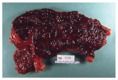

Misunderstanding Toxic Colitis

When the syndrome of toxic colitis (not toxic megacolon) persists beyond a few days, an immediate decision must be made either to try cyclosporine or infliximab, or to proceed directly to colectomy.


Choosing the Wrong Goals of Therapy

Coming from an internationally recognized gastroenterologist, Sachar’s thoughts on this subject are worth quoting verbatim, even though in this text primarily for surgeons, he would be truly preaching to the choir:


We too readily accept as a criterion of success the ability to keep our patients from surgery. Somehow the internist tends to view surgery as a “last resort” or as an indication of “failure” of medical therapy. In adopting such an attitude, we render our patients a terrible disservice. The object of treatment should not be simply “the avoidance of surgery,” but rather to make our patients well. To be sure, if we can accomplish this purpose with our panoply of pills, powders, and potions, well and good. But if we can restore patients to good health and well-being more swiftly, safely, and surely with surgery, then we should not hesitate to do so. Making people better is, after all, the name of the game.

And it is obviously the name of the game for every one of us—internist, gastroenterologist, and surgeon.


▶ SURGICAL MANAGEMENT


Preparation of the Patient

Preparation of the patient for elective surgery is not significantly different whether the procedure is resection for IBD or surgery for cancer. It is a wise idea, however, to limit the amount of laxative administered. In fact, if a patient is troubled by diarrhea, a preoperative cathartic should be avoided. On the morning of surgery, an enema may be carefully administered until the returns are clear. This is the only mechanical preparation advised for patients with severe bowel frequency problems. Those who are to undergo small bowel resection for Crohn’s disease do not require a mechanical preparation unless the possibility of colonic resection also exists. Intravenous antibiotics are administered as described in Chapter 23.

Only gold members can continue reading. Log In or Register to continue

Stay updated, free articles. Join our Telegram channel

Jul 17, 2016 | Posted by in GASTROENTEROLOGY | Comments Off on Ulcerative Colitis

Full access? Get Clinical Tree

Get Clinical Tree app for offline access