Carcinoma of the Rectum


Carcinoma of the Rectum


E. Leslie Bokey


Doctors without anatomy are like moles: they work in the dark and their daily tasks are mole hills.

—FRIEDRICH TIEDEMANN (1781-1861)

The management of patients with rectal cancer is very much a team effort, and I would like to thank three members of my team who have assisted me with the preparation of this chapter: Professor Pierre Chapuis, who has maintained our database; our statistician, Professor Owen Dent; and senior lecturer, Dr. Scott MacKenzie.

Colorectal cancer is the second most common cancer among women worldwide and the third most common among men.173 In high-incidence countries, approximately 25% of colorectal cancers are located in the rectum; 21% among women and 30% among men. Surgical technique is important in the treatment of rectal cancer, and a clear understanding of the anatomy of the rectum and its fascial relationships is therefore essential.

Over the past 30 years or so, many advances have been made in the management of rectal cancer. These include earlier diagnosis, improved preoperative assessment, implementation of adjuvant therapy, better surgical technique, enhanced operating room lighting, and upgraded instruments. A better understanding of fecal continence and the importance of the levator muscle in that regard has enabled a profound increase in sphincter-saving procedures, whereas in those patients requiring a permanent colostomy signal advances in enterostomal care have improved their quality of life.


▶ SYMPTOMS AND SIGNS OF RECTAL CANCER

Many patients with rectal cancer are asymptomatic and are discovered after having a positive fecal occult blood test. Some lesions are found on “routine” endoscopy, whereas others are identified while undergoing a screening colonoscopy for a family history of colorectal cancer or a history of inflammatory bowel disease. Occasionally, a patient may
present with a CT scan or an ultrasound, which may have been performed for another reason, showing an abnormality in the pelvis or liver or other site of metastases.

The most common symptoms of rectal cancer include bleeding per rectum, mucous discharge, unexplained weight loss, a feeling of incomplete evacuation from the rectum, or a persisting change in bowel habit, the last often taking the form of spurious diarrhea. Rectal or pelvic pain that radiates is often a symptom of a locally advanced tumor.


▶ TRENDS IN THE MANAGEMENT OF RECTAL CANCER

The history of the surgical management of rectal cancer will be outlined later. Briefly, there has been a trend away from abdominoperineal resection (APR) of the rectum toward that of restorative procedures. Furthermore, there has been a trend for these procedures to be performed by specialist colorectal surgeons because it has become clear that surgical technique65,248 and volume of work12,460 can influence outcome.

There is good evidence that neoadjuvant radiotherapy and chemoradiotherapy can reduce the risk of local recurrence. Therefore, close collaboration with medical and radiation oncologists is essential in the management of each individual patient.298 In fact, a wider range of experts is now required to treat patients with rectal cancer. These include enterostomal therapists, psychologists, radiologists, anatomical pathologists, as well as radiation and medical oncologists. Most centers now conduct regular multidisciplinary meetings to canvas opinions and to plan treatment options for each patient.

In this chapter, I will deal mainly with open surgery, but I will also include current views on laparoscopic and robotically assisted surgery for rectal cancer. Although there is evidence that laparoscopic surgery is as satisfactory as open surgery for colon cancer,26,378 the evidence for its value with rectal cancer is not yet at hand.503,530


▶ DECIDING THE BEST OPTIONS FOR PATIENTS WITH RECTAL CANCER

There are many options that can be considered in the management of patients with rectal cancer. In many individuals, a decision must be made whether to recommend a restorative or a nonrestorative procedure and whether to recommend preoperative (neoadjuvant) therapy. In a smaller number of patients, other procedures may need to be considered, such as local operations and that of palliative care. Before taking a final decision, several clinical and histopathologic features, as well as certain specific investigations, can be used to help both the surgeon and the patient to decide on the best option. I believe that treating a patient with rectal cancer is analogous to ordering a hand-tailored suit rather than accepting an off-the-rack product.


▶ INVESTIGATIONS

If the entire colon cannot be visualized at colonoscopy, then occasionally a contrast study may be useful in order to exclude synchronous lesions, provided there is no obstruction.

A CT scan of the chest and abdomen should also be performed to document distant metastases. A pelvic MRI is now considered essential in assessing the extent of local tumor invasion and, in particular, in assessing whether the tumor extends beyond the perirectal fascia to potentially involve a postoperative circumferential margin (CRM). This information is important when the patient is a candidate for preoperative chemoradiotherapy. Logically, preoperative MRI cannot validly predict an involved circumferential margin, but it should guide the surgeon in determining patients at risk and in choosing the appropriate operation. Indeed, a patient with a positive MRI margin, if properly managed, should have a negative histologic CRM.144

Endoluminal ultrasound assessment of low tumors is also useful in determining local invasion and is described in full later. Other investigations include a complete blood count, liver and renal function tests, baseline carcinoembryonic antigen (CEA), and cardiac and respiratory assessment.


▶ FACTORS INFLUENCING THE CHOICE OF OPERATION

To save or not to save the sphincter is a perennial question. Is there a level below which an anastomosis should not be attempted? When is an APR inappropriate or an anterior resection the operation of choice? Unfortunately, there is no reliable answer to these questions. Some may even advise with the simple adage, “if you can feel the lesion you should not perform a sphincter-saving operation”—the rule of the index finger. However, this is much too simplistic an approach and may prejudice the surgeon to embark upon an inappropriate proctectomy.

Conversely, surgeons, in a zealous effort to avoid a colostomy and to reestablish intestinal continuity, may compromise the margins of resection. The consequences can be tragic: recurrent disease, anastomotic obstruction, unremitting pelvic pain, and the requirement for subsequent surgery, including a colostomy. An important factor that may influence the choice of operation is the experience of the surgeon. In most developed countries, there are centers that specialize in colorectal surgery. Moreover, there is evidence that volume of work12,460 and technique65,248 are independent factors that affect outcomes. Variables that are helpful in determining the choice of operation for cancer in the rectum are summarized as follows:


  • Level of the tumor


  • Macroscopic appearance (ulcerated, polypoid)


  • Extent of circumferential involvement


  • Fixity


  • Degree of differentiation (histologic appearance)


  • Endorectal ultrasound


  • CT scanning


  • Magnetic resonance imaging


  • Positron emission tomography (PET) scanning


  • Presacral adenopathy


  • Body habitus


  • Gender


  • Age


  • Metastatic disease


  • Other systemic disease


  • Other conditions that may affect one’s ability to manage a colostomy (e.g., blindness, severe arthritis, mental incapacity)


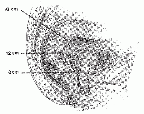
FIGURE 24-1. Distance of tumor from anal verge measured by rigid sigmoidoscopy.

The reader is referred to a publication by the American Society of Colon and Rectal Surgeons. This organization has established practice parameters for the preoperative evaluation and treatment of rectal cancer.611


Level of the Lesion

The distance of the lower edge of the tumor from the anal verge is probably the single most important variable that aids the surgeon in the choice of operation. This distance should be carefully measured using the rigid proctosigmoidoscope, and the result should be recorded (Figure 24-1). The flexible sigmoidoscope is not as accurate for this determination. When measuring, care must be taken to spread the buttocks, so that the instrument can be seen emerging from the anus, not from the contour of the buttocks. The preconceived notion that a tumor 7 cm from the anal verge requires APR but one at 8 cm can be treated by anterior resection is erroneous. Other factors may prove the opposite to be true in both cases (e.g., fixity, size, pelvic anatomy).


Macroscopic Appearance and Level of the Tumor

Ascertaining the appearance of the lesion, whether ulcerated, scirrhous (infiltrative), or polypoid, is very helpful in aiding the surgeon in the choice of operation (Figure 24-2). Generally, the distal margin of resection should be at least 1 cm; but for infiltrative carcinomas, 1 cm may not be sufficient to avoid local recurrence, even if the tumor is at or above 7 cm from the anal verge. The length of distal intramural spread of tumor in resected specimens is extremely variable, with three-fourths of the rectal tumors in one study demonstrated to have no intramural spread.655 A small, exophytic, well-differentiated lesion may be adequately removed with a 1-cm cuff of normal distal bowel.


Extent of Circumferential Involvement

Usually, more highly aggressive tumors tend to involve a greater circumference of the bowel wall. Anteriorly, located lesions have a higher incidence of local recurrence,94 and this should be taken into account when deciding whether or not to include neoadjuvant therapy in the management plan. The depth of local anterior invasion also has to be considered when planning the extent of surgical excision. For example, in females, one may have to include an en bloc hysterectomy or an en bloc disk excision of the posterior wall of the vagina. In selected male patients with anterior invasion of the prostate gland, consideration may be given to prostatectomy, and in those with invasion of the bladder, consideration should be given to partial or total cystectomy after consultation with and seeking the assistance of a specialist urologist.


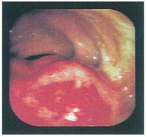
FIGURE 24-2. Endoscopy demonstrates macroscopic appearance of exophytic malignant tumor.


Fixity

Fixity is best assessed by an examination under anesthesia (EUA). Fixity of the tumor in the pelvis implies locally advanced disease, a greater likelihood of residual tumor following resection, and hence local recurrence. Under these circumstances, an APR may offer a better chance of avoiding local recurrence. The presence of a fixed tumor usually signals the need for neoadjuvant therapy (see later).


Histologic Appearance

A biopsy is, of course, mandatory and is done routinely, usually at the time of initial discovery of the lesion. Ideally, the material obtained should be from the edge of the lesion because much useful information can be achieved. For example, an “expanding” margin implies that the area of invasion is pushing or reasonably well circumscribed, whereas an “infiltrating” margin suggests diffuse or widespread penetration of normal tissue.289 All too often, however, the surgeon pays scant attention to the details of the report, except for noting whether the tumor is indeed malignant. However, it is important to be aware of the specific histologic appearance of a malignant tumor. Is it poorly differentiated, moderately well-differentiated, or well-differentiated (see Figures 23-19,23-20,23-21 and 23-22)? Generally, tumors regarded as poorly differentiated have highly irregular glands or no glandular differentiation.289 The more anaplastic, the more aggressive is the lesion; the more aggressive, the greater is the resection margin that would be required. The chance of local recurrence is much higher with a poorly differentiated cancer than with one that is well-differentiated. It is axiomatic that one must choose the most favorable cancers for performing less than a radical resection (see later). Therefore, one cannot overestimate the importance of degree of differentiation (Broders’ classification), the depth of penetration, and the
presence or absence of venous or perineural invasion (PNI) in making the appropriate choice. Shirouzu and colleagues evaluated whether PNI is an independent prognostic factor in individuals who underwent curative surgery.566 There was a significant difference in local recurrence rates between those individuals with stage III lesions who were found to have PNI and those without PNI. In addition, the investigators found that patients with PNI and stage III lesions had a significantly lower survival rate. Nevertheless, the importance of PNI as an independent prognostic factor needs further evaluation (see also Chapter 23).


Gagliardi and colleagues studied the effect of microacinar growth patterns on survival following radical surgery for rectal cancer in 138 consecutive patients.192 They found that acinar size (whether microacinar or macroacinar) had independent prognostic value. Patients with microacinar tumors had a significantly reduced 5-year survival rate compared with those with macroacinar lesions. Saclarides and colleagues attempted to determine which features were predictors of nodal metastases.536 They used 9 histologic and morphologic features of 62 radically excised rectal cancers to determine which were associated with nodal disease. Statistically significant variables were worsening differentiation, increasing depth of penetration, microtubular configuration of 20% or more, the presence of venous invasion or PNI, and, of course, lymphatic invasion. Exophytic tumor morphology, mitotic count, and tumor size were not significant predictors.536 In an analysis of all the variables or combination of factors, Broders’ classification was the strongest predictor of nodal disease.

A particularly useful microscopic variable to identify is the extent of lymphocytic infiltration at the border of the tumor.288 Jass and colleagues regarded this observation as “conspicuous” when there is a distinctive and delicate connective tissue mantle or cap at the invasive margin of the growth.289 Patients who harbor tumors that demonstrate pronounced lymphocytic infiltration have a better prognosis than those who do not. If the pathologist fails to supply this information, he or she should be asked to review the slides and to amend the report. Optimally, the surgeon should view the histologic evidence himself or herself in order to make the most reasoned recommendation to the patient.


Presacral Adenopathy

By careful palpation of the rectum with the patient in the knee-chest position, the surgeon can occasionally identify a hard lesion outside the rectal wall, presumably a lymph node with metastasis. This should be taken into account when formulating a treatment plan. Endorectal ultrasound is much more accurate for identifying enlarged and presumably involved lymph nodes in this area, but this does not diminish the importance of careful palpation.


Imaging Techniques

Several imaging modalities exist today for preoperative staging of rectal cancer, including endorectal ultrasonography, CT, MRI, and PET.451,576,662 All of these modalities are complementary, and each has a specific role.


Computed Tomography

CT is of great value in the diagnosis of metastatic disease, especially in the liver (see Figure 23-17) and elsewhere within the abdomen (Figure 24-3) or the chest.


Magnetic Resonance Imaging

MRI is used for preoperative assessment of local invasion. It allows accurate assessment of size and relationship of the tumor to the perirectal fascia.477 Brown and coworkers used high-resolution MRI and compared their stagings with the pathologic specimens.77 There was a 94% weighted agreement between MRI and pathology assessment of T stage.

An improvement in MRI technique is the use of powerful gradient coil systems and high-resolution surface coils to allow a higher definition of obtainable image with a smaller field of view.317


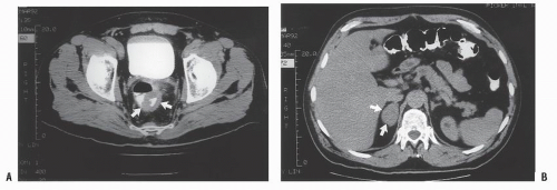
FIGURE 24-3. A: Computed tomography demonstrates a tumor in the wall of the rectum (arrows). B: Metastatic tumor to the adrenal gland in this same patient can be appreciated (arrows).


Endorectal or Transrectal Ultrasound Examination

Endorectal ultrasound (Brüel & Kjær Instruments, Inc., North Billerica, MA) (see Figure 7-18) was first described by Beynon in 1986.51 Since then, both the technique and the equipment have improved, and the method has developed into an extremely useful tool for the preoperative assessment of patients with rectal cancer.40,619 After a small enema is administered, the probe is introduced into the rectum beyond the tumor (Figure 24-4). A balloon is filled with approximately 50 mL of water, and an acoustic contact is produced between the rotating part of the transducer and the rectal wall.257 During withdrawal, the monitor is observed and the findings recorded. Each of the layers of the rectum can be sonographically visualized, with a tumor usually appearing as a hypoechoic disruption of the rectal wall. The procedure may also reveal whether underlying lymph nodes are affected. Detection of invasive carcinoma within an otherwise villous lesion can be achieved with this technique.5,337 In the experience of Hildebrandt and colleagues, lymph node metastases can be predicted with an accuracy of 72% and inflammatory lymph nodes with a specificity of 83%.258 Many investigators, however, have expressed concern about the lack of specificity in distinguishing benign from malignant nodes.293,529 The Memorial Sloan-Kettering Cancer Center group observed that the overall risk of undetected and untreated (if a local procedure is embarked upon) is 15%.64


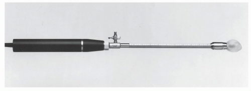
FIGURE 24-4. Ultrasound probe. (Courtesy of Brüel & Kjær Instruments, Inc., North Billerica, MA.)

Accurate preoperative assessment of local invasion with endorectal ultrasound implies the patients may be selected for a less than radical operation (see Other Local Procedures for Rectal Cancer).206 There is uniformity of agreement that optimal results can only be obtained if there is consistency in technique and in interpretation. Certainly, accuracy improves considerably with experience.469 MacKay and colleagues emphasized that one needs to undertake 50 or more ultrasound procedures before optimal accuracy is achieved.382 It is therefore preferable for the surgeon to be the individual responsible for performing and evaluating the study. Some investigators have commented that tumors of the lower rectum are incorrectly assessed much more frequently than those of the middle and upper rectum.253


Technique

The late Douglas Wong was regarded as an international authority on TRUS and in its interpretation. He previously had been kind enough to provide me a narrative of his technique, and I thought it would be useful to reproduce it here:

The patient is instructed to take a small volume enema 1 hour prior to the examination. The procedure is
carefully explained to the patient and pertinent questions answered. The assistant enters the demographic data in the ultrasound computer as well as the frequency of the ultrasound probe and its focal range. The patient is placed in the left lateral position on the examining table. The study is preceded by a digital rectal examination to evaluate the size, fixation, location, and morphology of the rectal lesion. In most instances the use of a large-bore proctoscope serves several purposes. It allows visual examination of the rectal tumor with exact determination of its location both with respect to circumferential involvement of the rectal wall and the distance from the anal verge. Secondly, it allows suctioning of any residual stool or enema fluid that might interfere with the acoustic pathways of the ultrasound waves that may distort the image. Most important, however, it allows easy passage of the probe above the tumor to ensure that the transducer is advanced above the rectal lesion to allow complete imaging. This is significant since the lower border of a rectal cancer can differ in the depth of invasion than the center or upper portions of the cancer and lymph nodes in the perirectal region are often seen just above the level of the tumor. They will be missed if complete imaging is not obtained. Small distal lesions can be adequately imaged with the ultrasound inserted blindly and advanced above the lesion, but for most midrectal, bulky tumors the use of a proctoscope will facilitate the passage of the transducer.


The probe is prepared by placing a condom over the transducer head followed by a metal ring or rubber band that secures the base of the balloon. The assistant holds the probe with the balloon in the most dependent position and fills the balloon with about 50 mL of water via the connector at the base of the metal shaft. Any air in the system is aspirated through the syringe and expelled. A water-soluble lubricant is liberally applied to the outside of the balloon. The probe is now ready for insertion. The proctoscope is then advanced above the rectal lesion, and water-soluble lubricant is inserted into the lumen of the proctoscope to facilitate passage of the probe. The probe is then gently introduced through the proctoscope and advanced such that the transducer is sited above the rectal cancer. Once the 20 cm mark on the shaft of the probe is at the proximal end of the proctoscope, the proctoscope is then pulled back on the probe as far as possible, thus exposing the transducer for 7 cm beyond the end of the instrument and positioned about the rectal cancer. The balloon is then instilled with 30 to 60 mL of water; this is the volume required to obtain optimal imaging. The transducer is activated by depressing the button on the proximal end of the probe, and the image on the screen is visualized.

When the connector for introducing the water into the balloon is pointing upwards (towards the ceiling), by convention the anterior aspect of the rectum will be on the superior part of the screen, the right lateral rectum will be on the examiner’s left, the left lateral wall will be on the examiner’s right, and the posterior rectum will be in the lower screen. The tip of the ultrasound probe should be maintained in the center
of the rectal lumen in order to achieve optimal imaging of the rectal wall and perirectal structures. Some adjustments may have to be made on the gain of the ultrasound unit in order to provide better imaging. Occasionally, it is possible to perfectly depict all five layers of the rectum circumferentially, but usually only a portion of the rectal wall at a time will be optimally imaged. Minor adjustments will have to be made in the location of the probe relative to the rectal wall at various locations to optimally image all five layers clearly. Once optimal imaging of the rectal wall and surrounding structures has been achieved, the ultrasound probe is gradually withdrawn while carefully observing the screen and the images obtained. Several hard-copy images should be obtained for future reference through the use of a Polaroid image recorder. These images can be obtained by stopping the rotation of the transducer by depressing the activation/deactivation button and holding it longer, thus activating the recorder.

When a critical area of the tumor needs to be visualized at a higher magnification, the window can be activated which gives a large image of the area being examined. The entire length of the rectal tumor is evaluated. It is not uncommon for one to perform several passes along the full length of the tumor in order to acquire all the relevant information.


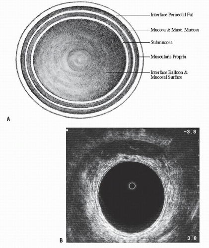
FIGURE 24-5. Endorectal ultrasound. A: Schematic diagram of normal rectal wall anatomic layers. B: Endorectal ultrasound imaging of normal rectum.

Attention must also be focused on the perirectal tissues in order to search for potentially involved lymph nodes. In general, normal lymph nodes are not visualized with the ultrasound, and, therefore, any hypoechoic structure in the surrounding perirectal tissue should be suspected of harboring metastatic disease. Lymph nodes often exhibit hypoechoic echogenicity comparable to that of the primary tumor, are more often round than oval, and are frequently irregular in appearance. Lymph nodes can be distinguished from blood vessels that are also circular hypoechoic areas, but when followed distally and proximally, they seem to extend further and may be seen to elongate and to branch. Once the study is completed, the balloon is deflated, and the probe is removed. Figure 24-5 illustrates the five layers of the rectal wall seen schematically and in the TRUS image of the normal rectum. The inner white
line represents the interface of the balloon with the mucosal surface of the rectal wall. The inner black line represents the mucosa and muscularis mucosa, whereas the middle white line corresponds to the submucosa. It is this middle white line that is the most crucial layer to visualize in order to determine whether the tumor is invasive.292 The outer black line corresponds to the muscularis propria, whereas the outer white line represents the interface between the muscularis propria and the perirectal fat. Once it has been ascertained that the middle white line is broken, then the presence of an invasive tumor has been confirmed. It is then a matter of determining the depth of invasion. Figure 24-6 demonstrates an ultrasound of a minimally invasive cancer with the corresponding resected, microscopic area. Figure 24-7 shows transmural involvement by tumor.


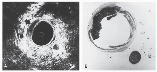
FIGURE 24-6. Endorectal ultrasound. A: Sonogram of a rectal cancer confined to wall of the bowel (arrow). The black cavity is the waterfilled balloon with the white circle in the center corresponding to the transducer. A hyperechoic lymph node can be seen in the lower right. B: Corresponding photomicrograph of the excised specimen confirms the depth of invasion by tumor. The lymph node in the lower right was free of tumor. (Courtesy of Ulrich Hildebrandt, MD.)


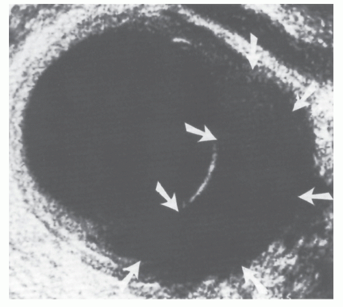
FIGURE 24-7. Endorectal ultrasound. Sonogram of a rectal cancer that penetrates into perirectal fat. Note the hypoechoic area (arrows). The outermost layer of the bowel wall has been interrupted. (Courtesy of Ulrich Hildebrandt, MD.)

The TNM (tumor, node, metastasis) classification is used with a u modifier to describe depth of invasion and the presence or absence of metastatic lymph nodes as described by Beynon and coworkers51,53:


T Stage


Ultrasound Characteristics


uT0


Noninvasive lesion. Hyperechoic submucosal interface is intact.


uT1


Invasion of submucosa only. Hyperechoic middle white line is stippled and irregular but not disrupted.


uT2


Breaching of hyperechoic middle white lineindicates invasion of hypoechoic muscularis propria (Figure 24-8). Outer white line is intact. Deep uT2 lesions have a scalloped appearance.


uT3


Invasion through muscularis propria into perirectal fat (Figure 24-7). Outer (hyperechoic) white line (junction of muscularis propria with perirectal fat) is disrupted.


uT4


Extension into adjacent organ or structure (e.g., vagina, prostate, bladder, cervix, seminal vesicles). Plane between any of these structures and the rectum is obliterated.


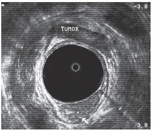
FIGURE 24-8. Endorectal ultrasound scan of a uT2 tumor. Note disruption of the middle white line with the intact outer white line.

In the foregoing, uN0 represents evidence of lymph nodes, uN1 represents lymph nodes positive for tumor, MX indicates metastatic tumor status unknown, M0 represents no evidence of metastatic tumor, and M1 means that metastatic tumor is present.

In addition to the cited indications, this technique offers the opportunity for clear visualization of the full thickness of an anastomotic area in those patients who have undergone restoration of rectal continuity, especially with respect to the possibility of early detection of recurrent cancer and for assessing the effects of preoperative radiotherapy.52,102,296,400,454,519 Further information can be obtained by means of ultrasonographically guided biopsy and histologic determination of lymph node status.562,664


Published Experience and Comparative Results

The Creighton University group in Omaha, Nebraska, performed preoperative ultrasound staging on 107 patients with rectal cancer.4 TRUS identified 18 of 19 patients with uT3 tumors and 44 of 51 individuals with less invasion than uT3 (86.3%). Garcia-Aguilar and colleagues reported the University of Minnesota group’s experience of 1,184 patients with rectal adenocarcinoma or villous adenoma who underwent TRUS, comparing their assessment with pathologic specimens obtained by either resection or transanal excision without neoadjuvant treatment.198 Somewhat disappointingly, overall accuracy in assessing the degree of rectal wall invasion was only 69%, with 18% overstaged and 13% understaged. Overall accuracy in assessing nodal involvement in those who underwent radical surgery (238 patients) was 64%, with 25% overstaged and 11% understaged. The primary benefits appear to have been in determining which tumors were benign and those that either went completely through the rectal wall and those that did not.198


Positron Emission Tomography

As yet, there is no evidence base for preoperative PET “T and N” staging of rectal cancer, and the technique is not routinely used for this purpose.451,662 However, PET may assist in identifying systemic distant metastases when CT results are equivocal.565


Rectal Endoscopic Lymphoscintigraphy

Endoscopic lymphoscintigraphy is not in routine practice and is still being evaluated.42 Arnaud and colleagues performed preoperative evaluation of patients with known rectal cancer in order to identify evidence of lymphatic spread.13 The procedure involves the endoscopic injection of 0.1 mL of radiocolloid (rhenium sulfur marked with 99mTc) into the submucosa of the extraperitoneal rectum bilaterally. The diffusion of the tracer along the lymphatics was registered by means of a computerized gamma camera. In 10 control subjects and in a series of 85 patients with rectal cancer, the technique was found to have a sensitivity rate of 85%, a specificity of 68%, an overall accuracy of 76%, a positive predictive value of 71%, and a negative predictive value of 71%. The authors concluded that rectal endoscopic lymphoscintigraphy represented the only currently available method for evaluating lymphatic spread in rectal cancer.13


Other Factors That May Influence Management Plan and Surgical Procedure

The previously discussed methods of evaluation concern the tumor itself. The following factors are patient related and may also influence the choice of operation.


Body Habitus

A rectal resection carried out on an asthenic patient usually permits a technically lower anastomosis than does an operation for the same level of lesion in an obese individual. Body habitus is an important factor to consider when deciding whether or not a restorative procedure is possible. As a rule, however, I try to make that decision with the patient before the operation rather than at the time of surgery.


Gender

An anastomotic procedure is more likely to be possible in women than in men. A broad pelvis, furthermore, usually permits a wider resection, whereas a narrow pelvis tends to impede dissection, potentially limiting the adequacy of tumor margins and the use of conventional anastomotic techniques. This is especially true when one performs a low anterior resection.


Age

Age is not a contraindication to a restorative procedure. However, one has to take into account continence and bowel dysfunction in an elderly patient after a low or ultralow restorative procedure. In some elderly and frail patients, it may be more prudent to perform an APR or even a low Hartmann procedure rather than a restorative operation. A radical resection is not contraindicated in patients solely because of advanced age. However, some elderly patients may be better suited to a local approach to treatment.


Metastatic Disease

If metastatic disease is present at the time of diagnosis, then the extent has to be carefully evaluated. This is preferentially accomplished by means of PET scanning. A multidisciplinary team approach is very useful. If there is significant intra-abdominal metastatic disease, a restorative procedure aimed at avoiding a colostomy may not be the ideal solution because there is a high risk of developing bowel obstruction that may require further operation and a stoma. In some patients with extensive metastatic disease at the time of operation, a Hartmann procedure or even a defunctioning loop stoma may be preferable. Alternatively, a local procedure or other nonsurgical form of therapy may suffice to palliate the individual’s symptoms.


Comorbidities at the Time of Surgery

Patients, regardless of age, are at increased surgical risk if comorbidities are present (e.g., cardiovascular, pulmonary, renal). In those with significant or multiple comorbidities, a procedure that does not involve an anastomosis may be considered safer.

Ondrula and colleagues analyzed the predictive value of a number of preoperative risk factors on operative outcomes in 825 patients.466 Those factors that were found to be statistically significant in predicting a greater risk were emergency operation, age greater than or equal to 75 years, congestive heart failure, prior abdominal or pelvic radiation therapy, corticosteroid use, serum albumin less than 2.7 g/dL, chronic obstructive pulmonary disease, prior myocardial infarction, diabetes, cirrhosis, and renal insufficiency. The authors assigned a “risk score” for each category in order to determine a strategy for management based on the sum total of the risks.


Other Conditions

Avoiding a colostomy in a patient who cannot cope with an appliance or a stoma is an unusual, albeit legitimate, reason for choosing an alternative procedure. This can happen if the individual is blind, has severe impairment in the use of hands (e.g., arthritis), or cannot be taught, or if there is no family or community support.


Preoperative Assessment Under Anesthesia

Examination under anesthesia is very useful for assessing patients with a low rectal cancer. Valuable information can be gathered about the circumferential location of the tumor, the distance of the tumor from the anal verge, and the presence and extent of tumor tethering or fixity. All of these are important in determining the choice of operation and whether preoperative adjuvant therapy is appropriate.


Multidisciplinary Meetings

Although such meetings are not mandatory, they are very useful in canvassing the opinions of other specialist nursing and medical staff. The final decision, however, should remain between the surgeon and patient.420,429 One of the main advantages of multidisciplinary meetings is the opportunity to discuss the appropriateness of neoadjuvant therapy.


Neoadjuvant Therapy

Over the past 30 years, there has been considerable debate over the use of neoadjuvant therapy for rectal cancer. In particular, there has been debate whether radiotherapy is useful at all, whether it should be given preoperatively or postoperatively, and whether it is applicable for all rectal cancers or for only those in the lower third of the rectum and for those that are locally advanced. There has been debate whether a long course is superior to that of short-course preoperative radiotherapy82,425,432 and whether adjuvant chemotherapy is useful for rectal cancer.114,447

In this section, I include a brief historical review of the subject and conclude with a summary of current practice.


Historical Review

The rationale for neoadjuvant radiation therapy in the management of rectal cancer is to alter the viability of cancer cells so that they are no longer capable of local implantation.570 When the concept was first proposed, however, there was concern that delay in initiating surgical treatment could increase the risk of tumor spread, but there is no evidence today to suggest that this is in fact the case. The primary issue is identifying those patients in advance who would most likely benefit from neoadjuvant radiotherapy. How can one make this determination? Clinical assessment is probably the gold standard, but endorectal ultrasound, CT, and MRI may also be quite helpful. Staging laparotomy has been suggested in order to determine mobility, resectability, staging, and the possibility of constructing a diversionary stoma before embarking on radiotherapy.81 Some authors believe that every rectal cancer should be treated by preoperative radiation, but most surgeons are selective in their approach. The following variables are generally considered appropriate indications for performing preoperative radiation:


  • Fixed tumor


  • Evidence of ureteric obstruction


  • Invasion of adjacent structures (e.g., bladder, seminal vesicles, vagina)


  • Presacral adenopathy


  • Anal canal invasion


  • Ultrasound uT3 or uT4 lesion


  • Poorly differentiated histology

The impetus for the application of preoperative radiotherapy can be attributed to the reports by Jean Papillon in Lyon483 and by Quan and Stearns and their colleagues at the Memorial Sloan-Kettering Cancer Center in New York.346,582,585,586 and 587 Although their initial studies indicated improved survival, a prospective evaluation demonstrated that the overall survival rate was not better, but that the incidence of failure because of local recurrence was reduced.587 At that institution, Stearns and colleagues used external radiation through opposing anterior and posterior portals, 2.5 Gy daily to 20 Gy. The resection was carried out 2 days to 6 weeks following treatment.

The classic article that evaluated preoperative radiation was reported by Dwight and Higgins and their colleagues from the Veterans Administration (VA).157 This study randomly allocated 700 men either to surgery or to preoperative radiotherapy plus surgery. These investigators found a statistically significant decreased incidence of positive nodes in the irradiated group and a lower incidence of recurrent disease in those who died. Roswit and associates used 20 Gy over 2 weeks, with a booster dose of 5 Gy if the tumor was less than 9 cm from the anal verge.527

Since these initial reports, there have been numerous papers that attest to the success of radiotherapy in reducing the size of the tumor, downstaging the degree of invasion, and decreasing
the risk of local recurrence.69,73,84,101,118,119 and 120,128,152,174,178,194,197,200,201,224,254,280,286,303,322,399,413,414,415,416,417 and 418,427,474,483,521,524,526,571,574,577,615,626 Some have stated that survival rates are better.181,605 Kandioler and coworkers observe that a tumor with a normal p53 genotype is predictive for response to preoperative short-term radiotherapy and increased patient survival.300 However, there remains disagreement whether preoperative radiation therapy has any effect on survival.204


Methods

Minsky commented about the weaknesses of the prospective, randomized trials.523 He stated that none uses standard dosages of radiation therapy. He also opined that the interval between the completion of radiation and surgery is generally considered to be inadequate. The Memorial Sloan-Kettering Cancer Center group demonstrated a trend toward an increased pathologic response rate and downstaging when an interval between completing the radiotherapy and surgery is at least 44 days.434 Most recommend 4 to 6 weeks following completion of the treatment in order to achieve maximum downstaging and tissue recovery. Moreover, he affirmed that using anteriorposterior/posterior-anterior portals, rather than multiple-field techniques, predisposes to increased morbidity associated with the radiation.424 Others confirmed that the morbidity and mortality of both preoperative and postoperative radiotherapy are higher when two-portal rather than three-portal or four-portal radiation technique is employed.467 This is especially true for elderly patients who may have an increased risk of impairment for blood supply. Stein and colleagues concluded in the analysis of their patients that a longer time interval (beyond 8 weeks) between completion of neoadjuvant chemoradiation and surgical resection did not increase the tumor response rate or reduce the morbidity associated with the surgery.591

There is considerable controversy as to what the optimal dose for preoperative radiation treatment should be. Some have recommended a short course of high-dose therapy, whereas most centers in the United States suggest 40 to 45 Gy, delivered in 4 to 6 weeks. Surgery is recommended approximately 4 to 6 weeks after the completion of the treatment because tumoricidal benefits may continue for some time. Data show no increased morbidity or mortality associated with supplementary treatment.155,504


▶ SURGICAL MANAGEMENT OF RECTAL CANCER


Historical Notes

Colostomy as a diverting procedure has its origins in antiquity. Praxagoras (c. 400 bc) was alleged to have employed some form of decompression maneuver for ileus. Alexis Littre (1710) is usually credited with the concept
of ultimately performing a colostomy when he undertook a postmortem examination on an infant who died with an imperforate anus. He is quoted as stating, “… it would be necessary to make an incision into the belly, open the two ends of the closed bowel,… bring the bowel to the surface of the body wall where it would never close, but perform the function of an anus.”360 Colostomy, however, did not achieve an important role until Amussat (1839),9 a French surgeon, urged that it be the routine procedure for obstructing rectal cancer.386 For most of the 19th and into the 20th century, the stoma was placed in the inguinal or iliac region. This avoided entering the peritoneal cavity. Luke,379 however, was an exception. He was the first to bring the bowel out in the area of the rectus muscle, whereas Deaver134 was an advocate of lumbar colostomy.


The treatment of carcinoma of the rectum by some form of excisional or amputative procedure dates back more than 250 years, but it was not until 1826 that Lisfranc successfully
excised the rectum for this condition.359 His was a transanal approach and, as such, was of necessity, used only for low-lying rectal lesions. This procedure and merely supportive care were the only available options. Modifications were introduced by von Volkmann,631 Cripps,116 and others, but the results of the perineal operation were poor. There was frequent incontinence, a high recurrence rate, and a high mortality rate.512


In 1885, Kraske removed the coccyx and part of the sacrum (a maneuver that had been accomplished many times as an extension of the perineal proctectomy), but he preserved the anus and sphincters to effect an anastomosis.325,326 Often, however, continuity of the bowel could not be restored, either because of too much tension on the upper segment, impairment of the blood supply, or both, and the procedure was often completed by the establishment of a sacral anus. The operation was quite popular for a time but ultimately fell into disrepute because of the complications of sepsis, anastomotic leak, and recurrent disease.


In 1894, Czerny was unable to remove a rectal cancer and combined the extirpation with an abdominal operation, thus
becoming the first person to perform an APR.127 In 1908, Miles described his modification of Czerny’s operation, placing emphasis on meticulous dissection and removing the zone of upward spread of the cancer (Figure 24-9).421 He concluded as follows:


(1) that an abdominal anus is a necessity; (2) that the whole of the pelvic colon, with the exception of the part from which the colostomy is made, must be removed because its blood-supply is contained in the zone of upward spread; (3) that the whole of the pelvic mesocolon below the point where it crosses the common iliac artery, together with a strip of peritoneum at least one-inch wide on either side of it, must be cleared away; (4) that the group of lymph nodes situated over the bifurcation of the common iliac artery are in all instances to be removed; and lastly (5) that the perineal portion of the operation should be carried out as widely as possible so that the lateral and downward zones of spread may be effectively extirpated.


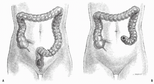
FIGURE 24-9. Carcinoma of the rectum. A: Extent of removal in classic abdominoperineal resection. B: The sigmoid colostomy is created in the left iliac fossa.

Although initially presenting 12 patients, with an operative mortality of 42%, Miles believed that with improved technique and further experience, the operation could be performed relatively safely. Later, as performed by him, it was a most impressive display of operative technique—one of the noted sites of London surgery in the 1920s and 1930s.210 The abdominal phase, carried out with the patient lying flat on the table and
in a steep Trendelenburg tilt, seldom took more than 35 to 40 minutes. Miles was quite prescient: the Miles resection has become the standard operation for the treatment of cancers of the low rectum. A presumably less radical but perhaps safer procedure was the attitude taken by Miles’ rival, Percy Lockhart-Mummery (see Biography, Chapter 12), who favored a perineal excision, preceded 2 or 3 weeks earlier by a minilaparotomy to determine that the growth was resectable.210 When this was the case, a loop-iliac colostomy was established.


Another option was proposed by Gabriel, an assistant to Lockhart-Mummery for many years.188 Gabriel started out as a perineal excisionist, but after careful study of the lymphatic spread of rectal cancer, he switched to his version of proctectomy, the perineoabdominal excision.

In 1939, Lloyd-Davies361 reaffirmed the value of the synchronous-combined (two-team) APR that had been originally proposed by Mayo as early as 1904.408 Mayo suggested that the synchronous-combined approach be considered “if the
surgeon has a good assistant.” By 1963, the Lloyd-Davies technique was the most commonly employed alternative at the St. Mark’s Hospital, and the operative mortality had been reduced to less than 3%.439 It is particularly helpful when one is confronted with a bulky or fixed tumor or a patient with a narrow pelvis. With all appropriate deference to Miles and to his statement that the operation takes no more than 1 hour and that his patients suffer “no more shock than after an ordinary perineal excision,”421 blood loss can be reduced and operative time decreased by using a two-team method. Interestingly, Miles’ resection as performed by Richard Cattell of the Lahey Clinic in Boston (see Biography, Chapter 31) was called by his assistants “the hour of charm.”


Others (e.g., Lahey) believed that if the operation were divided into two stages, it could be better tolerated by the patient.339 The first stage consisted of making a median incision, dividing the sigmoid colon, and creating a left iliac colostomy with a mucous fistula of the distal segment delivered through the lower part of the abdominal incision. Care was taken to preserve the superior hemorrhoidal artery to the distal bowel. In the second stage, the proctectomy was carried out. An alternative operation for treatment of cancer of the middle to upper rectum or of the rectosigmoid was proposed by Hartmann in 1923.240 This procedure succeeded in removing the tumor following establishment of a colostomy, but avoided the perineal dissection. However, the operation was useful for higher lesions only and, of course, was not designed for eventual reestablishment of intestinal continuity, although the Hartmann resection is frequently applied today in the initial surgical management of complicated sigmoid diverticulitis. The original article probably is worth reproducing in translation. It must represent a record for the briefest paper leading to one’s eponymous immortality because Hartmann’s fame is based on only two paragraphs of narrative:


It is the rule that, in order to remove cancers of the distal pelvic colon, it is necessary to perform a very serious operation when removing the rectum by means of an abdominoperineal excision. In two patients who underwent colostomy for intestinal obstruction, at the second operation I limited resection to the intermediate portion of the colon between the artificial anus and the rectum, including the corresponding area of innervation. Following this, I closed the upper end of the rectum and reperitonealized it, without reconstructing the perineal floor. Following the operation both cases
were as uneventful as an operation for a cold appendix. The conservation of a small cul-de-sac of the rectum above the sphincters did not present a particular problem, and follow-up 9 and 10 months later revealed the patients to be quite well.240


The first documented attempt at abdominal resection with restoration of continuity is generally attributed to Reybard of Lyons (see Biography, Chapter 23).515 He performed a partial sigmoid resection; the patient survived approximately 10 months. The fear of sepsis and anastomotic leak inspired Murphy, in 1892, to create his “button” (see Biography, Chapter 23; see Figure 23-35).450 The same year, Maunsell407 reported a technique using an anastomotic method employed by Hochenegg263 in what has come to be called the “pull-through” procedure. A more practical modification of this approach was suggested by Weir644 in 1901.

During the first half of the 20th century, restoration of continuity by means of primary anastomosis evolved through abdominosacral resection and the familiar operation of anterior resection. The introduction of newer suture materials, the advocacy by some of interrupted suture technique, and the application of the stapling devices all indicate that the operation can nevertheless be improved and that the risk of complications can still be diminished.


The Anatomy of the Rectum and Its Fascial Relationships

Surgical technique is an important predictor of outcome for rectal cancer and is intimately related to a clear understanding of the anatomy of the rectum and its fascial relationships. Therefore, before proceeding with a description of operative details, a précis of this subject is presented.


Anatomists generally agree that the rectum commences opposite the sacral promontory and is approximately 15 cm in length, extending from the rectosigmoid junction proximally to the palpable, lower, anorectal ring. If one includes the length of the anal canal (2 to 3 cm), then the rectosigmoid junction is identified approximately at 16 cm from the anal verge when examining an anesthetized patient in the left lateral position with a rigid sigmoidoscope.275 The rectum is usually described as having three segments: an upper, a middle, and a lower.

Although there are excellent contemporary reviews of the surgical anatomy of the rectum and its fascial planes,58,106,199,542,667 the description by the little-known Romanian surgeon-anatomist, Thoma Ionescu (Thomas Jonnesco), is generally considered the “most graphic and complete.”30 In particular, his original description in French of the perirectal fascia in the first edition of Poirier and Charpy’s Traité d’Anatomie Humaine in 1896 has not been surpassed. His description was adopted by Waldeyer in 1899 in his classic text, Das Becken, and remains essentially unchanged in
contemporary anatomy textbooks.96 Knowledge and understanding of this anatomy is the key to identifying the correct plane of mobilization when resecting the rectum for cancer. Indeed, this anatomy has been rediscovered by many surgeons over the past century who were unaware of Ionescu’s singular contribution. For example, Heald rediscovered this anatomy for himself and renamed and popularized the “retrorectal space” and plane as the “holy plane” in his operation of total mesorectal excision or TME.248 However, some would describe Heald’s use of the term “mesorectum” as a misnomer438 or simply incorrect.621 These authors reinforce the key feature, as described by Ionescu, that the perirectal fascia per se takes its origin from the endopelvic parietal fascia off the pelvic side walls to encapsulate the rectum and not from the peritoneum.621 In this manner, surrounded by loose lobulated fat, the rectum is cushioned and cocooned in a serofibrous fascial envelope, together with its lymphatics, vessels, and nodes. Superiorly, the perirectal fascia is continuous with the peritoneum of the pelvic mesocolon at the level of S3 and where there is abundant fat enclosed within. Inferiorly, at its lower third, there is only minimal fat present, and a plane exists between the perirectal fascia and Denonvilliers’ fascia, separating the rectum from the prostate and seminal vesicles. This plane must be recognized in order to avoid unnecessary bleeding from the prostatic venous plexus or damage to the cavernous nerves when mobilizing the rectum anteriorly.358 The key is to dissect behind Denonvilliers’ fascia whenever possible, unless there is strong suspicion of direct involvement by tumor.358


Very likely the term “mesorectum” was introduced by Maunsell in 1892 (see earlier Biography) when describing his pull-through operation407 and was probably based on a misunderstanding of the term “meso” as used by Ionescu in his thesis on the pelvic colon that was published in same year.297 It should be noted that the term “meso” in gross anatomy pertains to two layers of peritoneum that suspend an organ, but as Ionescu quite rightly argued, because the rectum is not suspended but rather lies nestled and closely applied to the sacral hollow, there is no need for “meso,” and the term is therefore inappropriate. For this reason, it does not appear in Nomina Anatomica but is restricted to the Nomina Embryologica. More important, the proper emphasis should be on the precise recognition of the perirectal fascia to mobilize the rectum in the appropriate anatomical plane rather than to perform a TME when resecting the rectum.


Brief Description of the Anatomy of the Pelvic Nerves

In a recent review of the subject, Moszkowicz and colleagues present a summary of the innervation of the pelvis based on anatomy and highlighting where pelvic nerve injury occurs.448 Pelvic innervation is described as comprising supralevator and infralevator compartments. The supralevator pathways consist of the superior hypogastric (SHP) and inferior hypogastric plexuses (IHP). The infralevator compartment is essentially the pudendal nerve. The supralevator compartment consists of the superior hypogastric plexus and hypogastric nerves. These are a continuation of the preaortic sympathetic trunks. They can be readily identified and preserved as has been discussed in Operative Technique.

The parasympathetic nerves (pelvic splanchnic or erectile nerves), however, are not as easily identifiable as the hypogastric nerves. They arise from the ventral roots of S2-4, and they enter the pelvis through the sacral foramina. Small branches of these nerves have been reported in the lateral ligaments.548 Moszkowicz describes the inferior hypogastric plexus as a network of sympathetic and parasympathetic fibers that lie outside the fascia propria. It is located laterally and retroperitoneally on either side of the rectum. Damage to this plexus can cause urogenital and sexual dysfunction.


▶ RESTORATIVE PROCEDURE: ANTERIOR RESECTION


Open Anterior Resection


Technique

The technique that I use has been developed over many years (since 1980). Although it differs from the traditional method of mobilizing the rectum in that I start the dissection on the right side of the mid-pelvis, it is essentially a separation of the rectum along well-defined and previously described anatomical planes. I choose this approach because at that level the retrorectal space is totally avascular, and the risk of obscuring the view with minor bleeding is minimal. The technique incorporates many elements adapted from laparoscopic colorectal surgery (see Chapter 19).


Instruments

The instruments that I commonly use are listed next and are shown in Figure 24-10.


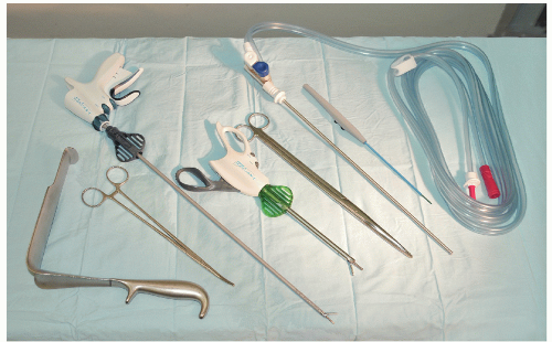
FIGURE 24-10. Instrument tray for anterior resection. From left to right: St. Mark’s retractor, Zenker forceps, Harmonic scalpel, Wave Harmonic scalpel, Abel scissors, sucker irrigation, and long diathermy.


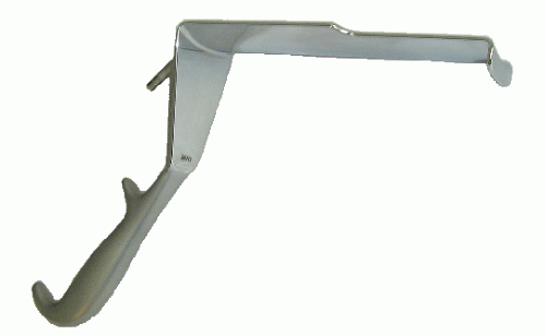
FIGURE 24-11. St. Mark’s pattern retractor.


Long Sucker/Irrigator

I prefer this instrument to the traditional Yankauer suction because of its length and also because I frequently use high-pressure water irrigation to open up planes, especially in redo surgery. Once an incision is made in the correct plane, water irrigation can develop the plane with ease. Furthermore, the long sucker does not have a bend in it. I find that an angled sucker can interfere with the line of vision deep in the pelvis, whereas straight suction permits a clear line of view.


Long Diathermy

A long diathermy is essential because sharp dissection is used throughout the procedure. I do not use hands or fingers for mobilizing.


Harmonic Scalpel

For the past 3 years, I have increasingly adapted the use of different energy sources for open as well as for laparoscopic surgery. I have found the long Harmonic scalpel and the Wave Harmonic scalpel particularly useful; they have complemented, and in some steps of the operation, they have replaced the use of diathermy.


St. Mark’s Pattern Retractor

This is essential for dissection in the pelvis (Figure 24-11).


Renal Vein Retractor

This retractor has the same shape as a St. Mark’s retractor but is much narrower (Figure 24-12). It is very useful in patients with a narrow pelvis or in those with a bulky tumor in whom it may be difficult to accommodate the width of a St. Mark’s retractor.


Lighting

Good lighting is essential for pelvic surgery. There are numerous battery-operated, fiberoptic, cable-connected headlights that provide excellent illumination.


Assistant

Teamwork is essential. Therefore, I strongly recommend having an experienced surgeon or a senior trainee assist with this operation.


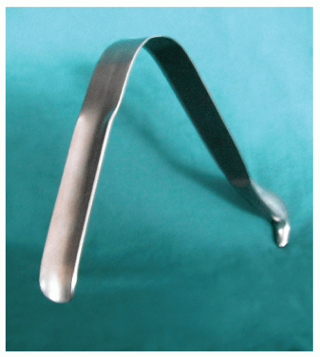
FIGURE 24-12. Wylie renal vein retractor.


Anterior Resection Technique (Total Anatomical Dissection of the Rectum)

The basic principles underpinning a successful restorative procedure for rectal cancer are as follows:


  • A total anatomical dissection (TAD) with mobilization along anatomical planes65


  • Achieving a distal clearance margin of at least 1 cm


  • Mobilizing the proximal colon, including the splenic flexure, and division and ligation of the inferior mesenteric vein at the lower border of the pancreas to ensure a tension-free anastomosis


  • Ensuring a good arterial blood supply to the segment of proximal colon that will be anastomosed to the rectum


  • Ligation and division of the inferior mesenteric artery close to its origin from the aorta


  • An anastomotic technique that ensures circumferential seromuscular bites


Positioning the Patient

The patient is placed in the Yellowfins stirrups (Allen Medical Systems, Acton, MA—Figures 24-13 and 24-14). Calf compressors are used to minimize the risk of deep
vein thrombosis. The abdomen is shaved. A urinary catheter is inserted, and when indicated, cystoscopy with insertion of ureteric catheters is performed by a urologist. The surgeon stands on the left side and the assistant stands opposite. A second assistant may stand between the legs.


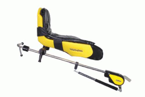
FIGURE 24-13. Yellowfins stirrups. (Courtesy of Allen Medical Systems, Acton, MA.)


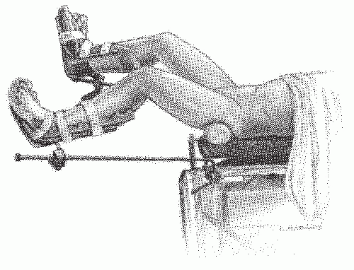
FIGURE 24-14. Perineolithotomy position with Allen’s stirrups (earlier version). The thighs are abducted and extended.

A midline incision is made (Figure 24-15), and after an exploratory laparotomy is performed, the small bowel is exteriorized in a wet pack (Figure 24-16). The technique used for mobilizing the rectum differs from that which is traditionally described, in that the dissection starts in the mid-pelvis on the right side rather than by mobilizing the sigmoid colon. Figure 24-17 shows a panoramic view of the pelvis in preparation for mobilization. A small diathermy incision is made strictly in the line of reflection of the visceral peritoneum covering the right anterolateral aspect of the rectum and carried onto the parietal peritoneum lining the right pelvic wall (Figure 24-18A). As soon as this is made, air is allowed into the retrorectal space as described by Ionescu earlier (Figure 24-18B). Once the retrorectal space is entered, the right branch of the hypogastric nerve is identified (Figure 24-19). This nerve is an important landmark. It lies anterior to the parietal fascia that lines the sacrum and lateral pelvic wall. The plane of dissection must stay anterior and medial to the nerve. Sharp diathermy or Harmonic scalpel is used to continue the dissection into the pelvis along the retrorectal space down to the floor of the pelvis (Figure 24-20). By remaining in that space, the surgeon avoids injury both to the nerve and to the presacral venous plexus.


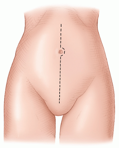
FIGURE 24-15. Midline incision.

A St. Mark’s retractor is used to retract the rectum anteriorly. Sharp dissection from right to left along the retrorectal space separates the rectum (encapsulated in its intact fascial envelope) from the hypogastric nerves and the parietal fascia covering the anterior surface of the sacrum Figure 24-21).

Attention is then directed to the sigmoid colon. The assistant holds the sigmoid colon up. The peritoneal attachments of the mesocolon to the lateral wall are dissected by sharp diathermy dissection (Figure 24-22). A diathermy incision is then made in the line of reflection of the peritoneum covering the mesocolon and the posterolateral abdominal wall peritoneum. The gonadal vessels are identified, and medial to them is the left ureter (Figure 24-23). The sigmoid colon is retracted anteriorly, and the areolar plane between the posterior aspect of the superior rectal artery and branches of the hypogastric nerves is identified. That plane is dissected by diathermy into the pelvis to join the retrorectal space.


Figure 24-24 shows the retrorectal dissection completed; the left and right hypogastric nerves are clearly demonstrated, and the perirectal fat is seen anteriorly, surrounded by an intact fascial layer.


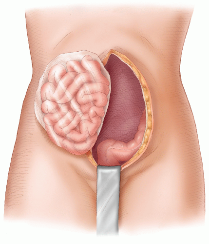
FIGURE 24-16. Small bowel exteriorized in a wet pack.


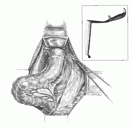
FIGURE 24-17. Panoramic view of the pelvis. St. Mark’s retractor is seen anteriorly.


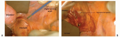
FIGURE 24-18. An incision is made along the line of reflection of visceral onto parietal peritoneum in the right mid-pelvis (A). Air is introduced into the retrorectal space (B).


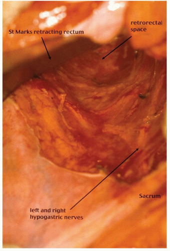
FIGURE 24-19. The hypogastric nerves are identified.


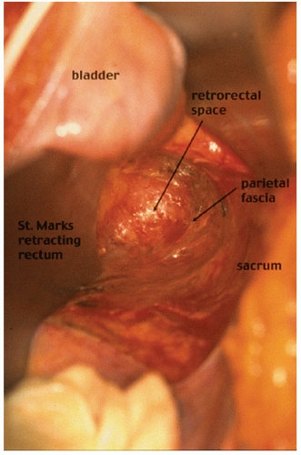
FIGURE 24-20. Dissection in the retrorectal space down to the pelvic floor.


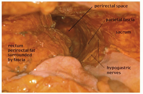
FIGURE 24-21. Pelvic dissection. Hypogastric nerves are clearly seen. Rectum, perirectal fat (with intact perirectal fascia) are separated from sacral parietal fascia.


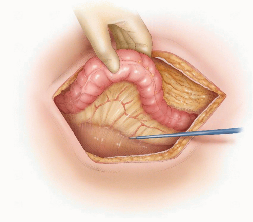
FIGURE 24-22. Congenital adhesions between the sigmoid mesocolon and left lateral wall are dissected.

Attention is then directed to the anterior part of the dissection. A small wet sponge under the left hand is used to retract the rectum upward and backward. The headlight is directed to the reflection of the visceral peritoneum onto the back of the vagina or prostate, and a small diathermy incision is made strictly in that line of reflection (Figure 24-25). This will open up the areolar avascular plane posterior to Denonvilliers’ fascia. A St. Mark’s retractor or a renal vein retractor is used to elevate the prostate or vagina, and the areolar plane is further developed using either diathermy or Harmonic scalpel. The anterior plane is developed as low as possible to enable a full mobilization of the rectum down to its lower third, where it is devoid of perirectal fat and consists only of a tubular muscular structure. Attention is then directed to the lateral ligaments and the middle hemorrhoidal and neurovascular bundles on the left and right anterolateral aspects of the midrectum. The left hand or a St. Mark’s retractor is used to retract the rectum anteriorly and to the left. This puts the lateral ligaments and neurovascular bundles under tension. The lateral ligaments are often avascular and are not ligaments at all. However, the neurovascular bundles containing the middle hemorrhoidal vessels and branches of the inferior hypogastric plexus need to be specifically attended to. This step of the dissection is important because injury to that plexus of sympathetic and parasympathetic nerves can result in urinary and sexual dysfunction. Injury can occur when the tissue in which it is contained is dissected too close to the parietal pelvic fascia. There is no need to do so; the dissection can remain close to the anterior and posterolateral aspects of the fascia enveloping the rectum. The Harmonic scalpel is used for this part of the dissection. It can seal vessels up to 7 mm in diameter and has simplified this step of the procedure (Figure 24-26A).

On the left side, the assistant retracts the rectum anterolaterally to the right and the same step is performed (Figure 24-26B). Once this is accomplished, there is usually more mobilization to be performed posteriorly. It is important to note that in the lower third of the pelvis, the parietal peritoneum covering the lower segments of the sacrum is reflected anteriorly to fuse with the encapsulating perirectal fascia. This fusion can be demonstrated by lifting the lower rectum upward and forward using a lipped St. Mark’s retractor. The fused fascia is divided by the Harmonic scalpel, and the dissection continues posteriorly to the levator muscle.


Transection of the Rectum


Tumors of the Upper Third of the Rectum

If the tumor is in the upper third of the rectum, the perirectal fat is divided 3 to 5 cm distal to the lower end of the tumor. This is usually straightforward in thin patients. However, in obese individuals, this can be a difficult step, and I have found the following technique useful and reproducible.


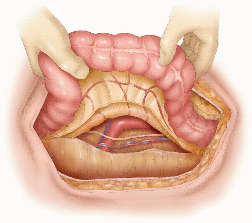
FIGURE 24-23. Gonadal vessels and left ureter identified.


FIGURE 24-24. Anterior dissection. Incision is made strictly along the line of reflection of the rectal visceral peritoneum onto the peritoneum, covering posterior vagina or prostate.


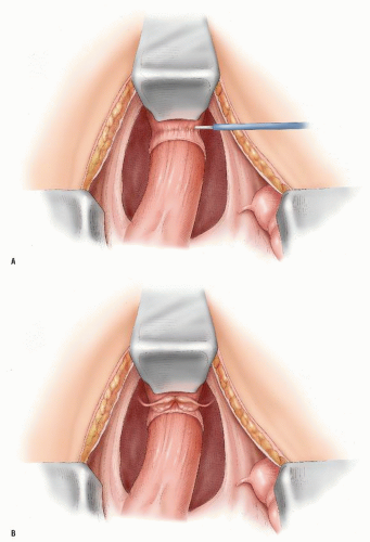
FIGURE 24-25. Anterior dissection behind the vagina (A) or prostate (B) posterior to Denonvilliers’ fascia.


FIGURE 24-26. Harmonic scalpel is used to divide the “lateral ligaments” and neurovascular bundles on the right (A) and on the left (B). (continued)


FIGURE 24-26. (continued)

The visceral peritoneum is divided by diathermy on either side of the rectum at the required level. The rectum is held up between the left index finger and thumb to palpate the antimesenteric border of the rectum. While keeping the index finger in place, the thumb is replaced by a long pair of straight scissors and pushed through from left to right. This opens up the plane between the posterior longitudinal muscle wall of the rectum and the perirectal fat. The perirectal fat is taken in two portions, the larger of which contains the superior rectal artery. Straight Kocher clamps are used for that purpose, and ligation is performed using a heavy suture ligature (Figure 24-27). The back of the rectum can then be inspected to ensure that there is an intact layer of longitudinal muscle, and the rectum is stapled and divided between two double rows of staples with a stapling device. I prefer to use the Contour stapler because its curved shape fits well into the pelvis and also because the rectum can be divided between two rows of staples on either side, thus avoiding any spillage (Figure 24-28). Alternatively, the rectum can be divided between a crushing clamp and a Furness clamp, which can then be used to apply a purse string to the rectum (Figure 24-29). I no longer use the Furness clamp because in a narrow pelvis,
there is the risk of a straight needle injuring structures on the lateral walls as it enters or exits the clamp. Alternatively, a hand-sewn purse string can be used (Figure 24-30).


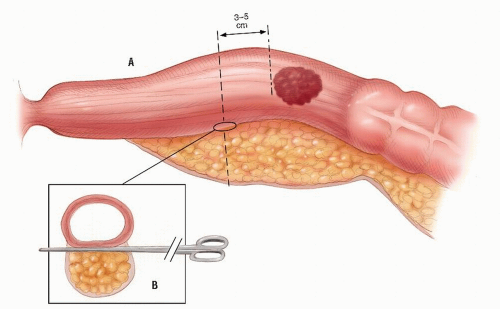
FIGURE 24-27. Division of perirectal fat for a tumor in the upper third of rectum. Inset: Opening the plane between the rectum and the perirectal fat.


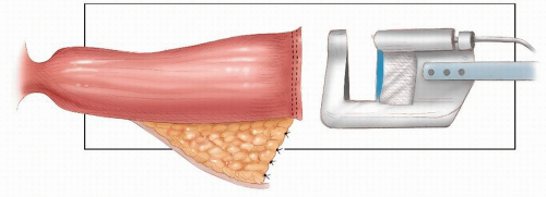
FIGURE 24-28. Transecting the rectum with Contour stapling device.


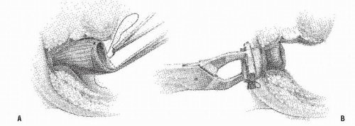
FIGURE 24-29. Circular-stapled anastomosis. A: Hand-sewn purse-string suture placed using “weaving” technique. B: Purse-string applicator employed on an inverted rectal stump.


FIGURE 24-30. Circular-stapled anastomosis. A hand-sewn purse-string suture is placed in the rectal stump.

If a hand-sewn anastomosis is performed, then a rightangled soft bowel clamp is applied to the rectum, and the specimen is divided between that clamp and a Kocher’s clamp. An assistant then irrigates the rectum until the return is clear. The rectal stump is surrounded with packs, and the soft bowel clamp is removed. Four stay sutures, two posteriorly and two anteriorly on either side of the midline, are applied to the rectal stump in preparation for a hand-sewn anastomosis as described later (Figure 24-31).


FIGURE 24-31. Four stay sutures applied to rectal stump in preparation for hand-sewn anastomosis.


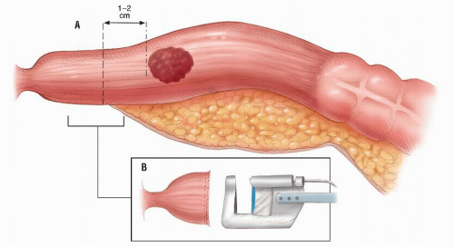
FIGURE 24-32. Tumor in the lower third of the rectum transected with Contour.


Tumors of the Middle or Lower Thirds of the Rectum

When the tumor is in the midportion or lower portion of the rectum, the dissection has to proceed in the retrorectal space to the pelvic floor, and the anterior plane of dissection should be as low as possible. The tubular lower end of the rectum is thus fully mobilized. There is little or no perirectal fat at this level, and the rectum can be transected using the Contour stapling device (Figure 24-32).

In obese patients and males with narrow pelvis, it is sometimes useful to ask an assistant to apply pressure to the perineum. This can often push the lower rectum into the pelvis and facilitate stapling and transection (Figure 24-33).


Dividing the Inferior Mesenteric Artery

Once the rectum has been transected, the inferior mesenteric artery is doubly ligated and divided (Figure 24-34). I usually perform a high ligation close to the origin of the artery on the aorta. Before doing so, the position of the ureter is again noted to avoid injury.


Mobilization of the Splenic Flexure

Several factors contribute to anastomotic leakage. These include poor blood supply, tension, and inadequate seromuscular apposition. Many patients in Western societies are obese and have concomitant sigmoid diverticular disease with a fat laden and shortened mesentery. In these individuals, the proximal descending colon must be mobilized to reach the lower rectum. Tension is avoidable by performing the following three steps:


  • Mobilizing the splenic flexure


  • Ligating and dividing the inferior mesenteric vein at the lower border of the pancreas


  • Unfolding the splenic mesentery

The method of mobilizing the splenic flexure must be reproducible and has to protect against injuries to the spleen. In order to minimize the risk of splenic injury, the plane of dissection has to be strictly in the lesser sac.

The surgeon stands on the patient’s right with an assistant between the patient’s legs and another retracting the left abdominal wall. The head of the operating table is elevated, and the table is rotated to the right. The surgeon wears a headlight. The dissection is performed by diathermy, or a long Harmonic scalpel is used.

The greater omentum is retracted vertically out of the wound and lifted caudally. The surgeon should resist the temptation of retracting the greater omentum across to the right
side. This is a dangerous maneuver that can tear fine adhesions between the omentum and the splenic capsule, resulting in injury to the spleen. The assistant gently retracts the distal transverse colon downward, and the greater omentum is retracted caudally (Figure 24-35). Sharp dissection then allows access into the lesser sac. The dissection continues toward the splenic flexure strictly in that avascular plane (Figure 24-36). Attention is then directed to the upper descending colon, which is retracted with the left hand (Figure 24-37). With diathermy in the right hand, the adhesions between the colon and the lateral abdominal wall are divided to meet the line of dissection of the distal transverse colon. The descending colon is thus mobilized to the midline.


FIGURE 24-33. Pressure on the perineum by the fist or some other blunt object facilitates the placement of sutures into the rectal remnant.


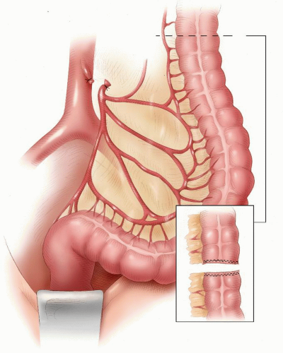
FIGURE 24-34. Author’s preference of ligating the inferior mesenteric artery on the aorta.


Dividing the Inferior Mesenteric Vein

It is important to understand that even after the flexure has been fully mobilized, the mesentery is still not sufficiently free. This is because it is held back by the inferior mesenteric vein at the inferior border of the pancreas (Figure 24-38). The vein should be ligated and divided at that level.


Unfolding the Splenic Flexure Mesentery

Once the flexure is mobilized, further length can be obtained by unfolding the splenic flexure mesentery to the marginal vessels as shown in Figure 24-39. The colon is now fully mobilized and can reach the low rectum deep in the pelvis without tension.


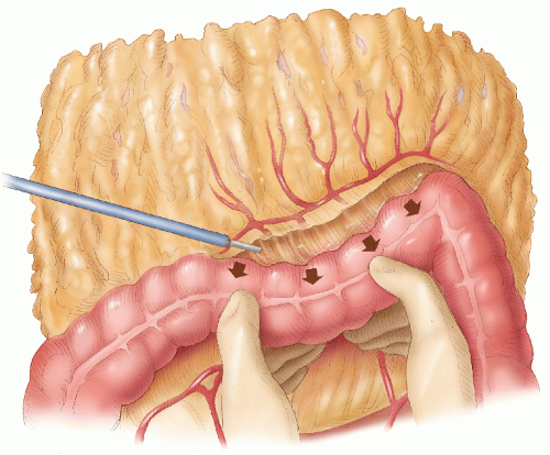
FIGURE 24-35. Mobilization of splenic flexure. The assistant holds the distal transverse colon down, and the lesser sac is entered.


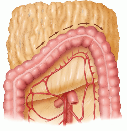
FIGURE 24-36. Line of dissection during mobilization of splenic flexure.


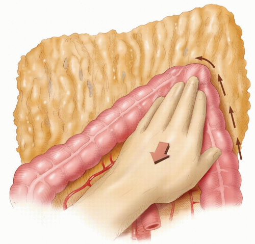
FIGURE 24-37. Mobilization of upper descending colon.


Testing the Blood Supply of the Proximal Colon

The proximal colon is now ready to be divided at an appropriate level after ligating and dividing the marginal vessels. It is advisable to test the blood supply of the proximal colon by observing pulsatile bleeding from the marginal artery prior to ligation.

The colon is then divided. If a stapled anastomosis is to be performed (and that is the preferred method), a purse string is applied before the colon is divided. This can either be performed by a disposable purse-string applicator (Figure 24-40), a Furness clamp, or a hand-sewn technique. I prefer to use a disposable applicator because I have found it to be accurate and less time consuming. The anvil of stapling instrument (Figures 24-41 and 24-42) is now introduced into the colon, and the purse string is tied.


Staple-on-Staple (Double-Stapled) Anastomosis

This is my preferred method of reestablishing continuity. The rectum has been stapled off as described earlier, and a purse string has been applied to the proximal colon.


Insertion of Stapling Instrument into the Rectal Stump

An assistant then washes out the rectal stump and inserts the stapling instrument. This step can at times be difficult, especially if there is anal stenosis or if the patient is obese. If there is anal stenosis, then gentle dilatation may be performed with the fingers or with dilators (Figure 24-43). If the patient is obese, it may be difficult to introduce the stapling instrument. The perianal skin can get caught by the lip of the instrument and pushed in, making insertion even more difficult. Occasionally, the rectum can be injured if an inexperienced assistant forces the instrument in.

Whenever there is difficulty in inserting the stapling instrument, I take charge of the situation and perform the insertion myself. I have used a simple technique to facilita te insertion under these circumstances. A Lone Star retractor is used to retract the anal skin and open the anal canal as shown in Figure 24-44. I have found that this is particularly helpful in obese patients.

Once the stapler is inserted, the instrument is carefully guided into the rectal stump. The rectum is gently gloved over the instrument, and the instrument is fully opened. I prefer to
advance the trocar tip through the staple line (Figure 24-45), but some surgeons like to penetrate the adjacent bowel. It is critical, however, to incorporate the linear staple line.


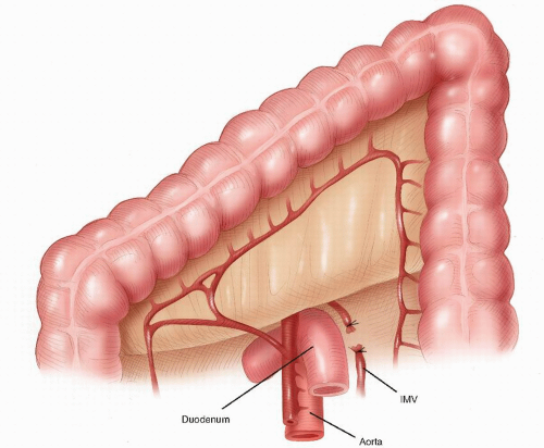
FIGURE 24-38. Inferior mesenteric vein divided at the inferior border of the pancreas.


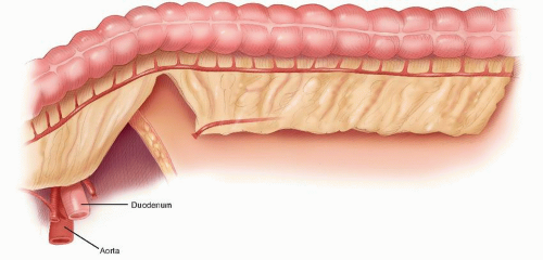
FIGURE 24-39. Splenic flexure unfolded to avoid tension on anastomosis.


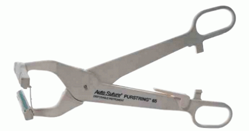
FIGURE 24-40. The Purstring 65 single-use instrument places a circumferential strand of 2-0 MONOSOF nonabsorbable monofilament nylon surgical suture (United States Pharmacopeia [USP]) held in place by stainless steel staples. (Courtesy of Covidien, Inc., Norwalk, CT.)

The proximal colon with the anvil is then pulled down into the pelvis. Care is taken to ensure that the mesentery is not twisted. The proximal anvil is pushed into the pin, and the assistant closes the mechanism so that the anvil and head are fully approximated (Figure 24-46). After the staples are fired, the instrument is opened three turns, and the stapler is gently withdrawn. The tissue rings (donuts) are carefully assessed for completeness and for an intact muscle coating.

Following this, the anastomosis is tested for leakage (Figure 24-47). Provided there is no leak, I use a drain in the retrorectal space, and the abdomen is then closed.


Temporary Proximal Loop Stoma

The decision whether to construct a proximal stoma is one that the surgeon must consider at surgery. It is preferable to forewarn the patient that a temporary stoma may be necessary and to always site a stoma preoperatively (see Chapter 31). As a rule, the lower the anastomosis, the more likely it is that a temporary stoma will be advisable. Other relative indications include a technically difficult procedure, unexpected intraoperative pelvic hemorrhage, significant comorbidities, obesity, and those in whom the donuts were incomplete even though there was no leakage on air testing. If there was a leak on testing, even though the site of leakage may have been identified and oversewn, this is another possible indication for fecal diversion.

If a decision is made to construct a temporary stoma, a temporary loop ileostomy rather than a loop colostomy is preferred. There are three reasons for this: no tension is
put on the proximal colon by exteriorizing the transverse colon; there is no risk of injuring the vital marginal vessels of the proximal colon either when the colostomy is being constructed or when it is closed; and if a planned temporary stoma needs to be permanent, a loop ileostomy is easier to manage. Moreover, there is a lesser incidence of prolapse and parastomal herniation with a loop ileostomy.


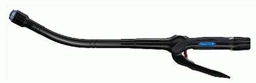
FIGURE 24-41. Proximate ILS curved intraluminal stapler with detachable head and with low-profile anvil. This is available in sizes 21,25,29, and 33 mm. (Courtesy of Ethicon Endo-Surgery, Inc., Cincinnati, OH.)


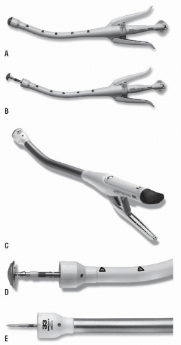
FIGURE 24-42. Multiple options are available in the circular instruments. Disposable curved CEEA (end-to-end circular stapler) with integrated flip-top anvil. This permits a low profile for ease of insertion and removal. A: Closed instrument. B: Opened instrument. C: End-to-end anastomosis (EEA) Open/XL—available in two shaft lengths, ergonomic handle and knob, and one-handed firing. D: Close-up view of open instrument. E: Distal anvil removed. (Courtesy of Covidien, Inc., Norwalk, CT.)


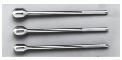
FIGURE 24-43. Sizers in three diameters. (Courtesy of Covidien, Inc., Norwalk, CT.)


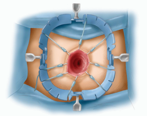
FIGURE 24-44. Lone Star retractor used to expose the anus and facilitate insertion of the stapling instrument.


Hand-Sewn Anastomosis

Although I prefer to use a staple-on-staple (double-stapled) anastomosis as described earlier, occasionally one needs to perform a hand-sewn anastomosis. When doing so, the rectum is transected as described, and four stay sutures are inserted in the rectal stump (see Figure 24-31). I use a single layer of interrupted 2-0 Vicryl sutures. The posterior layer is inserted first, including at least 1 cm of the muscle layer (Figure 24-48). The corners are turned, and then an interrupted seromuscular sutures are applied to the anterior row of the anastomosis (Figure 24-49).


Total Mesorectal Excision

This term was coined by Heald in the late 1970s (see earlier Biography).246 Heald and others163 had rediscovered the retrorectal space that Heald renamed the “holy plane.” Furthermore, he described the perirectal fat as the mesorectum even though the rectum has no true mesentery. He also came to understand that the rectum and its perirectal fat were covered by an intact layer of fascia, and that the specimen could be removed within an intact fascial envelope to reduce the risk of local recurrence by ensuring clear surgical margins.

When the term TME was first introduced, many colorectal surgeons were already mobilizing the rectum along welldefined and well-described anatomical planes, preserving an intact perirectal fascialenvelope163,259,312,358,442,616; so there was some confusion as to what exactly was meant by the term TME.

Many general surgeons who performed few rectal dissections per year were keen to adopt this technique and
took the word “total” in TME quite literally, performing unnecessary low and ultralow anterior resections when a high anterior resection would have sufficed. This resulted in a number of avoidable complications after low anastomoses, of which anastomotic leakage was the most worrisome. Karanjia and colleagues reported on the Basingstoke experience of 219 patients who underwent a TME for a tumor between 3.5 and 15.0 cm from the anal verge; an anastomotic leak rate of 17.4% resulted.302 Hainsworth and coworkers questioned the use of TME for upper third tumors in view of the high morbidity rate following this operation in their patients.233 The term TME has, however, taken hold. Today, it tends to refer to dissection of the rectum along the same anatomical planes as were originally described by Jonnesco (see earlier Biography), although many would now question the need to perform a TME for cancers of the upper rectum or rectosigmoid.352,372 From this concern has arisen the terms “partial” or “tailored” TME, wherein the mesorectum is divided 3 to 5 cm below the tumor. This shifts the emphasis to clearly identifying the retrorectal space and the perirectal fascia and not to the complete removal of the perirectal fat or “mesorectum” for tumors at all levels of the rectum.


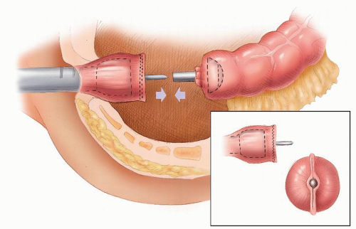
FIGURE 24-45. Tip of stapling instrument is opened through the linear staple line.


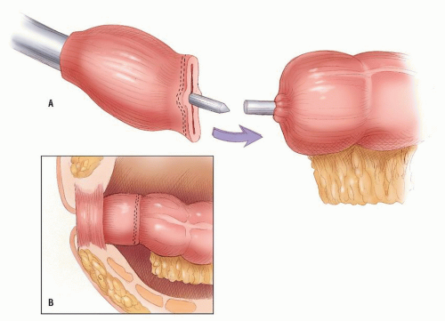
FIGURE 24-46. The anvil and head are approximated, and the staples are fired to form the anastomosis (inset).


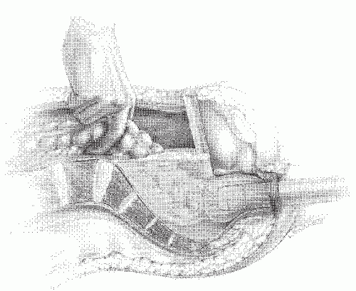
FIGURE 24-47. Method of testing for integrity of rectal anastomosis. Inspection can be achieved by means of the rigid sigmoidoscope. By compression of the proximal bowel with the fingers or a noncrushing clamp and by the insufflation of air with saline in the pelvis, bubbles may be seen to escape from a leak. The site can then be identified and possibly repaired.


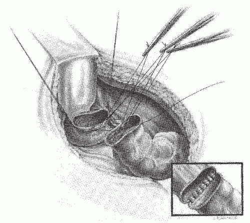
FIGURE 24-48. Hand-sewn rectal anastomosis. Sutures are initially placed in the posterior row but not secured. The inset demonstrates the posterior row completed.


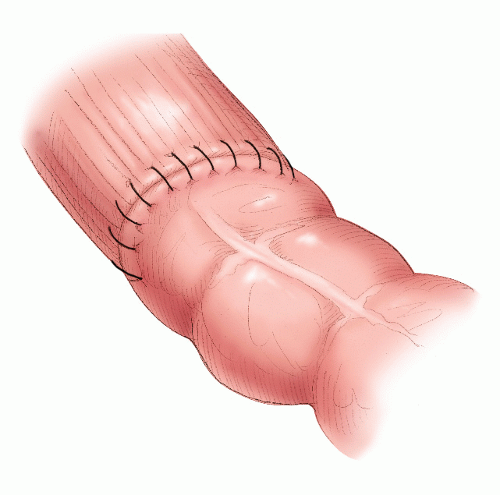
FIGURE 24-49. Hand-sewn rectal anastomosis. Interrupted sutures applied to anterior row.

In a study of 622 patients who underwent an anterior resection for cancer at the Queen Mary Hospital in Hong Kong, the authors compared the results of patients who had a TME for middle and lower third tumors with those who had a partial mesorectal excision (PME) for tumors in the upper rectum (this group constituted, in our experience, 30% of rectal cancers).342 There was no difference in local recurrence or survival between the two groups, but those who had a TME had a higher rate of complications, including anastomotic leakage. This study supports the view that what really matters in rectal cancer surgery is the anatomical nature of the mobilization rather than the total excision of the “mesorectum.” Moreover, it is noteworthy that TME has never been subjected to rigorous testing by a randomized, controlled trial. Indeed, some would argue that the recurrence rates claimed by Heald are the results of patient selection and/or case mix and the method of statistical analysis used to interpret the data.282,283 and 284 Nevertheless, the concept of TME has contributed to a greater awareness of the need for precise anatomical dissection for rectal cancer surgery.


Side-to-End and Side-to-Side Anastomoses

Baker advocated side-to-end anastomosis of the colon to the rectum in order to deal with the disparity between the two lumina.28 Others have also found this to be a useful technique.310 In my opinion, the anastomosis can be more readily accomplished in an end-to-end fashion, with the circular stapling device.


Coloanal Hand-Sewn Anastomosis

An alternative technique for reestablishing intestinal continuity is the coloanal anastomosis. Pulling the colon through the anus was first described by Cutait and Turnbull in 1961 (see later), and in 1972, Parks described a hand-sewn coloanal anastomosis.124,487,622 Circumstances where it is indicated include very low rectal cancers and on occasions when there has been a failure of a very low-stapled anastomosis.


Author’s Technique for Coloanal Hand-Sewn Technique

It is essential that the proximal colon be completely mobilized as described earlier. There must be no tension at all on the anastomosis when the colon is pulled through. As a rule, if the proximal colon can reach the patient’s left knee, it will be easy to pull it through without tension. The colon is prepared by stapling it with a linear device. A small incision is made in the middle of the staple line, and a Foley catheter is inserted into the colon; the catheter balloon is inflated. The catheter will be used to pull the colon down through the anal canal.


When the rectum is fully mobilized to the pelvic floor, it is transected by diathermy at least 1 cm below the tumor. A Lone Star retractor is then used for exposure, and eight sutures (at regular round-the-clock intervals) are inserted into it using 2-0 Vicryl on a 26 needle. The sutures are placed from outside in. The needles are not cut off, and the sutures are pulled out and clipped to the rim of the retractor (Figure 24-50). The proximal colon is then pulled through using the Foley catheter as a guide and for traction (Figure 24-51). The staple line is excised, and the colon is sutured to the anal canal by each of the individual sutures that had been preserved. After this has been accomplished, each suture is tied and divided. The suture line disappears out of sight into the anal canal.


Alternative Techniques for a Coloanal Hand-Sewn Anastomosis

A Parks’ self-retaining, three-bladed anal retractor (Figure 24-52); Gelpi retractors place at right angles to each other (Figure 24-53); a Lone Star retractor (Figure 24-54); or a Bookwalter rectal kit (Figure 24-55) can facilitate exposure for a hand-sewn transanal anastomosis (Figure 24-56). A 5/8-circle needle is particularly helpful for placing the sutures—for example, a round-bodied modification of a Turner-Warwick urethroplasty needle or a longterm absorbable suture. This is the technique employed for reestablishing continuity after colectomy, proctectomy, and ileal pouch for inflammatory bowel disease (see Chapter 29). The sutures incorporate the full thickness of the colon with the anal canal and the underlying internal sphincter. An alternative approach is to use a double-stapling technique.


Handling and Examination of the Surgical Specimen and Tumor Staging

The information provided by the surgeon to the patient must include details of the operative findings as well as details of the histopathologic features of the tumor. Without this information, the patient’s prognosis and the potential need for adjuvant therapy cannot be meaningfully discussed. Accurate reporting (classification and tumor staging) is essential for quality patient care when dealing with rectal cancer. To achieve this, there is an onus on the surgeon and the pathologist to collaborate closely to produce a comprehensive
report incorporating key clinical and pathologic features from the findings at operation and the examination of the surgical specimen as a basis for appropriate multimodality treatment for the individual patient.98


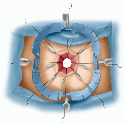
FIGURE 24-50. Lone Star retracting anus. Sutures are applied from outside in, and needles are kept.

The traditional method of staging rectal cancer is that of Dukes (see Chapter 23). This classification is based on the extent of sequential direct spread of tumor through successive layers of the rectal wall and the presence or absence of locoregional lymph node metastases in the operative specimen.154 In this strictly pathologic approach, there is no information provided that addresses the issue of “residual tumor,” whether local due to tumor transection (involved margin) or the presence of known distant metastases outside the operative field. This information is only available when tumors are staged using a clinicopathological system, now internationally adopted as a preferred method of cancer staging.97 I have adopted this approach, and I support the use of generic reporting to ensure that all relevant information is captured using a standardized, structured template.71,531 Although the majority of my published results have been reported using the Australian Clinicopathological System, one of the six internationally
recognized staging classifications,133 for the purpose of this chapter, the results tabulated are summarized using the TNM system.143


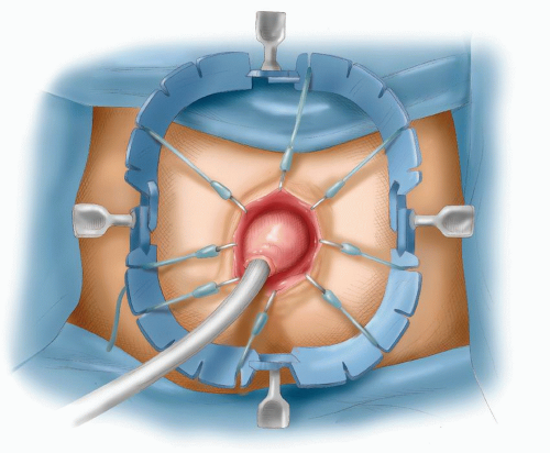
FIGURE 24-51. Foley catheter in proximal colon with balloon inflated, pulling colon down to anus to complete the coloanal anastomosis.

The surgeon must also take responsibility for prompt delivery of the correctly labeled and orientated specimen, preferably transported fresh and unopened to the laboratory. The pathologist should be provided with a precise but clinically relevant history, including a brief description of the important observations made at laparotomy. In particular, the surgeon should provide information about the type of resection, the site of the tumor, and the presence or otherwise of distant metastases or local residual tumor in order to enable the pathologist to determine the final clinicopathologic stage. In turn, the pathologist is responsible for preparing a thorough report, noting all key diagnostic (macroscopic and microscopic) and prognostic indicators. These should include the presence of extramural venous invasion, serosal surface involvement, clearance of all resection margins (proximal, distal, and circumferential), and presence of perforation in the specimen.97 Certainly, specimen handling, sampling, and dissection should be standardized to allow meaningful comparisons between treatment centers and for entering patients into clinical trials.507 Ideally, the final report should include representative images of key macroscopic and histologic features of the tumor. This will greatly assist discussion of prognosis and in counseling patients and their relatives. In this regard, the essential standardized protocol favored by the author over many years is that recommended by the Australian National Health and Medical Research Council.456


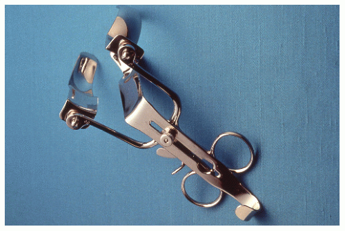
FIGURE 24-52. Parks retractor.
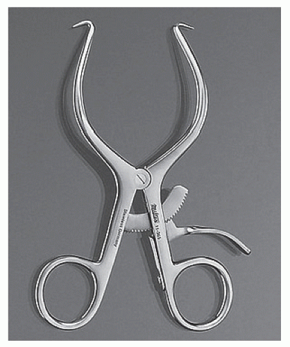

Abdominoperineal Resection


Technique

I favor a combined synchronous abdominal and perineal resection.


Abdominal Procedure

The mobilization of the rectum is the same as has been described earlier for a low anterior resection. The rectum is mobilized along anatomical planes all the way down to the pelvic floor. I aim to perform as much of the dissection as possible during the abdominal dissection before proceeding with the perineal operation. The inferior mesenteric artery is ligated and divided as previously described, and the sigmoid mesentery is divided at an appropriately selected segment (see Figure 24-34). I always test the marginal artery before ligation and division to ensure a good blood supply. Once the mesentery is divided, the colon is transected with a linear stapling device.


Perineal Dissection

General Comments. Various techniques have been described for the perineal dissection. In the first instance, the “traditional” method of dissection will be described. Other techniques that involve a wider and more radical approach will be dealt with later in this text.

I consider the perineal dissection as important, if not more important, than the abdominal procedure. It can be technically demanding, and I strongly recommended that it should be performed either by the principal surgeon or by another experienced colorectal surgeon or by senior-level resident or a fellow under direct supervision. It should not be left to a junior or inexperienced resident. The perineum is prepared and draped. I prefer to have instruments, including diathermy and the Wave Harmonic scalpel, placed on a small perineal table. There should be no leads dangling off the patient’s leg drapes; they get in the way and obstruct the view. The table is tilted head down and the stirrups are elevated. A purse-string suture is applied to the anus to prevent fecal contamination. An elliptical incision is made with the diathermy (Figure 24-57), and a Lone Star retractor is used to retract the skin. In the past, the rest of the dissection was performed using diathermy; however, over the past 3 years, I have used the Wave Harmonic scalpel. This instrument is useful in preventing bleeding during the procedure and an almost bloodless dissection can be achieved (Figure 24-58). The Wave Harmonic scalpel is used to deepen the incision through the ischiorectal adipose tissue. The index finger of the left hand is then used as a guide to locate the tip of the coccyx, and the anococcygeal ligament is divided (Figure 24-59). The finger is then inserted anterior to the coccyx. The abdominal operator lifts the rectum in his left hand and inserts his right hand deep into the retrorectal space until both operators can feel each other’s fingers as separated by Waldeyer’s fascia (Figure 24-60). The perineal surgeon then uses a pair of Abel scissors to break through this layer into the retrorectal space (see also Figure 24-60). Usually, a small amount of blood that has collected at the lower end of that space needs to be sucked out. Attention is then directed to dividing the iliococcygeus part of the levator muscles on either side. The left index finger is inserted into the opening, which has been made in the presacral space, and the finger is hooked under the left levator muscle. The Wave Harmonic scalpel is used to
divide the muscle (Figure 24-61). The left index finger is similarly used to identify the right levator muscle, and the wave instrument is used to divide the muscle on the right side. Attention is then directed to the pubococcygeus and the puborectalis parts of the levator muscle. The muscle is divided with the Wave Harmonic scalpel from lateral to medial on either side. Branches of the inferior hemorrhoidal vessels are in close proximity to the puborectalis and can cause troublesome bleeding. Simply dividing the muscle with diathermy usually does not achieve adequate hemostasis. However, I have found that this can be achieved with the Wave Harmonic scalpel (Figure 24-62). Dissection between the rectum and prostate or vagina can now begin. The abdominal surgeon’s fingers can be used to guide the perineal surgeon. The perineal surgeon’s left hand is used to retract the rectum down, and the diathermy is used to dissect in the plane between the rectum and the prostate in males. The rectourethralis muscle on either side is divided (Figure 24-63); the dissection continues until the abdominal surgeon’s fingers are met, posterior to Denonvilliers’ fascia; and the dissection breaks into the pelvic cavity. By remaining in this plane, injury to the urethra is avoided. Another method of performing the anterior part of the dissection is to deliver the specimen through the perineum; once the posterolateral part of the dissection is performed, the specimen can be passed to the perineal surgeon who then retracts the specimen downward to better identify the anterior plane of dissection (Figure 24-64). In females, the anterior plane of dissection is between the rectum and the posterior wall of the vagina. If the tumor has invaded the posterior wall of the vagina, then the Wave Harmonic scalpel can be used to excise en bloc a portion of the vaginal wall. If the Harmonic scalpel is not available, then the vaginal wall has to be oversewn with an interlocking suture to prevent hemorrhage (Figure 24-65). The wound is closed in layers and drained through the vagina (Figure 24-66).


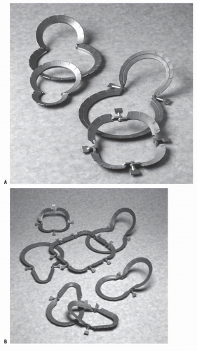
FIGURE 24-54. Loan Star retractors.


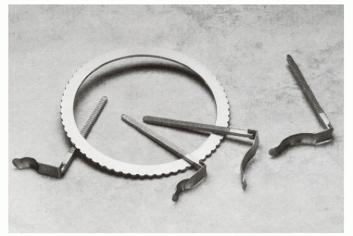
FIGURE 24-55. Bookwalter rectal kit. (Courtesy of Codman & Shurtleff, Inc., Raynham, MA.)


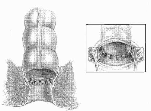
FIGURE 24-56. A Parks retractor facilitates the insertion of sutures (inset) in creating a coloanal anastomosis.


FIGURE 24-57. Perineal dissection. An elliptical incision is made outside the anus.


FIGURE 24-58. Perineal dissection. Lone Star retractor in place. Wave Harmonic scalpel is used to deepen incision.


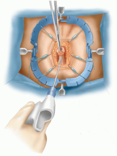
FIGURE 24-59. Perineal dissection. Division of anococcygeal ligament.


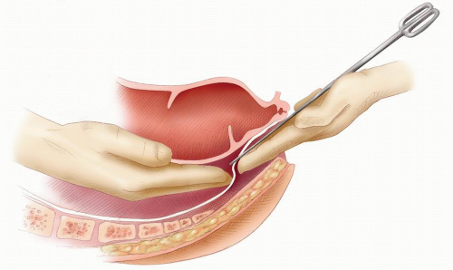
FIGURE 24-60. Perineal dissection. Identifying Waldeyer’s fascia and confirmation of the plane of dissection as the two surgeons’ fingers touch. The scissors facilitates their joining.


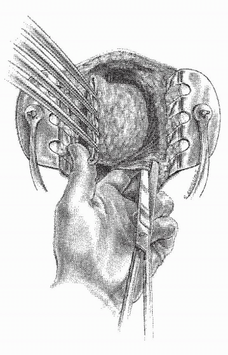
FIGURE 24-61. Perineal dissection. Left index finger hooked under right levator muscle, which is divided by Wave Harmonic scalpel (the illustration shows it being accomplished with scissors—not my personal preference).

The specimen is then delivered through the perineum, although sometimes if the specimen is bulky and the pelvis is narrow, then the specimen can be delivered through the abdomen. Once the specimen has been delivered, the perineal wound is carefully examined to ensure hemostasis, especially from the posterior aspect of the prostate or vagina. Because I have started to use the Wave Harmonic scalpel to perform the perineal dissection, there has been a noticeable reduction in blood loss. Once hemostasis is secured, the wound is closed in layers around a drain (Figure 24-67). Occasionally, if there is a bulky tumor with a significant anterior component, the perineal dissection can be performed with the patient in the jackknife position. In order to do so, the colostomy must be exteriorized and the abdomen closed
before the patient is turned over for the perineal dissection. Although repositioning the patient is cumbersome, this position can achieve better exposure and access to the perineal anatomy. It is also more comfortable for both surgeon and assistant. Although I have occasionally used this technique, it is not routine in my practice.


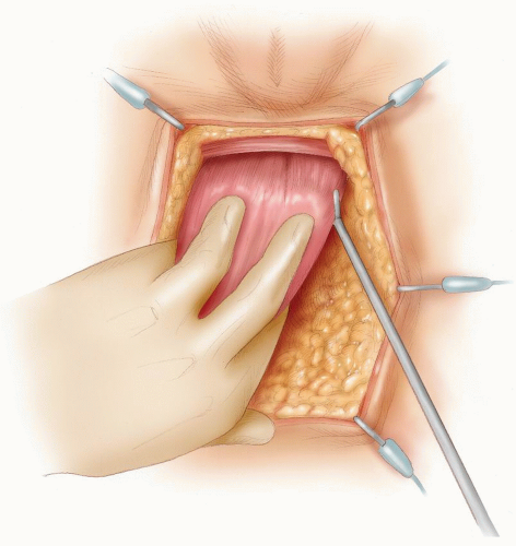
FIGURE 24-62. Perineal dissection. Division of puborectalis muscle.


▶ EXTERIORIZING THE COLOSTOMY

The colostomy site would have been carefully selected and marked preoperatively by an enterostomal therapist. A disk of skin is excised and deepened to the anterior rectus sheath (Figure 24-68). The sheath is divided transversely and to a lesser extent vertically. The assistant retracts the skin as well as anterior and posterior rectus sheaths to the right of the incision. With hand protected by a sponge, the assistant pushes the posterior rectus sheath forward. The surgeon exposes the underlying rectus muscle, its fibers are separated, and the posterior rectus sheath is divided by diathermy onto the assistant’s fingers, which are protected by the sponge. The opening is enlarged to admit two fingers. The colostomy exit site is carefully inspected to ensure that there is no bleeding from the inferior epigastric vessels. These are suture ligated if they have been torn. It is best to inspect for bleeding from the posterior aspect of the colostomy exit wound as shown in Figure 24-69. The stapled sigmoid colon is then exteriorized through the opening, and when the abdomen is closed, the stoma is matured (Figure 24-70).


Closing the Pelvic Floor

Some surgeons advocate closure of the pelvic floor by mobilizing the peritoneum on either side and suturing the
two leaves together in the midline (Figure 24-71). It is thought that closure reduces the risk of small bowel obstruction and that it may be useful in patients in whom postoperative radiotherapy may be contemplated. There is, however, no clear evidence to support this. On the contrary, there is a theoretical risk that a gap can occur in the suture line through which a loop of small bowel may herniate and cause obstruction. Furthermore, during mobilization of the parietal peritoneum from the lateral pelvic wall, the ureters could be injured. However, in the few patients in whom postoperative radiotherapy is contemplated, there is a potential risk of radiotherapy injury to the small intestine if it were to fall into the dissected pelvis. For this reason, several techniques have been proposed to reconstruct the pelvic floor. In view of the fact that adjuvant therapy is now mainly given preoperatively, closing the pelvic floor is not essential nor is it practiced routinely.


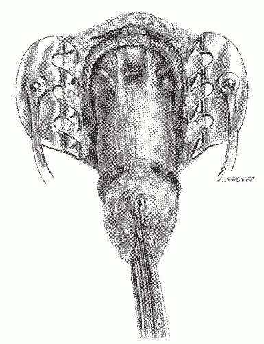
FIGURE 24-63. Perineal dissection. The rectourethralis muscle has been divided, exposing the prostate. With the division of the fascia of Denonvilliers, the peritoneal cavity can be entered anteriorly.

They are described here, although I do not use them in my practice. These include suturing the terminal ileum and its mesentery around the linea terminalis632; construction of an omental envelope; the use of the rectus abdominis muscle; and placement of a synthetic absorbable or nonabsorbable mesh sling, a breast prosthesis, and a synthetic polymermold.79,139,146,147 and 148,156,166,220,225,324,347,560,601 The possibility of infection with nonabsorbable material is, of course, a concern, as well as the inconvenience if one must remove it.


Technique

The use of polyglycolic acid mesh (either Dexon or Vicryl mesh) for this procedure can be considered whenever postoperative radiation is being entertained following either APR or even anterior resection. Because the mesh that is provided is not usually of sufficient size to create the sling, two are used and sutured together. Commencing at the level of the sacral promontory, the mesh is anchored (Figure 24-72). Using a continuous, locking suture technique, one anchors the mesh laterally on each side to the peritoneum (Figure 24-73). The mesh is then brought to the anterior abdominal wall and secured in place, creating a halter or sling that keeps the small bowel out of the pelvis (Figure 24-74).


Extralevator Abdominoperineal Resection

In recent years, several authors have questioned the perineal technique described earlier. There have been concerns that the perineal dissection may not be sufficiently wide. It is thought that this can result in a higher rate of involvement of the circumferential margins, a higher rate of perforation, and, subsequently, a higher rate of local recurrence and ultimately poorer survival. These surgeons advocate a wider excision, an extralevator abdominoperineal resection (ELAPR). The theory is that this operation will result in a cylindrical specimen rather than a “waisted” one.646,647 The philosophy behind ELAPR is that the wider the resection margins, the better the outcome. Some surgeons have recommended the inclusion of the coccyx as part of the wider excision.37 ELAPR is not dissimilar to the original procedure that Miles described in 1908.421 He recommended that “… the perineal portion of the operation should be carried out as widely as possible so that the lateral and downward zones of spread may be effectively extirpated.” To date, there is no randomized trial evidence that an ELAPR is any better than a conventional APR, and the evidence advanced by its proponents is based
on limited, selected patient series. Furthermore, it has to be recognized that ELAPR, with or without sacrectomy, results in a larger perineal wound. This may require various plastic surgical procedures to close, potentially leading to a higher risk of perineal wound complications.37,268


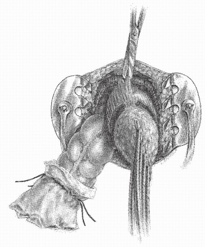
FIGURE 24-64. Perineal dissection. The proximal colon has been delivered through the pelvic defect. The rectourethralis muscle and the visceral fascia are the only structures remaining to be divided.


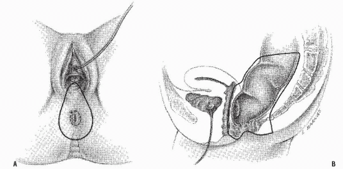
FIGURE 24-65. Perineal dissection in women. A: Outline of the incision for excising the posterior vaginal wall. B: Lateral view showing the extent of removal.


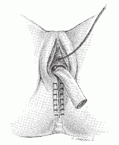
FIGURE 24-66. Perineal dissection in women. The skin wound is completely closed, and the perineal body is reconstructed. The drain is placed in the pelvis and brought out through the defect in the posterior vaginal wall.


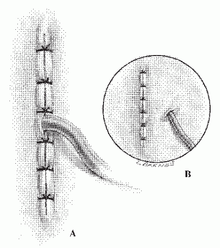
FIGURE 24-67. Perineal dissection. The wound is closed primarily, and the pelvis is drained, either through the incision (A) or through a stab wound (B).


FIGURE 24-68. Abdominoperineal resection. Creating the abdominal wall opening for the colostomy. A: A disk of skin is excised. B: A cruciate incision is made in the anterior rectus fascia. C: The rectus muscle is split longitudinally. D: The completed abdominal wall opening.


FIGURE 24-69. Abdominoperineal resection. Exposure of the epigastric vessels by sponge retraction.


FIGURE 24-70. Abdominoperineal resection. The wound is closed and the colostomy matured.


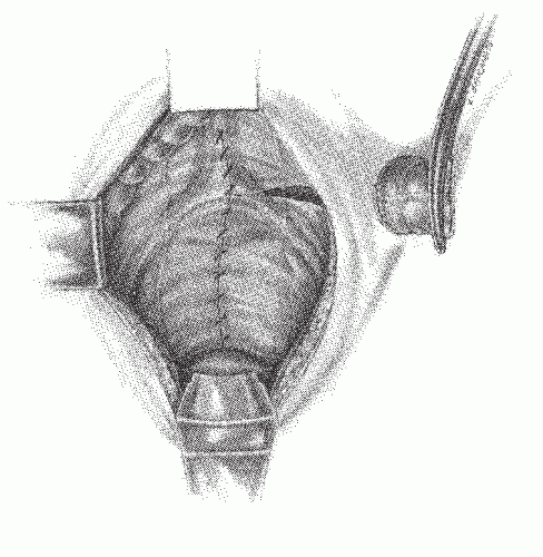
FIGURE 24-71. Abdominoperineal resection. The floor of the pelvis has been reconstituted.


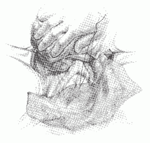
FIGURE 24-72. Implantation of mesh for postoperative radiation. The mesh is anchored to the sacral promontory.


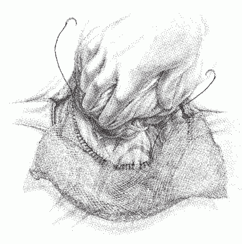
FIGURE 24-73. Implantation of mesh for postoperative radiation. Using a continuous, interlocking suture technique, the mesh is anchored to the posterior and lateral peritoneal surfaces. Two pieces of mesh may be required to effect this maneuver.


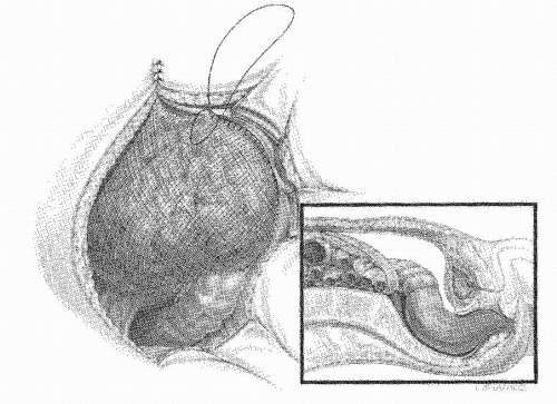
FIGURE 24-74. Implantation of mesh for postoperative radiation. The completed suspension requires the mesh to be anchored to the anterior abdominal wall. Care must be taken not to impede the exit of the bowel for the stoma. The abdominal wound should be closed without incorporating the mesh, but anterior fixation can be achieved to the peritoneum. Final position of the fixation in an individual who has undergone an anterior resection (inset).


Before adopting ELAPR as a standard, it would be prudent to conduct a randomized study to determine among other things whether the procedure is beneficial in terms of local recurrence and survival.


▶ LAPAROSCOPIC SURGERY FOR RECTAL CANCER (SEE CHAPTER 19)


Other Procedures That May Accompany Resection of the Rectum


En Bloc Resection of Adjacent Organs

It is not unusual for another organ to be involved with a rectal cancer mass. Most commonly, the bladder, the vagina, the uterus, or a loop of small bowel are involved. Occasionally, the sigmoid colon, especially if it is on a long mesentery, may fall into the pelvis and become involved. It is sometimes difficult to know at the time of operation whether these structures are adherent to the cancer by an inflammation or by the tumor invasion. Whenever possible, the involved organ (or part of it) should be resected en bloc. Some of these procedures are described next.


Incidental Appendectomy

Incidental appendectomy is unnecessary and not indicated at the time of rectal resection, unless the appendix is involved in the tumor mass.


Bladder Resection

The urinary bladder is not uncommonly directly invaded by rectal cancer. If the dome of the bladder is invaded, an en bloc resection is recommended. It is difficult to know exactly whether the involvement is neoplastic invasion or inflammatory. It is better to assume that it is neoplastic and proceed with en bloc resection of the dome. However, if the trigone is involved, then a specialist urologist should be invited to attend and assist. Under these circumstances, the ureters must be protected from injury, and complex ureteric reconstruction may be required. Depending on the site of invasion, a total cystectomy may be necessary.

Carne and colleagues analyzed 53 patients who underwent en bloc bladder resection for colorectal cancer in New Zealand.87 Forty-five had a partial cystectomy. All who did not have en bloc resection developed local recurrence. The authors noted that the decision whether to perform partial or total cystectomy depends on the site of the bladder invasion.


Sacrectomy and Pelvic Exenteration

Total pelvic exenteration is defined as the removal of the distal colon and rectum, along with the lower ureters, bladder, internal reproductive organs, perineum, draining lymph nodes, and pelvic peritoneum.370 In a highly selected series from the Ellis Fischel State Cancer Center, Columbia, Missouri involving 24 patients over a 30-year period, Lopez and colleagues reported an operative mortality of about 20% (9% during the last decade).370 The overall survival rate was a remarkable 42%. A review by Williams and colleagues summarizing the results of several series concluded that the procedure can be carried out with a mortality rate of less than 10%.654

Sugarbaker has advocated en bloc excision of rectal cancer with sacrectomy for lesions that are fixed posteriorly.600 In reporting his experience with six patients, he noted that four survived more than 3 years. Pearlman and colleagues carried out 12 pelvic and 7 sacropelvic exenterations.493 Of the 15 patients without extrapelvic disease, there was 1 operative death, 2 died free of disease, 3 died of cancer, 1 was alive with recurrence, and the remainder were free of disease (none had achieved 5 years). Shirouzu and coworkers performed total pelvic exenteration on 26 patients for locally advanced colorectal cancer.567 The operative mortality was 8%. In those with stage II primary disease, the recurrence rate after curative surgery was three of seven, but the mean survival time was 58 months, with a 5-year survival of 71%. Those with stage IV disease had a mean survival time of only 5 months. Others have demonstrated that pelvic exenteration (APR and cystectomy) in selected patients may produce a 25% to 50% 5-year survival.117,281,316

Only gold members can continue reading. Log In or Register to continue

Stay updated, free articles. Join our Telegram channel

Jul 17, 2016 | Posted by in GASTROENTEROLOGY | Comments Off on Carcinoma of the Rectum

Full access? Get Clinical Tree

Get Clinical Tree app for offline access