The Clinical Evaluation of Pelvic Organ Prolapse


The Clinical Evaluation of Pelvic Organ Prolapse


Geoffrey W. Cundiff


INTRODUCTION

There are several key principles that inform the clinical evaluation of pelvic organ prolapse (POP). Firstly, vaginal support defects occur with and without symptoms. Secondly, many of the symptoms attributed to POP can result from other etiologies. Consequently, the clinical evaluation focuses on eliciting the patient’s complaints, defining and quantifying the location and severity of support defects, and establishing a relationship between the symptoms and the support defects, through elimination of other etiologies of pelvic floor symptomatology.


EARLY EFFORTS

The earliest attempts to quantify POP objectified the degree of bulge by comparing it to a known volume, such as a “hen’s egg” (1). These descriptive systems were imprecise and were used inconsistently. The absence of a standardized objective system frustrated many surgeons, as was clearly expressed by Friedman and Little in 1961: “Specious and misleading discrepancies exist with reference to classification of the extent of descent of the uterus in disorders involving fascial relaxation” (2). Thereafter, several grading systems were introduced that helped to define the important facets of evaluating POP. Beecham recognized the importance of evaluating the vaginal apex and anterior and posterior walls independently, although his system was limited by the prescribed absence of straining by the patient during the examination (3). Baden and Walker also recommended a site-specific system initially in 1968, with later modifications that evolved into the “halfway” system (4). The system was widely used for many years. While it provided a means to quantify the amount of prolapse at six vaginal sites, it provided only an estimate and not an exact measurement of descent of the prolapsing structure relative to the hymen (Table 25.1).

In September 1993, a subcommittee of the International Continence Society (ICS) met in Rome to draft a system to enable accurate quantitative description of pelvic support findings. The subcommittee completed a final draft of their recommendations that was distributed to members of the ICS, the American Urogynecologic Society (AUGS), and the Society of Gynecologic Surgeons (SGS) in late 1994 and early 1995. This quantification system, the Pelvic Organ Prolapse Quantification (POPQ) system, was formally adopted by the ICS in October 1995, the AUGS in January 1996, and the SGS in March 1996 (5). The system is an adaptation of Baden and Walker’s site-specific system that requires measuring eight sites to create a tandem vaginal profile before assigning site-specific ordinal stages.

The subcommittee report also addressed the presence of functional symptoms related to the presence of POP. Specifically, the report acknowledged four functional symptom groups, including urinary, bowel, sexual, and other local symptoms, and emphasized the importance of systematically assessing associated symptoms.


SYMPTOMS ASSOCIATED WITH PELVIC ORGAN PROLAPSE

Recent studies have sought to define the symptoms associated with POP. Ellerkmann et al investigated symptoms commonly attributed to POP, categorizing symptoms according to both prolapse severity
and associated anatomic compartment (6). Pelvic pressure and discomfort along with visualization of prolapse were strongly associated with worsening stages of POP in all compartments. Impairment of sexual relations, including dyspareunia, and urinary incontinence associated with coitus, as well as duration of abstinence were also strongly associated with worsening POP. Defecatory dysfunction, including incomplete evacuation and digital manipulation, was weakly associated with worsening posterior POP. Similarly, in a multicenter, cross-sectional study, 1,004 women attending routine gynecologic health care underwent POPQ measurements and were surveyed regarding symptoms of disordered defecation (7). Most associations between bowel symptoms and vaginal or pelvic organ descent were weak, although after controlling for important covariates, straining at stool remained associated with anterior vaginal wall and perineal descent. Weber et al also described defecatory dysfunction in association with posterior POP (8). The majority of the sample in this study had stage I or greater posterior POP. While most (92%) reported normal stool frequency, 74% reported straining and 24% strained usually or always. Similarly, 31% required splinting of the posterior vaginal wall or digitation of the rectum during bowel movement, and 16% reported fecal incontinence. Not surprisingly, on a 10-point “bother” scale, the impact of bowel function was 5 or more in 50% and 8 or more in 28%. While these symptoms occur with posterior POP, they also result from other forms of defecatory dysfunction.


TABLE 25.1 Halfway System for Grading Relaxations


Urethrocele, cystocele, uterine prolapse, culdocele, or rectocele: patient strains firmly. Grade descent of desired sites. Grade posterior urethral descent, lowest part other sites.


Grade 0: normal position for each respective site


Grade 1: descent halfway to the hymen


Grade 2: descent to the hymen


Grade 3: descent halfway past the hymen


Grade 4: maximum possible descent for each site


Anterior perineal laceration: grade with patient holding


Grade 0: normal; superficial epithelial laceration


Grade 1: laceration halfway to the anal sphincter


Grade 2: laceration to the anal sphincter


Grade 3: laceration involves anal sphincter


Grade 4: laceration involves rectal mucosa


When choosing between two grades, use the greater grade (i.e., if there is a question as to grade 2 or 3 cystocele, use cystocele, grade 3). Grade still in doubt? Regrade with patient standing. Grade worst site, worst segment, or vaginal canal PRN. Grades are interchanged with mild to severe and degrees methods.


From Baden W, Walker T. Surgical repair of vaginal defects. Philadelphia: JB Lippincott, 1992:14, with permission.


Appropriate treatment of posterior prolapse, therefore, requires the pelvic surgeon treating posterior POP to understand and apply the differential diagnosis of defecatory dysfunction. The same is true for treatment of symptomatic POP in the apical and anterior compartments (Table 25.2). Validated condition-specific questionnaires are available that help to elicit symptoms associated with POP (9). Generally, symptoms related to protrusion are the most reliably associated with POP, while urinary, defecatory, and sexual symptoms demand a careful investigation for other possible etiologies.


ELEMENTS OF THE PELVIC EXAMINATION

The goals of the pelvic examination are to objectively define the degree of prolapse and to determine the integrity of the connective tissue and muscular support of the pelvic organs. The POPQ staging system reliably objectifies the degree of POP, while the evaluation of the integrity of the
connective tissue and muscular supports is more subjective.


TABLE 25.2 Differential Diagnosis for Prolapse-Associated Symptoms


Symptom Group


Symptom


Other Aspects of Differential Diagnosis


Herniation symptoms


Pelvic pressure


Rectal prolapse


Vaginal protrusion


Voiding symptoms


Urinary hesitancy


Detrusor dysfunction


Incomplete emptying


Detrusor sphincter dyssynergia


Splinting to complete urination


Behavioral voiding disorders


Lower urinary tract symptoms


Urinary frequency


Overactive bladder


Urinary urgency


Excessive fluid intake


Dysuria


Interstitial cystitis


Urinary tract infection


Urinary incontinence


Urinary incontinence


Stress incontinence


Detrusor overactivity


Defecatory dysfunction


Dyschezia


Irritable bowel syndrome


Incomplete defecation


Colonic inertia


Splinting to complete defecation


Anismus


Fecal incontinence


Fecal urgency


Irritable bowel syndrome


Fecal incontinence


Diarrhea


External anal sphincter dysfunction


Sexual dysfunction


Dyspareunia


Levator ani syndrome


Decreased sensation


Libido dysfunction


While patients are generally most symptomatic when standing or sitting, the pelvic examination is usually performed in the dorsal lithotomy position, which has the potential to mask the severity of prolapse. It is, therefore, important that the patient confirms maximal protrusion at the time of examination. This may require further examination on a commode or in the standing position. Valsalva with hard straining facilitates maximal protrusion, and the patient can use a hand mirror to confirm maximal protrusion (4).

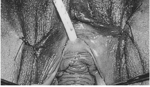
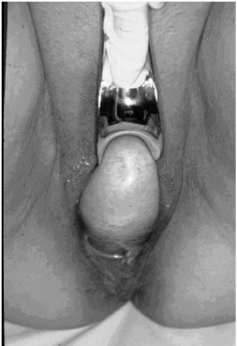
Vaginal support should be evaluated independently at all sites, including the vaginal apex, the anterior wall, and the posterior wall. After the maximal extent of POP is noted without a speculum, the support of the apex is evaluated with a bivalved speculum. Gradually removing the open speculum permits the examiner to assess apical support isolated from the anterior and posterior vaginal walls. The anterior wall is then assessed while supporting the vaginal apex and posterior wall with a Sims speculum or with a disarticulated posterior blade of a Graves speculum (Fig. 25.1). Similarly, the single speculum blade supports the vaginal apex and anterior wall while evaluating the posterior wall (Fig. 25.2). This permits the examiner to focus on the support defects in each compartment.


FIGURE 25.1 ● Support of the posterior vaginal wall and vaginal apex with a single-blade speculum permits an isolated evaluation of the support of the anterior vaginal wall. Note the normal rugated epithelium of the anterior vaginal wall. (From Baggish MS, Karram MM. Atlas of pelvic anatomy and gynecologic surgery. Singapore: WB Saunders, 2001:382, with permission.)


FIGURE 25.2 ● Support of the anterior vaginal wall and vaginal apex with a single-blade speculum permits an isolated evaluation of the support of the posterior vaginal wall.


STAGING PELVIC ORGAN PROLAPSE

It is important to objectively document the extent of prolapse, both before and after interventions. There are a number of ordinal staging systems to describe the degree of descent, although the POPQ examination is the most widely accepted.


Pelvic Organ Prolapse Quantification System

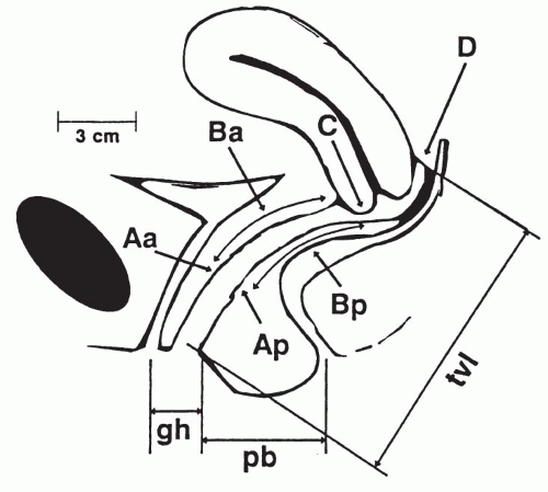
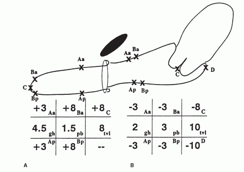
This standardized system was published in the American Journal of Obstetrics and Gynecology in July 1996 (4). The system measures eight sites to create a tandem vaginal profile before assigning site-specific ordinal stages. Keys to this classification scheme are specifically defined points of measurement and use of a defined anatomic landmark as a fixed point of reference. The hymen is the fixed point by which measurements of six vaginal points are referenced. The report discourages the use of imprecise terms such as introitus. Points of measurement within the vaginal canal are defined for the anterior and posterior vaginal wall and vaginal apex. Anteriorly, the two points of reference include a point 3 cm proximal to the external urethral meatus (point Aa) and a point Ba that represents the most distal or dependent portion of the anterior vaginal wall proximal to Aa (Fig. 25.3). Posteriorly, the points of reference are similar by use of a midline posterior point 3 cm proximal to the hymen (point Ap) and a point Bp that represents the most distal or dependent position of the posterior vaginal wall proximal to point Ap (see Fig. 25.3). The vaginal apex is defined by two points: the most distal edge of the cervix or vaginal cuff scar (point C) and the location of the posterior fornix or pouch of Douglas (point D; see Fig. 25.3). This last point is omitted in patients who have no cervix. Measurements of the genital hiatus, perineal body, and total vaginal length are also included in this classification scheme (see Fig. 25.3). A grid or line diagram may be used to describe normal support as well as support defects of the vaginal cuff and anterior and posterior vaginal walls (Figs. 25.4 and 25.5).


FIGURE 25.3 ● Six sites (points Aa, Ba, C, D, Bp, and Ap), genital hiatus (gh), perineal body (pb), and total vaginal length (tvl) used for pelvic organ support quantitation. (From Bump RC, Mattiasson A, Bø K, et al. The standardization of terminology of female pelvic organ prolapse and pelvic floor dysfunction. Am J Obstet Gynecol 1996;175:10-17, with permission.)

All measurements are made in centimeters and expressed as above (proximal) or below (distal) the hymen and designated negative or positive, respectively. The numbers may then be recorded as a simple line of numbers (tandem profile) or as a three-by-three grid. In addition, the report establishes an ordinal staging system to be used after the quantitative description is completed (Table 25.3).

The committee acknowledges the arbitrary nature of such a staging system but concludes that it
is necessary as staging allows for description and comparison of populations of patients, correlation of symptoms with severity of prolapse, and assessment of treatment outcomes. Unfortunately, the staging system does not predict women who will be symptomatic. For example, a retrospective cross-sectional study assessing prolapse in 905 women using the POPQ examination found no discrete stage that discriminated between symptomatic and nonsymptomatic prolapse (10).


FIGURE 25.4(A) Grid and line diagram of complete eversion of vagina. Most distal point of anterior wall (point Ba), vaginal cuff scar (point C), and most distal point of the posterior wall (point Bp) are all at same position (+8), and points Aa and Ap are maximally distal (both at +3). Because total vaginal length equals maximum protrusion, this is stage IV prolapse. (B) Normal support. Points Aa and Ba and points Ap and Bp are all −3 because there is no anterior or posterior wall descent. Lowest point of the cervix is 8 cm above hymen (−8) and posterior fornix is 2 cm above this (−10). Vaginal length is 10 cm, and genital hiatus and perineal body measure 2 and 3 cm, respectively. This represents stage 0 support. (From Bump RC, Mattiasson A, Bø K, et al. The standardization of terminology of female pelvic organ prolapse and pelvic floor dysfunction. Am J Obstet Gynecol 1996;175:10-17, with permission.)

The subcommittee’s efforts in creating this classification scheme and incorporating objective criteria for the description of pelvic organ prolapse were a first step toward establishing a standard, reliable, and validated description of pelvic anatomy and function. They acknowledged the need for studies designed to evaluate and validate the descriptions and definitions they propose. In 1996, Hall et al evaluated the interobserver and intraobserver reliability of the POPQ system (11). The reproducibilities of the nine site-specific measurements and the summary stage and substage were evaluated. There was substantial and highly significant correlation between measurements for both interobserver and intraobserver examinations. Although it took new POPQ examiners an average of 1.7 minutes longer than experienced POPQ examiners to complete the examination, the reliability did not vary between the groups.

Reports suggest that the degree of prolapse observed varies by patient position, with an increase in prolapse with the patient in a sitting “45% upright” position in a birthing chair as compared with the patient in a dorsal lithotomy position (11, 12, 13). This difference did not seem to be related to other patient characteristics, including age, race, parity,
weight, or the prolapse stage or genital hiatus measurement in the lithotomy position (13). POPQ measurements have also been compared by Swift and Herring in the standing and dorsal lithotomy positions (14). A high degree of correlation of measurements in the two positions was found, and it has been postulated that this was related to differences in pelvic tilt produced by standing and dorsal lithotomy positions as compared with a sitting position. As with the McRoberts maneuver, maximum hip flexion is likely to occur in the birthing chair, which results in opening of the pelvic outlet (13,14).

Only gold members can continue reading. Log In or Register to continue

Stay updated, free articles. Join our Telegram channel

Jul 24, 2016 | Posted by in UROLOGY | Comments Off on The Clinical Evaluation of Pelvic Organ Prolapse

Full access? Get Clinical Tree

Get Clinical Tree app for offline access