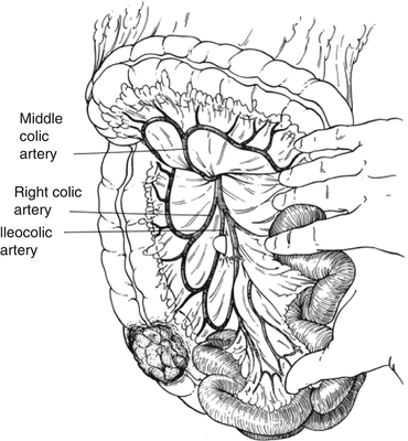
Fig. 41.1
The drawing demonstrates the incision made at the root of the right colon mesentery just caudal to the third portion of the duodenum to the right of the superior mesenteric artery
Third, the superior approach enters the retroperitoneum by opening the lesser sac and incising the peritoneum at the hepatic flexure.
Finally, with the medial to lateral approach, the ileocolic pedicle is grasped and elevated. The peritoneum on the caudal side of the pedicle is incised and the retroperitoneum is entered.
Regardless of how the retroperitoneum is accessed, the principles of the resection are the same. The right colon mesentery is elevated off the retroperitoneum and the duodenum is identified. The lateral attachments are incised, and the hepatic flexure is fully mobilized.
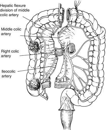
The ileocolic (Fig. 41.2) and right or hepatic branch of the middle colic vessels are ligated at their origins. The terminal ileum should be divided 10–15 cm proximal to the ileocecal valve to allow for good vascular supply (see Fig. 41.3 for extent of resection). The transverse colon is divided just to the right of the main trunk of the middle colic artery. The ileocolic anastomosis can be fashioned according to the desire of the operating surgeon. The authors prefer to divide the ileum and colon with linear staplers and perform a functional end-to-end anastomosis by anastomosing the antimesenteric surfaces of the bowel segments with a linear stapler and closing the remaining colostomy with a linear stapler or sutures.


Fig. 41.2
The vessel(s) is elevated off the retroperitoneum, and a proximal ligation is performed at the origin of the superior mesenteric artery. The surgeon’s finger is used to demonstrate the vascular origin for accurate placement of the ligation

Fig. 41.3
The drawing demonstrates the appropriate levels for vascular ligation and colonic transition for a right hemicolectomy. Notably, the transverse colon is divided just to the right of the main trunk of the MCA, although the right branch of the MCA may be taken, if required. The middle colic vessels are demonstrated and may be ligated during the performance of an extended right hemicolectomy. This leaves the descending colon in place supported by the left colic artery
Extended Right Colectomy
An extended right colectomy may be performed for any lesion involving the transverse colon.
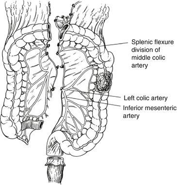
The operation proceeds in similar fashion as the right colectomy described above. However, rather than proceeding through the transverse colon mesentery to ligate and divide the right branch of the middle colic artery, dissection continues in the retroperitoneal plane to identify the main middle colic arterial trunk anterior to the pancreas. This vessel is ligated and divided.
The splenic flexure is released and the bowel with its mesentery is divided just proximal to the left colic artery, which is preserved for right-sided lesions.
The left colic may be sacrificed for left transverse colon lesions, where a more distal colonic anastomosis is desired.
Left Colectomy
The left colon can be mobilized in either a lateral to medial or medial to lateral approach.
Resection of proximal left colon lesions may require division of the middle colic artery to allow the right transverse colon to reach the rectal stump for an anastomosis.
A subtotal colectomy and ileosigmoid or ileorectal anastomosis may be preferable if there is any concern related to the blood supply. Another alternative is to perform a retroileal right colon to rectum anastomosis if maintenance of the right colon is desired (Fig. 41.4).


