Fig. 5.1
Buie anoscope
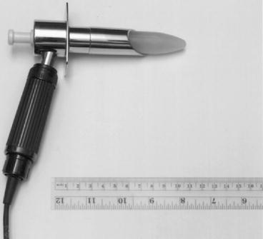
Fig. 5.2
Lighted Welch-Allyn anoscope
Slotted or side opening: Vernon-David scope with Hirschman handle (Fig. 5.3) and Hinkel-James anoscope (Fig. 5.4)
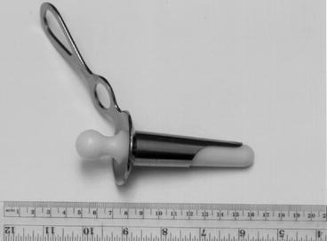
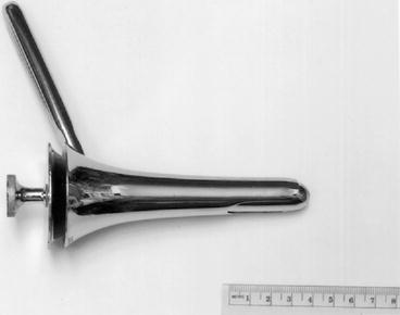
Fig. 5.3
Vernon-David with Hirschman handle anoscope
Fig. 5.4
Hinkel-James anoscope
Indications
Any anal and perianal diseases or conditions require a full examination of the anal canal. These include anal fissures, anal fistulas, anal Crohn’s disease, anal tumors, hemorrhoids, anal condyloma, bright red rectal bleeding, and pruritus ani.
Anoscopy is frequently used in conjunction with other endoscopic examinations.
Contraindications
Anal stricture or severe anal stenosis.
Patients with severe anal pain (acute anal fissure or a perianal or intersphincteric abscess) may not tolerate the examination. In general, if a patient can tolerate a digital examination, anoscopy can usually be done. A 2 % lidocaine jelly may be used in patients with anal pain.
Preparation
No preparation is required.
Positioning
A prone jackknife position gives the best exposure. An alternative is the left-lateral recumbent (Sims) position.
Technique
Inspect the anal area and then gently spread the buttock cheeks with good lighting to gain exposure.
Look for skin tags, excoriation, and change in color or thickness of the anal verge and perianal skin.
A scarred, patulous, or irregularly shaped anus may give clues to the cause of anal incontinence. Particularly in multiparous women, the anal verge may be pushed down too far during straining – a feature of the perineal descent syndrome.
A digital examination is performed with a well-lubricated index finger. The finger is pressed on the anal aperture to “warn” the patient and gradually inserted and swept all around the anal canal to detect any mass or induration. In men, the prostate should be felt. In women, the posterior vaginal wall should be pushed anteriorly to detect any evidence of a rectocele.
Anal tone (normal, tight, or loose) is estimated and the canal is assessed for any stricture or narrowing or for a defect in the internal or external sphincters.
A fibrous cord or induration in the anal area and the anal canal may indicate a fistulous tract. The external sphincter, puborectalis, and levator ani muscles can also be appreciated by digital examination.
When the puborectalis is pulled in the posterior quadrant, the anus will gape but will close immediately when the traction is released. Persistence of the gaping indicates an abnormal reflex pathway in the thoracolumbar region frequently seen in paraplegic patients.
The finger should press gently on these muscles for signs of tenderness. With good anal function, the examiner not only feels the squeeze of the muscle on the examining finger but also feels the finger pulled forward by the puborectalis muscle when the patient is asked to contract the muscles.
Insertion of the anoscope should always be done with the obturator in place. The obturator is removed during examination and reinserted to rotate the instrument to another area. In patients with redundant mucosa, reinsertion of the obturator may cause discomfort if the mucosa gets trapped between the obturator and the anoscope. However, if the beveled type of endoscope is used, the endoscope can be rotated without having to reinsert the obturator.
If an inverted (jackknife) position is used, the examination table need not be tipped down more than 10–15°. If a left-lateral position is used, an assistant needs to pull up the right cheek of the buttock for exposure.
During examination, the patient is asked to strain with the anoscope sliding out to detect any prolapse of the rectal mucosa and the anal cushion. Excoriation, metaplastic changes, and friable mucosa indicate a prolapsed hemorrhoid.
Biopsies may be taken via the anoscope. Local anesthesia may be necessary for biopsies in the sensate area distal to the dentate line.
Complications
Anal tear, especially at the posterior midline, can occur in patients with anal stenosis.
Friable hemorrhoids may bleed from trauma from the anoscope.
Rigid Proctosigmoidoscopy
Three sizes of rigid proctosigmoidoscope are available (Fig. 5.5):
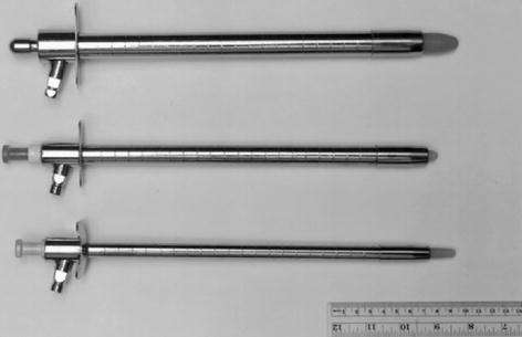
Fig. 5.5
Rigid proctosigmoidoscope. Top 19 mm × 25 cm, middle 15 mm × 25 cm, bottom 11 mm × 25 cm
A 19 mm × 25 cm scope is the standard size for a general examination and for polypectomy or electrocoagulation. A disposable standard-size rigid proctosigmoidoscope is available.
A 15 mm × 25 cm endoscope is an ideal size for general examination. It is much better tolerated by the patient, causing less spasm of the rectum and thus, minimal air insufflation, yet enables as adequate an examination as the standard-size endoscope.
An 11 mm × 25 cm endoscope should be available for examining the patient who has anal or rectal stricture, such as Crohn’s disease.
Indications
Rigid proctosigmoidoscopy has largely been replaced by flexible sigmoidoscopy but remains useful as it allows evacuation of blood clots or stool and can identify the precise site and size of rectal neoplasm.
Contraindications
Severe anal pain (from an acute fissure, thrombosed external hemorrhoids, and perianal abscess).
Anal stricture.
Patients with acute abdomen of any cause or a rectal or sigmoid anastomosis less than 2 weeks postoperatively should have a rigid proctosigmoidoscopy with caution.
Preparation
Two phosphate enemas should be given within 2 h of the examination. This is not necessary in a patient who has diarrhea or active bleeding. Sedation is unnecessary.
Positioning
Prone jackknife is preferred but the left-lateral position also gives an adequate examination and should be used in conditions such as pregnancy, severe hypertension, retinal detachment, or postoperative eye surgery and some apprehensive patients.
Technique
Although a standard proctosigmoidoscope is 25 cm in length, the average distance that the scope can be passed is 20 cm. In men, the scope can be passed to 21–25 cm half of the time, and in women, it can be passed that distance one-third of the time.
Rigid proctosigmoidoscopy is suitable only to examine the rectum and, in some patients, the distal sigmoid colon.
Properly performed, rigid proctosigmoidoscopy produces only mild discomfort. Some patients are fearful of the examination because of past bad experience with the procedure or from what they have heard. A few words of reassurance will be helpful. Pain is produced by excessive stretching of the mesentery by direct scope pressure or by air insufflation.
With the obturator in place and held steady with the right thumb, the well-lubricated rigid proctosigmoidoscope is gently inserted into the anal canal, aiming toward the umbilicus for a distance of about 4–5 cm. Then the endoscope is angled toward the sacrum and advanced another 4–5 cm into the rectum. The obturator is removed and the bowel lumen is negotiated under direct vision.
Air insufflation is limited to the amount necessary to open the lumen.
When an angle is encountered, the endoscope is withdrawn 3–4 cm and then readvanced. This may be repeated several times to straighten the angulation.
If further advancement is unsuccessful, the procedure is terminated. The length of insertion should be measured from the anal verge without stretching the bowel wall.
Careful examination is done as the instrument is withdrawn. The instrument should be rotated on withdrawal to ensure examination of the entire circumference, and it is usually necessary to insufflate a small amount of air for good visualization of the lumen.
The mucosal folds in the rectum (valves of Houston) can be flattened with the tip of the endoscope to see the area immediately proximal to them.
The appearance of the mucosa, depth of insertion, and the size, appearance, location, and level of any lesion should be accurately recorded.
If a biopsy is performed, the location, level, and number of biopsies and whether electrocoagulation is necessary should be noted.
A rigid cautery snare (Frankfelt snare) and cautery tip attachments are useful for excision or ablation of rectal neoplasms.
During the entire procedure, suction and water irrigation should be available.
Complications
The tip of the endoscope can tear the mucosa producing hemorrhage.
Perforation from diagnostic rigid proctosigmoidoscopy is extremely rare (1 in 8–20,000 examinations).
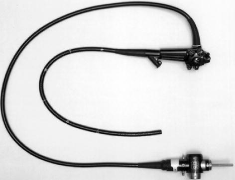
Flexible Sigmoidoscopy
The flexible videosigmoidoscope (FFS) is 60 cm in functional length (Fig. 5.6) and visualizes the entire sigmoid colon in some cases up to the splenic flexure.