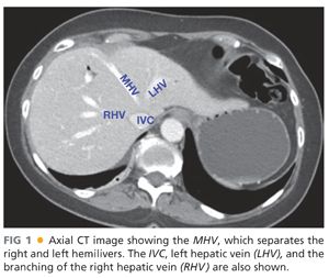
■ The right and left hemilivers are anatomically and spatially divided by Cantlie’s line, which runs from the fundus of the gallbladder to the suprahepatic inferior vena cava (IVC). Within the substance of the liver, the middle hepatic vein courses along this same path and is the true division of the right and left hemilivers (FIG 1).

COUINAUD’S SEGMENTS
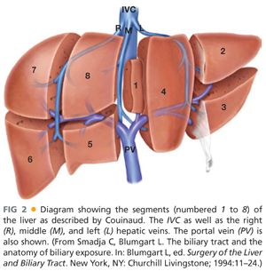
■ In 1954, Couinaud2 published his seminal work, Le Foie: Études Anatomiques Et Chirurgicales, in which he classified the liver into segments, each with their own inflow, outflow, and biliary drainage (FIG 2). He based his segments on the arborization of the portal vein within the liver. These segments begin with the caudate lobe (segment 1) and continue in a clockwise fashion from left to right.

■ Couinaud3 further divided the liver based on sectors, which are based on the arborization of the hepatic veins, each of which relies on a portal pedicle (FIG 3).

GOLDSMITH AND WOODBURNE CLASSIFICATION
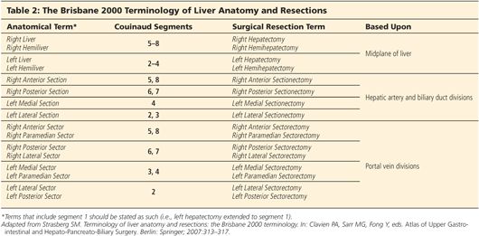
■ Goldsmith and Woodburne4 divided the liver into the caudate lobe and four distinct segments:
■ Left lateral (Couinaud segments 2 and 3)
■ Left medial (Couinaud segment 4)
■ Right anterior (Couinaud segments 5 and 8)
■ Right posterior (Couinaud segments 6 and 7)
BISMUTH CLASSIFICATION
■ Bismuth divided the liver based on fissures: three vertical lines corresponding to the divisions made by the three major hepatic veins.5
■ The fourth fissure, named the transverse fissure, is based on the level of the portal vein bifurcation within the liver and divides the “upper” liver from the “lower” liver.
■ Bismuth identified eight subsegments:
■ Caudate lobe (Couinaud segment 1)
■ Left superior subsegment (Couinaud segment 2)
■ Left inferior subsegment (Couinaud segment 3)
■ Left medial subsegment (Couinaud segment 4)
■ Right anterior inferior subsegment (Couinaud segment 5)
■ Right posterior subsegment (Couinaud segment 6)
■ Right posterior superior subsegment (Couinaud segment 7)
■ Right anterior superior subsegment (Couinaud segment 8)
THE BRISBANE TERMINOLOGY OF LIVER ANATOMY AND RESECTION
■ Due to the confusion between various classifications, the International Hepato-Pancreato-Biliary Association (IHPBA) formed a committee in 1998 to delineate an accepted and universal terminology to describe hepatic anatomy and, more specifically, hepatic resections.6 In 2000, their terminology was published. In it, a nomenclature for anatomic terminology and that of surgical resection were clearly delineated (Table 2).

CAUDATE LOBE
■ The caudate lobe is distinct in many ways. Because of its distinct inflow, outflow, and drainage, it is sometimes termed “the third lobe of the liver.”
■ Embryologically, it is derived from the right lobe,7 laying on the posterior surface of the right hemiliver, bordered on the left by the ligamentum venosus. It is bordered superiorly by the hepatic veins as they enter the IVC and posteriorly by the IVC itself. Inferiorly, it is bordered by the hilum of the liver and the bifurcation of the left and right portal veins.
■ Anatomically, there are three portions to the caudate lobe (FIG 4), each based on the portal inflow:8
■ The Spiegelian lobe (or Spiegel’s lobe)
■ The paracaval portion (or Couinaud’s segment 9)
■ The caudate process

■ Surgically, the caudate can be divided in two: segment 1 (Spiegel’s lobe) and so-called segment 9 (paracaval portion and caudate process) (FIG 5).

■ The use of terminology related to Couinaud segment 9 is often dropped from the literature and this term is neither universally accepted nor used.
■ The paracaval portion, which projects medially, can circumferentially wrap around the IVC.9 This can be misinterpreted on computed tomography (CT) scan as an enlarged lymph node, and the surgeon should be aware of this to avoid inappropriate preoperative staging of a patient.
■ The caudate derives its portal inflow from the left and right portal veins; venous drainage is directly into the IVC via multiple small caudate veins. Thus, in diseases of hepatic venous obstruction, such as Budd-Chiari syndrome, it will hypertrophy and provide alternate routes for obstructed hepatic venous flow.
SURFACE ANATOMY
■ The liver is covered by a thin membrane called Glisson’s capsule. This epithelial layer is contiguous with internal anatomy of the liver as sheaths around the portal veins, hepatic arteries, and bile ducts.10
■ Despite its rounded, pyramidal shape, the liver can be described as having two surfaces: visceral and diaphragmatic.
■ The visceral surface is inferior.
■ The diaphragmatic surface can be divided into anterior, posterior, superior, and lateral aspects.
■ The anterior aspect opposes the diaphragm and the posterior abdominal wall. It is generally smooth and has few if any impressions.
■ The posterior aspect contains the bare area of the liver as well as the confluence of the hepatic veins prior to their entry into the IVC.
■ The lateral aspect opposes the right ribs 7 to 11, which often leave impressions on the liver surface.
■ The superior aspect is a continuation of the anterior surface and ends at the coronary ligament. It contains the cardiac impression and extends medially and laterally, sitting below the left and right lungs and pleura. This is clinically important as neoplasms of the dome of the liver can manifest by sympathetic effusions and diaphragmatic splinting.
■ The majority of the surface of the liver is free of peritoneum, but there are several attachments and reflections of peritoneum—referred to as ligaments—that suspend the liver in its position.
■ Failing to properly understand these attachments can lead to injury to the associated visceral organs or the diaphragm.
■ The falciform ligament is a bileafed peritoneal reflection, which envelops the ligamentum teres.
■ The ligamentum teres, or round ligament, is the remnant of the umbilical vein.
■ After enveloping the ligamentum teres, the peritoneum attaches anteriorly and superiorly to the posterior abdominal wall, tethering the liver slightly to the right of the midline.
■ Posteriorly and inferiorly, the ligamentum teres enters the liver between segments 3 and 4, whereas, in utero, the umbilical vein drains into the left portal vein.
■ Although technically obliterated early in life, the umbilical vein recanalizes in cirrhotics and other patients with portal hypertension and must be carefully controlled when divided.
■ The ligamentum venosus, also known as Arantius’ ligament, in which the remnant of the ductus venosus lies, sits between the caudate lobe (segment 1) and the left lobe of the liver.
■ It forms a bridge between the left portal vein and the left hepatic vein, or sometimes to the junction of the left and middle hepatic veins. This can be very useful in locating the left hepatic vein during hepatic resection.
■ The coronary ligament is the reflection of the visceral peritoneum as it attaches to the diaphragm. This creates a fibrous peritoneal ring or crown (Latin: corona) around the caudal posterior surface of the right hemiliver, encircling the bare area of the liver (FIG 6).
■ The bare area is so named because there is nothing between the liver and the diaphragm other than loose areolar tissue.
■ As the coronary ligament makes a sharp turn at the right posterior lateral edge of the liver, it creates an acute angle and is called the right triangular ligament.
■
Stay updated, free articles. Join our Telegram channel

Full access? Get Clinical Tree


