Pelvic Floor Tension Myalgia


Pelvic Floor Tension Myalgia


Richard P. Marvel


INTRODUCTION

Pain has been defined by the International Association for the Study of Pain as “an unpleasant sensory and emotional experience associated with the actual or potential tissue damage, or described in terms of such damage” (1). It is always subjective, unpleasant, and an emotional experience. It cannot be confirmed or refuted by a physical test. Patients must be taken at their word that they are in pain.

Pelvic floor myalgia literally means muscular pain emanating from the muscles of the pelvic floor or in their attachments to the sacrum, coccyx, ischial tuberosity, and pubic rami. Pelvic floor tension myalgia is a chronic pain condition related to chronically increased tone and tenderness of one or several of the muscles that compose the pelvic floor. It is a poorly recognized, underdiagnosed, but common problem existing as a component of chronic pelvic pain. It has been recognized as a cause of pelvic pain in women for over a century (2), but only more recently in men (3).

Myofascial pain of the muscles of the pelvic floor has been referred to by several other names, including coccydynia, levator ani syndrome, and proctalgia fugax. Each of the terms have a unique definition appearing in the literature (Table 9.1). The most widely used terms related to pelvic floor dysfunction and pain come form the Rome diagnostic criteria for the functional bowel disorders. These criteria were developed by the Committee on Functional Bowel Disorders, Multinational Working Teams to Develop Diagnostic Criteria for Functional Gastrointestinal Disorders (4). The functional bowel disorders require that organic causes of the pain be ruled out and are based solely on the history of symptoms.

One of the cornerstones in the evaluation of women with chronic pelvic pain is the ability to recreate the pain on physical examination. This makes the functional gastrointestinal disorders less useful in evaluating women with chronic pelvic pain.


HISTORY

One of the first reports in the medical literature of pain involving the pelvic floor was by Sir J. Y. Simpson in 1859, when he described a case of a woman with unrelenting chronic pain after she was thrown from her horse. After recovering from her injuries, she developed severe pain with sitting. She continued with pain for 2 years and had a “miserable and wretched existence.” In 1855, after conservative treatments failed to bring relief, Simpson performed the first reported coccygectomy, which led to complete resolution of her pain. Simpson coined the term “coccygodynia” after the leading symptom of pain in the region of the coccyx, acknowledging that the pain was emanating from the pelvic floor muscles and their attachments. He noted that injuries such as a hard fall backwards or sitting down forcibly on a chair or angled body could bring about the painful syndrome. Patients suffering from coccygodynia were noted to have pain with sitting, reclining, and rising from the sitting position. He believed that the pain occurred from the action of the muscles causing motion of the coccyx, with possible inflammation (2).

The first concise description of the syndrome, including the muscular origin of the pain, is credited to Dr. George Thiele in 1936 (5). He described 38 cases with confirmed presence of tonic spasm of the levator ani and coccygeus muscles by his own examination. After pooling patients from
other colorectal surgeons, he reviewed 87 patients from nine practices. None had a history of recent injury of the coccyx or fracture. The duration of symptoms ranged from 3 days to 32 years, with an average of 2 years. Of these patients, 19 of 87 (21.8%) had a history of trauma, including “falls, parturition, and long automobile rides.” Tonic spasm was reported in 64 of 69 cases. He also described a group of 33 patients with piriformis spasm and tenderness and noted a consistent history of supragluteal pain and pain radiating down the posterior thigh(s). He developed the first recognized treatment, still known as Thiele’s massage (described later in this chapter).


TABLE 9.1 Definitions of Pelvic Floor Pain Disorders


Diagnostic Term


Definition


Pelvic floor dyssynergia (previously “anismus”)


Paradoxical contraction of the pelvic floor with defecation (39)


Levator ani syndrome


Chronic anal pain lasting more than 20 minutes in absence of organic disease (39)


Proctalgia fugax


Fleeting severe rectal pain lasting several seconds to minutes, especially at night (39)


Pelvic floor tension myalgia


Pain due to tension of the pelvic floor musculature with pain in the muscles themselves or emanating from the areas of attachment such as the sacrum, coccyx, ischial tuberosity, and pubic rami (24)


Dyspareunia


Recurrent or persistent genital pain associated with sexual intercourse, divided into entry and deep components (40)


Vaginismus


Recurrent or persistent involuntary spasm of the outer third of the vagina interfering with vaginal penetration and causing personal distress (40)


Levator syndrome


Symptom complex of pain, pressure, or discomfort in the region of the rectum, sacrum, and coccyx, with tenderness and spasm of the levator muscles (41)


Coccydynia


A symptom of pain in or around the coccyx, usually reproduced with palpation or movement of the coccyx (2,25)


CHRONIC PELVIC PAIN

Chronic pelvic pain is a common clinical condition encountered by gynecologists, urogynecologists, and primary care physicians. It was most recently defined as noncyclic pain of 6 or more months’ duration that localizes to the anatomic pelvis, anterior or abdominal wall at or below the umbilicus, the lumbosacral back, or the buttocks and is of sufficient severity to cause functional disability or lead to medical care (6). It is a common problem, especially among women in the reproductive years. In Great Britain, chronic pelvic pain was found to have a community-based prevalence of 3.8% in women aged 15 to 73 , higher than migraine headache (2.1%) and asthma (3.7%) (7). In a U.S. phone survey, a prevalence of 14.7% was reported over a 3-month time period (8). It accounts for 10% of referrals to gynecologists, 40% of gynecologic diagnostic laparoscopies, and 12% of all hysterectomies (9). Chronic pelvic pain has a significantly negative impact on quality-of-life factors, with 26% of women reporting being bedridden due to pain within a 2-week period of time (8).

The evaluation of chronic pain of any location is complex. In the acute pain model, the pain is usually due to an isolated factor that can be diagnosed and managed, leading to alleviation of the pain, such as in acute appendicitis. Chronic pain is very different. In chronic pelvic pain, the pain is usually due to a combination of painful stimuli that as a whole compose their pain syndrome. An exhaustive search for “the” cause of the pain is generally fruitless and frustrating for patient and
physician alike. A completely different approach is necessary using a rehabilitation model, improving each component of the pain over time to achieve the goal of improved function.

Pain is a very individual experience and is based on a multitude of factors, including past experiences, culture, genetics, injury, trauma, abuse, personality, and support systems. The vast majority of women have multiple components related to the etiology of their pain involving many aspects of the human mind and body. It is not uncommon for women with chronic pelvic pain to have endometriosis, interstitial cystitis, myofascial pain, peripheral neuropathy, depression, anxiety, a history of abuse (physical, emotional, or sexual), and poor coping skills. All of these and many undetermined factors play a part in their overall pain and illness. Diagnosing and treating one component (e.g., endometriosis) will many times be unsuccessful in alleviating the pain and suffering of this population of women. It is within this concept of the genesis of chronic pelvic pain that evaluation and management of all components, among them pelvic floor myalgia, is the cornerstone in helping women with chronic pelvic pain.


ANATOMY

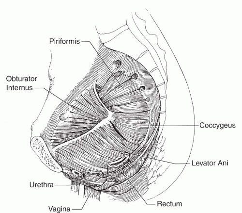
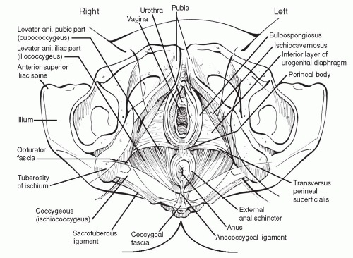
The pelvic floor is made up of a system of muscles and fascial attachments in the shape of a bowel inside the true pelvis (Fig. 9.1). The muscles of the pelvic floor include the levator ani, comprised of the puborectalis, iliococcygeus, pubococcygeus, and coccygeus. Some authors also include the muscles of the urogenital triangle, including the ischiocavernosus, bulbospongiosus, and superficial transverse perinei muscles, which lie inferior to the true pelvic floor (Fig. 9.2). It is important to realize that within this group of muscles, the boundaries of individual muscles are difficult to distinguish. Although a common cause of deep pelvic pain, the piriformis muscle is actually part of the posterior wall of the pelvis. The piriformis muscle originates from the anterior and lateral surfaces of the sacrum, portions of the ilium, and sacroiliac joint capsule. It forms the posterolateral border of the pelvis. It traverses the greater sciatic foramen, the main space-filling muscle, inserting into the medial side of the upper border of the greater trochanter of the femur. It is innervated from branches from the L5, S1, and S2 nerve roots. Its action is to laterally rotate the extended thigh and abduct the flexed thigh and is thus an external rotator of the thigh.


FIGURE 9.1 ● Muscles of the pelvic floor and sidewall, demonstrating the proximity to the vagina and rectum and slinglike support of the pelvic organs. (From Howard FM. Pelvic pain: Diagnosis and management. Philadelphia: Lippincott Williams & Wilkins, 2000:36.)

The coccygeus, sometimes referred to as the ischiococcygeus, attaches to the lateral border of the coccyx and lower sacrum, with the apex of this triangular muscle attached to the ischial spine. It may be mostly tendinous and fused with portions of the sacrospinous ligament, which it covers. Its function may have been related to movement of the residual tail of other species. It is innervated by branches from the S2, S3, and S4 nerve roots. The iliococcygeus is more inferior and medial to the ischiococcygeus. It originates from the white line (arcus tendineus fascia pelvis) of the pelvic sidewall and fascia of the obturator internus muscle. Most of the fibers attach in the midline to the anococcygeal ligament or raphe, thus fusing with muscles fibers from the contralateral side. The
pubococcygeus muscle originates from the posterior pubic ramus. Some fibers branch off from the main body of the muscles to encircle the urethra and vagina. These portions are sometimes referred to as the pubourethralis and pubovaginalis muscles. The main body of the muscle then passes posteriorly around the rectum, joining with fibers from the contralateral side. This forms a sling of muscle that maintains the rectoanal angle. This portion of the muscle pulls the rectum anteriorly towards the pubic symphysis and is important in maintaining the anorectal angle and fecal continence (10).


FIGURE 9.2 ● Pelvic floor muscles as seen from below in the supine female subject. (Illustration by Barbara D. Cummings. From Travell JG, Simons DG. Myofascial pain and dysfunction. The trigger point manual, volume 2: The lower extremities. Baltimore: Williams & Wilkins, 1983:113.)

The innervation of the pelvic floor has been debated. It is well accepted that branches of the pudendal nerve innervate the external anal sphincter, ischiocavernosus, bulbocavernosus, and external urethral sphincter. Some authorities believe that the puborectalis also has some innervation from the pudendal nerve (11). In a cadaveric study with histologic confirmation, Barber et al found no branches of the pudendal nerve leading to the puborectalis muscle. In fact, they found a separate nerve from either the S3 and S4 or the S4 and S5 nerve roots (not all three), which they named the levator ani nerve (12). The muscles of the urogenital diaphragm, superficial and deep perinei, bulbospongiosus, and ischiocavernosus, are innervated by branches of the pudendal nerve. The internal anal sphincter is innervated by the autonomic nervous system via the inferior hypogastric plexus, which occupies the 2 and 10 o’clock positions along the rectum and extends onto the lateral walls of the proximal and midvagina. The proximal urethral sphincter component is also innervated from this plexus, while the external urethral sphincter is innervated by a branch of the pudendal nerve (13).


PELVIC FLOOR FUNCTION

The pelvic floor serves a variety of functions. It is part of the support system for the pelvic organs, including the rectum, cervix, vagina, and bladder, helping to maintain normal anatomic relationships and actively participating in the storage function of these organs. The maintenance of some tone in the puborectalis is part of the anal continence mechanism,
specifically for solid stool. The puborectalis and external anal sphincter maintain a constant tone and relax at the time of defecation. The resting tone pulls the anorectal junction anteriorly to create a 90-degree angle between the rectal and anal canals. The musculature has a normal baseline tone eloquently described by Simmons (14).

Muscle tension depends on the viscoelastic properties of the tissues in the muscle as well as the degree of activation of the contractile apparatus of the muscle. Muscle stiffness, meaning the resistance to movement with palpation, is a combination of these two properties. A variety of factors, such as radiation, can alter the elasticity of the tissues, leading to increased stiffness, absent of significant increases in contractility of the muscle fibers themselves. Electromyographic recording identifies only the electrogenic contraction of the muscle (i.e., the contraction elicited by electrical activity of the motor nerve and muscle cell). Pelvic floor musculature has a baseline resting tone defined as the viscoelastic stiffness in the absence of contractile activity (motor unit activity and/or contracture).

Defecation is initiated by voluntary relaxation of the puborectalis. The puborectalis also functions in the prevention of incontinence. With an effort to prevent involuntary loss of stool or gas, the anal canal constricts concentrically and pulls in, the latter a function of the puborectalis. A reflexive contraction of the pelvic floor during Valsalva helps to maintain the bladder neck in an intra-abdominal position, helping to maintain urinary continence.

The pelvic floor also has a role in sexual function. Contraction of the pelvic floor plays an important role in the sensation of the female orgasmic response.


Emotional Control of Pelvic Floor Function

One of the more fascinating aspects of pelvic floor function is the emotional motor control via the limbic system. The emotional motor system consists of a medial and a lateral component. The lateral component of the emotional motor system consists of a set of cell groups in the fore and midbrain, involved in a number of specific motor activities generally related to survival mechanisms, including defensive postures, vocalization, mating, and continence (15). Holstege has demonstrated an output system in the feline in which the nucleus retroambiguus (NRA) projects to a distinct set of motoneuronal cell groups in the lumbosacral cord (15). These cell groups are thought to participate in the posturing necessary for mating. In females, the strength of this NRA motoneuronal projection pattern appeared to depend strongly on the estrous cycles and was almost nine times as strong in the estrous as in the nonestrous females.

A common, well-known example of an emotional motor component is seen in the canine. Tail wagging and other tail movements have a strong emotional basis. With happiness or elation, the tail wags back and forth, a maneuver of the sacrococcygeus ventralis muscle. When threatened, the tail is brought between the legs, protecting the genitals, a function of the coccygeus muscles, much more developed in canine species. These are emotionally driven conditioned motor responses well documented in animal species.

As some of the more important functions of the pelvic floor involve continence of urine and stool, it is logical that some control of the pelvic floor should be activated in the fight-or-flight response (i.e., when threatened). This aspect of pelvic floor function was investigated in studies of vaginismus. Vaginismus is an involuntary contraction of the muscles of the urogenital diaphragm and perhaps the puborectalis muscle in response to attempted penetration. Van der Velde et al investigated pelvic floor muscular activity in women who were exposed to several different film segments, namely threatening, erotic, neutral, or sexually threatening (16). They measured surface electromyographic activity during a baseline period of rest and during the film segments. Pelvic floor muscle activity was correlated with the threatening aspect of the film segments rather than the sexual content. In fact, the sexually threatening segment led to less pelvic floor activity than the threatening segment. They concluded that the increase in pelvic floor muscle activity was related to a generalized defense mechanism to a threatening situation rather to a sexually threatening situation. In addition, there was no difference between the vaginismistic subjects and the control subjects in response to the films, both groups tightening their pelvic floors similarly.

They subsequently repeated the study while measuring emotional responses to the film segments measured on a 7-point Likert scale, including enjoyment, fright, sexual arousal, disgust, relaxation, threat, and powerlessness. They concluded that the involuntary muscle contractions of vaginismus occur as an automatic defensive reaction in situations where conditioning of an emotion-symptom relationship had been established. Of the vaginismistic women, seven had a history of negative sexual experiences. This subgroup of women showed more muscle activity during the erotic segment. They concluded that in these women the
vaginismistic reactions may be explained by the fact that based on earlier experiences, erotic situations always have a threatening component (17). This evidence gives an anatomic and functional basis to an emotional component of pelvic floor dysfunction and myalgia. With a history of exposure to a consistently threatening environment, pelvic floor dysfunction, tension, and myalgia can develop due to a prolonged sustained guarding posture. Over time, this leads to continued shortening of the muscles, overload, hypoxia, muscle dysfunction, and pain. This necessitates psychotherapy as a component of therapy to alleviate the threat, break the guarding posture, and enable the pelvic floor to exist in a more relaxed state. This again substantiates the notion of an interdisciplinary approach to chronic pain syndromes including physical therapy, psychotherapy, medical and possibly surgical interventions.


PATHOGENESIS

Our current understanding of myofascial pain is largely based upon the work of Travell and Simmons (18). Spasticity is associated with hyperactive stretch reflexes and tendon jerks and is related to a loss of supraspinal inhibition. Muscle spasm is an involuntary muscle contraction caused by contractile activity and documented by electromyographic findings. Pain is due to muscle ischemia and the releasing of pain-producing substances due to the ischemia. If the muscle contracts at or above 30% of maximal contraction, compression of intramuscular blood vessels leads to further ischemia. In addition, the entire muscle need not be in spasm to cause pain: pain can emanate from only a segment of the muscle that is overloaded. This type of spasm is likely to occur when the muscle has remained for some time in the shortened position. It can be induced by voluntarily contracting the muscle when in the shortened position (14).

Only gold members can continue reading. Log In or Register to continue

Stay updated, free articles. Join our Telegram channel

Jul 24, 2016 | Posted by in UROLOGY | Comments Off on Pelvic Floor Tension Myalgia

Full access? Get Clinical Tree

Get Clinical Tree app for offline access