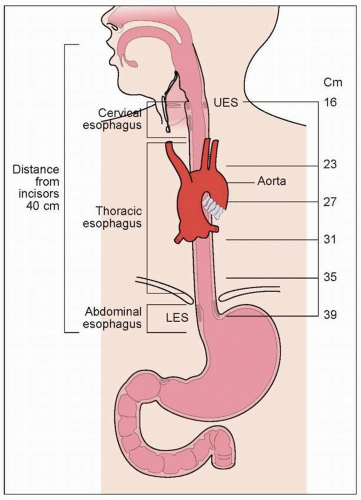
Normal esophageal anatomy and physiology
Gross anatomy
The esophagus is a muscular tube connecting the pharynx to the stomach, the proximal margin of which is the upper esophageal sphincter (UES). This is the functional unit that correlates anatomically with the junction of the inferior pharyngeal constrictor and cricopharyngeus muscles. The esophagus extends distally for 18-26 cm within the posterior mediastinum as a hollow muscular tube to the lower esophageal sphincter (LES) (1.1), which is a focus of tonically contracted, thickened, circular smooth muscle 2-4 cm long, that lies within the diaphragmatic hiatus.
Anatomy of the esophageal wall
The esophageal wall is comprised of four layers: mucosa, submucosa, muscularis propria, and adventitia; it has no serosa, making it unique to the rest of the gastrointestinal (GI) tract. Normal mucosa consists of stratified squamous epithelium, lamina propria, and muscularis mucosa, with lymphatic drainage beginning in the lamina propria (1.2).
The muscularis propria consists of both skeletal and smooth muscle: the proximal 5-33% is skeletal muscle, the middle 35-40% is mixed, and the distal 50-60% is smooth muscle. Muscles are arranged into inner circular and outer longitudinal layers (1.3).
 1.2 Wall layers and lymphatic drainage of the esophagus. Note the four wall layers: mucosa (stratified squamous epithelium, lamina propria, and muscularis mucosa), submucosa, muscularis propria, and adventitia. There is a rich lymphatic supply, which begins in the lamina propria.
Stay updated, free articles. Join our Telegram channel
Full access? Get Clinical Tree
 Get Clinical Tree app for offline access

|


