Esophageal testing


Esophageal testing


Endoscopy


Indications

Endoscopy is the technique of choice to detect structural abnormalities of the esophagus and to evaluate the mucosa. The most common indications include dysphagia, symptoms of gastroesophageal reflux disease (GERD), and gastrointestinal (GI) bleeding.


Equipment

Endoscopy allows direct visualization of the esophageal mucosa and detect structural abnormalities. Endoscopes use fiberoptic technology to capture and transmit the image from the distal end of the endoscope (2.1). Four-way tip deflection is permitted by the use of two control knobs, one with up/down movement and the other with right/left movement (2.2, 2.3 and 2.4). Endoscopes are equipped with internal channels for air, water, suction, and instruments (2.5, 2.6). The separate instrument channel allows the passage of biopsy forceps and other instruments used for treatment of upper GI disorders. Visualization is improved when air is used to insufflate the esophagus and stomach, which are normally compressed.


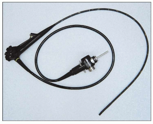
2.1 The typical forward-viewing endoscope used for examination of the upper GI tract in an adult. Standard endoscopes range from 8-11 mm in diameter, and are from 100-160 cm in length. Controls allow for manipulation of viewing direction, air insufflation, suction, water spray, and taking still photos or video.

Both small and large scopes are available (2.7, 2.8): the ‘therapeutic’ endoscope contains a larger instrument channel that permits passage of ‘jumbo’ biopsy forceps and larger coagulation devices, whereas ‘pediatric’ endoscopes may be as small as 4 mm and allow transnasal or transoral endoscopy without sedation.


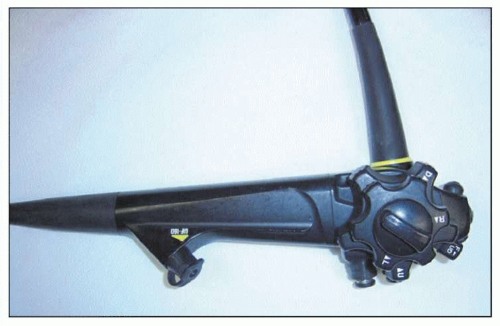
2.2 Side view close up. The two control knobs allow for four-way tip deflection. The outer/smaller dial is for right/left movement and the inner/larger dial for up/down movement. The biopsy channel allows for insertion of multiple instruments including biopsy forceps, brushes, electrocautery probes, snares, and sclerotherapy needles.


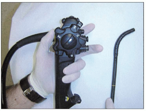
2.3 Rotating the inner dial up with the thumb causes the tip of the endoscope to deflect downward.


2.4 Rotating the inner dial down in the opposite direction causes the tip to deflect upward. Note the degree of deflection is not as great as when deflected downward.


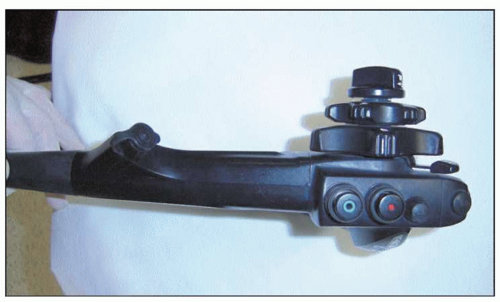
2.5 Front view of the endoscopic controls. The two buttons allow for suction, water spray, and air insufflation. The forefinger is used to press the top button for suction. The second finger is usually employed to cover the second button resulting in air insufflation. Pressing this button will result in water spray.


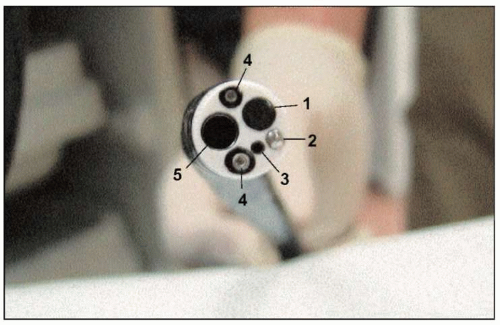
2.6 End view of the endoscope. Note the three channels for suction, insufflation, and instruments. Two light sources are also present. (1: video camera lens; 2: water flush nozzle for lens cleaning; 3: auxillary water channel; 4: light; 5: instrument channel.)


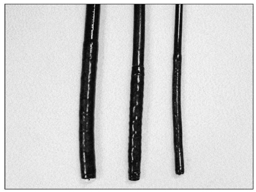
2.7 Upper GI endoscopes come in a variety of sizes. The small caliber endoscope (right), or ‘pediatric’ endoscope, has a diameter of 5-6 mm. It is ideal for use in children, but is also useful in adults with strictures or narrowings that the standard caliber endoscope cannot pass. These smaller instruments do have disadvantages, including decreased durability, poorer image quality, and smaller biopsy sizes. The larger ‘therapeutic’ scope (left) allows the passage of larger tools and more effective suction.


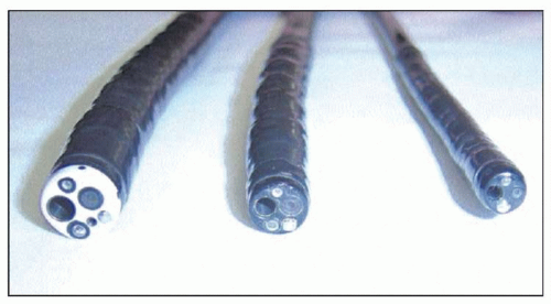
2.8 End view of instrument channel of different size endoscopes. Note the decrease in size of the channel from the therapeutic endoscope (left) to the small caliber endoscope (right).


Technique

In the United States, upper GI endoscopy is routinely performed under conscious sedation. Local anesthetic is sprayed on the posterior pharynx and intravenous sedation administered while the patient is in the left lateral decubitus position (2.9). The endoscope is inserted into the posterior pharynx where the pharynx and larynx can be examined for abnormalities (2.10). The endoscope is then advanced under direct vision into the tonically closed upper esophageal sphincter. The patient is asked to swallow to relax the upper esophageal sphincter (UES) and the endoscope is advanced to the proximal esophagus, where the mucosa should normally be smooth and light pink (2.11, 2.12).

The area of the gastroesophageal junction (GEJ) is carefully examined to identify specific landmarks (2.13), and is defined by the proximal margin of the gastric folds. The squamocolumnar junction (SCJ) can be recognized by the irregular Z-line demarcating the interface between the light pink esophageal squamous mucosa and the red columnar mucosa gastric mucosa (2.14).


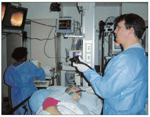
2.9 The patient is placed in left lateral decubitus position for proper positioning. The endoscopist stands directly in front of the patient’s mouth with the view screens located directly opposite. (Courtesy of John J Vargo, MD, Cleveland Clinic, Ohio, USA.)

The diaphragmatic hiatus can be identified by diaphragmatic contraction noted during patient respiration. The SCJ, the GEJ, and the diaphragmatic hiatus are normally located at the same level, unless pathology is present. In patients with Barrett’s esophagus, the SCJ is more proximal in the esophagus than the GEJ, whereas in patients with hiatal hernia, the GEJ is more proximal than the diaphragmatic indentation. While in the stomach, the endoscope is ‘retroflexed’ to look back at the GEJ; this yields a better view of the gastric side of the junction (2.15).


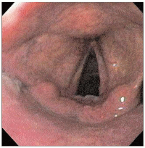
2.10 An endoscopic view of normal appearing vocal cords. This landmark is identified as an endoscopic exam begins, after the endoscope is passed through the incisors and over the tongue. Patients with extraesophageal reflux disease can present with symptoms, such as hoarseness, which can be caused by exposure of the vocal cords to gastric acid. Findings suggestive of reflux disease may include erythema, edema, granulomas, ulcerations, and laryngeal carcinoma.


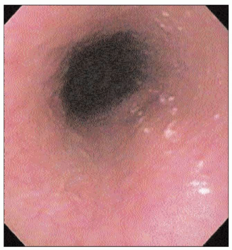
2.11 Endoscopic appearance of the mid-esophagus during a normal examination. The squamous mucosa is pale and pink, without ulceration or inflammation. The lumen is of uniform caliber, and expands uniformly with air insufflation. Landmarks seen as the esophagus is traversed include pulsation of left atrium and aorta along with indentation from left mainstem bronchus.


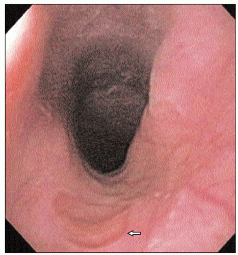
2.12 An esophageal inlet patch (arrow) is an area of heterotropic gastric epithelium found in the cervical esophagus. Note the darker red gastric mucosa, in contrast to the pale, pink esophageal mucosa. This is a common finding on upper endoscopy, and does not cause any symptoms in the patient. Inlet patches do not undergo malignant transformation, and usually no further follow-up is warranted.


2.13 Schematic diagram of the composition of the normal gastroesophageal junction. Note the relationship of the squamocolumnar junction to the diaphragm, which comprises the LES. The LES is the most important barrier protecting the esophagus from the regurgitation of gastric contents.


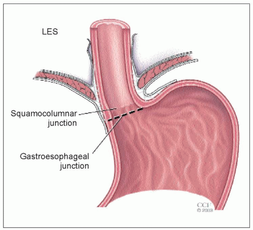
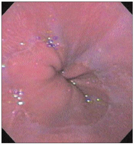
2.14 An endoscopic view of a normal SCJ. The more proximal pale pink esophageal squamous mucosa meets the darker red gastric mucosa. The junction is irregular, and is called the ‘Z-line’. The location of the transition point more than 2 cm above the diaphragm signifies hiatal hernia. This junction is usually found at 38-40 cm from the incisor teeth in adults.


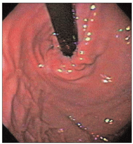
2.15 Gastric cardia on endoscopic retroflexion. Abnormalities to be noted include hiatal hernia, ulcers, gastric varices, and Mallory-Weiss tears.


Endoscopic ultrasound


Indications

Endoscopic ultrasound (EUS) provides images of the distinct layers of the esophageal wall and periluminal structures (such as lymph nodes). The primary indications for esophageal EUS are staging of esophageal cancer and evaluation of submucosal esophageal lesions.


Equipment

An endoscope is used that has ultrasound transmission and reception capability at the tip of the instrument (2.16, 2.17). Linear echoendoscopes scan in the same plane as the long axis of the endoscope, whereas radial echoendoscopes have a rotating mechanical ultrasound probe that scans in a circle at 90 degrees to the long axis of the endoscope.


Technique

The echoendoscope is inserted using a similar technique to a regular endoscope. Two video screens are used: one displays a standard endoscopic image, and the other the ultrasound image (2.18). The endosonographic layers of the esophagus can then be delineated as five alternating hyperechoic and hypoechoic bands that correspond to the histologic layers (Table 2.1, 2.19). Structures adjacent to the esophagus including the aorta, spine, left lobe of the liver, left atrium, lungs, and lymph nodes can also be visualized (2.20, 2.21, 2.22 and 2.23). With a linear instrument, fine-needle aspiration (FNA) can be performed of adjacent lymph nodes. This is critical for accurately staging esophageal cancer.


2.16 View of echoendoscopes. The radial echoendoscope (left) has a rotating mechanical ultrasound probe that scans in a circle 90 degrees to the long axis of the scope. The linear echoendoscope (right) scans in the long axis of the scope.


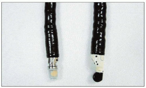
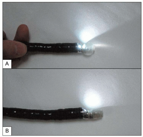
2.17 Balloon insufflation. A small latex balloon is placed over the ultrasound transmitter. With the balloon filled with water (A), better acoustic coupling can be achieved between the probe and the esophageal wall. This results in an improved ultrasound image. Note the light source is on which will provide an oblique endoscopic view.


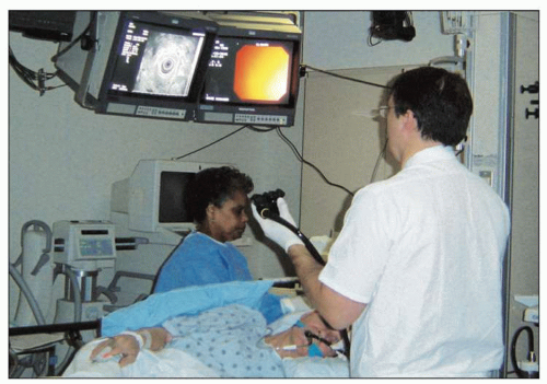
2.18 Two video screens are utilized: one screen shows the standard endoscopic image and the other the ultrasound image. (Courtesy of John J Vargo, MD, Cleveland Clinic, Ohio, USA.)


Table 2.1 Endosonographic appearance of esophageal wall layers


Esophageal wall
layer


Endosonographic
appearance


Superficial mucosa


Hyperechoic


Deep mucosa


Hypoechoic


Submucosa


Hyperechoic


Muscularis propria


Hypoechoic


Adventitia


Hyperechoic


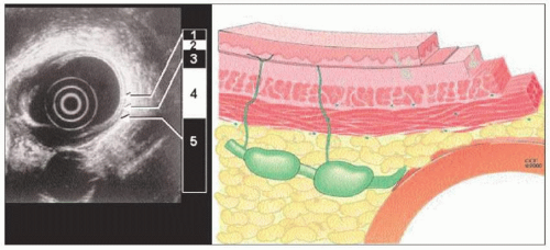
2.19 Schematic view of the esophageal wall and associated endoscopic ultrasound view. The endosonographic layers of the esophagus can be delineated as five alternating hyperechoic and hypoechoic bands that correspond to the histologic layers. The innermost layer is hyperechoic and corresponds to the interface between the ultrasound waves and the superficial mucosa. The second layer is hypoechoic and corresponds to the deep mucosa. The third layer is hyperechoic and corresponds to the submucosa. The fourth layer represents muscularis propria and is depicted as a hypoechoic band. The fifth layer is hyperechoic and represents the adventitia in the esophagus.


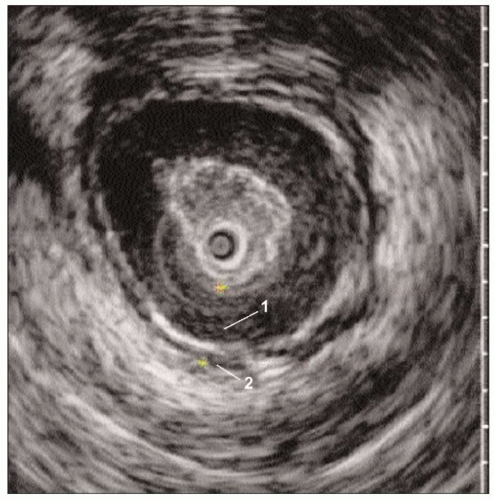
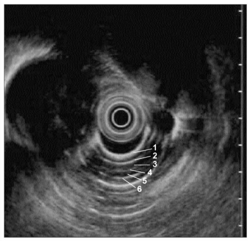
2.20 Normal endoscopic ultrasound, radial. (1: mucosa and muscularis mucosa; 2: submucosa; 3: inner circular smooth muscle; 4: intermuscular connective tissue; 5: outer longitudinal muscle; 6: adventitia.)


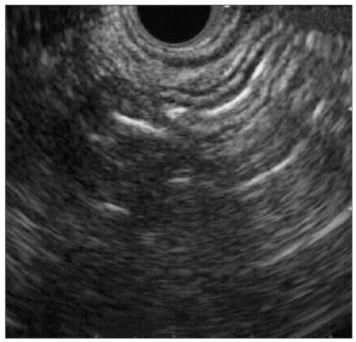
2.21 Normal endoscopic ultrasound, linear. (Courtesy of John J Vargo, MD, Cleveland Clinic, Ohio, USA.)


2.22 Endoscopic ultrasound of the LES. An echoendoprobe with higher frequency (higher detail, less penetration) can allow detailed visualization of the musculature of the LES. Note the hypoechoic inner circular and outer longitudinal layers separated by a thin band of hyperechoic material (1: inner circular smooth muscle; 2: outer longitudinal smooth muscle).


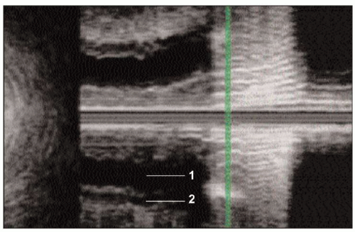
2.23 3-D Endoscopic ultrasound of the LES. Here the ultrasound image is reconstructed to a longitudinal view. Again note the two layers of the muscularis propria (1: inner circular smooth muscle; 2: outer longitudinal smooth muscle).


Barium swallow


Introduction

Contrast studies enable radiographic examination of the esophagus and can be done alone, in combination with oropharyngeal evaluation, or as a part of an upper GI series. The following techniques are used for routine examination:


  • Double contrast.


  • Full-column.


  • Mucosal relief.


  • Fluoroscopic observation.


Indications

Indications for a barium swallow include:


  • Dysphagia.


  • Odynophagia.


  • Esophageal strictures.


  • Esophageal motility disorders.


  • Esophageal function testing.


Double contrast

This can be achieved by coating the esophagus with dense barium and subsequently distending it with gas (2.24). Double contrast is a good test for evaluating the esophageal mucosal surface to detect small neoplasms, esophagitis, and diverticulosis. In some cases the gastroesophageal junction may not adequately distend, resulting in poor detection of hiatal hernias, lower esophageal mucosal rings, and peptic strictures.


Full-column

This involves rapid filling of the esophagus with barium (2.25) while patient is in prone position and is a good test for evaluating esophageal motility. It is also a useful technique to visualize hiatal hernias, lower esophageal mucosal rings, and peptic strictures (see figures from the appropriate sections), but may not detect small neoplasms, mild esophagitis, and esophageal varices.


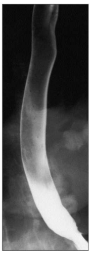
2.24 Double contrast barium swallow. Double contrast barium swallow is performed by coating the esophagus with a dense barium and distending it with gas. This allows simultaneous examination of the distended esophagus and its mucosal surface.


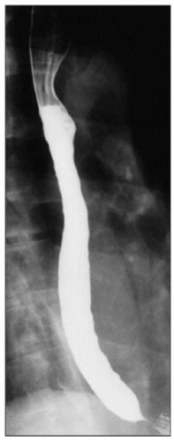
2.25 Full-column barium swallow, obtained by rapid filling of the esophagus with barium while the patient is in the prone position. Esophageal motility may be assessed with fluoroscopy in the prone position by observing multiple swallows of barium. It is a good test to visualize hiatal hernias, lower esophageal mucosal rings, and peptic strictures. However, it may not detect small neoplasms, mild esophagitis, and esophageal varices.


Mucosal relief

Imaging of the collapsed esophagus coated with dense barium allows good visualization of smooth, longitudinal esophageal folds. Irregularity of the thickening of these folds can be due to small neoplasms, esophagitis, and esophageal varices. Lesions that require esophageal distension are not seen well.


Fluoroscopic observation

Fluoroscopic observation provides a motion recording of esophageal function and motility with each barium swallow, which facilitates evaluation of esophageal and oropharyngeal functional disorders.


Esophageal manometry


Introduction

Manometry can be used as a diagnostic test to evaluate esophageal motor function.

It measures intraluminal pressures and coordination of the pressure activity of the three functional regions of the esophagus: the lower esophageal sphincter (LES), esophageal body, and UES. The manometry probe consists of a 4 mm polyvinyl catheter containing several small caliber lumens that are perfused with water from a low compliance perfusion device (2.26). When a catheter port is occluded by an esophageal contraction, water pressure builds within the catheter exerting a force, which is conveyed to an external transducer. The electrical signals from the transducers are transmitted to a computer, which produces a graphic record.


Indications

Indications for manometry are presented in Table 2.2, and include:


  • Evaluation of dysphagia in patients without evidence of mechanical obstruction, such as strictures, or in whom achalasia is suspected.


  • Defining the location of the LES for placement of intraluminal devices, such as a pH probe, which requires positioning relative to LES.


  • Pre-operative evaluation for anti-reflux surgery in patients suspected of having esophageal motility disorder such as achalasia.


  • Other possible indications are:


    • Evaluation of dysphagia in patients treated for achalasia or have undergone anti-reflux surgery.


    • Routine pre-operative assessment of esophageal peristalsis prior to anti-reflux surgery.


Manometry is not indicated for making or confirming the diagnosis of GERD, or as the initial test for non-cardiac chest pain.


Technique

Manometry is performed after an overnight fast using a round 4 mm polyvinyl catheter continuously perfused with distilled water at a rate of 0.5 ml/min by a low compliance, pneumohydraulic capillary infusion system (2.27). The stationary pull-through technique is used to determine the location and length of the LES and UES.


Table 2.2 Indications for esophageal manometry


Indicated


Possibly indicated


Not indicated


Evaluation of dysphagia in patients without evidence of mechanical obstruction or if achalasia is suspected


Evaluation of dysphagia in patients treated for achalasia or undergone anti-reflux surgery


As the initial test for non-cardiac chest pain


Pre-op evaluation for anti-reflux surgery in patients suspected of having esophageal motility disorder, such as achalasia


Routine pre-op assessment of esophageal peristalsis prior to anti-reflux surgery


Diagnosis of GERD


Defining the location of LES for placement of intraluminal devices, such as pH probe, which requires positioning relative to LES


GERD: gastroesophageal reflux disease; LES: lower esophageal sphincter

Only gold members can continue reading. Log In or Register to continue

Stay updated, free articles. Join our Telegram channel

Aug 1, 2016 | Posted by in GASTROENTEROLOGY | Comments Off on Esophageal testing

Full access? Get Clinical Tree

Get Clinical Tree app for offline access