Nephronophthisis—Medullary Cystic Kidney Disease


Nephronophthisis—Medullary Cystic Kidney Disease


Friedhelm Hildebrandt

Rannar Airik

John A. Sayer


A group of hereditary renal diseases is summarized under the term NPHP-MCKD complex,1,2 because the different disease entities share several features regarding (1) macroscopic pathology, (2) microscopic pathology, and (3) clinical symptoms (Table 15.1A). In this way the complex describes a distinct clinicopathologic entity.3 The term nephronophthisis (NPHP) is used for the autosomal recessive variants, which lead to end-stage renal disease (ESRD) in the first 3 decades of life, whereas the term medullary cystic kidney disease (MCKD) refers to the autosomal dominant forms, in which ESRD develops in the third to seventh decade of life. Extrarenal manifestations such as ocular motor apraxia, retinitis pigmentosa, hepatic fibrosis, skeletal defects, and cerebellar vermis aplasia have exclusively been described in association with juvenile nephronophthisis. The only extrarenal associations in MCKD are hyperuricemia and gout. The identification of causative recessive genes in nephronophthisis has implicated the function of primary cilia and centrosomes in its pathogenesis.


FEATURES SHARED AMONG DISEASES OF THE NEPHRONOPHTHISIS-MEDULLARY CYSTIC KIDNEY DISEASE COMPLEX


Macroscopic Pathology

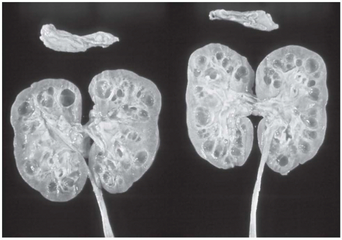
A major feature shared among the disease entities of the NPHP-MCKD complex (Table 15.1A[i]) is the appearance on macroscopic pathology as described in 27 patients with juvenile NPHP by Waldherr et al.1 Kidney size is normal or moderately reduced. Cysts primarily appear at the corticomedullary border of the kidneys (Fig. 15.1). This is quite distinct from autosomal dominant and recessive polycystic kidney disease, where kidneys become grossly enlarged as a result of cystic dilatation throughout the organ. From the external surface, the kidney is indistinguishable from the kidney affected by glomerulonephritis or pyelonephritis. The surface usually has a finely granular appearance, most likely due to the protrusion of dilated cortical collecting ducts. Calices and pelvis appear completely normal. There are from 5 to over 50 cysts of 1 to 15 mm in diameter located preferentially at the corticomedullary border (Fig. 15.1). The cysts primarily arise from the distal convoluted and medullary collecting tubules as shown by microdissection,4 but may also appear in the papilla. Cysts are not always present, but do occur in about 70% of autopsy cases. They apparently arise late in the course of the disease5 and do not seem to be important for disease progression to renal failure.6 Therefore, the presence of cysts is not a prerequisite for diagnosis.


Microscopic Pathology

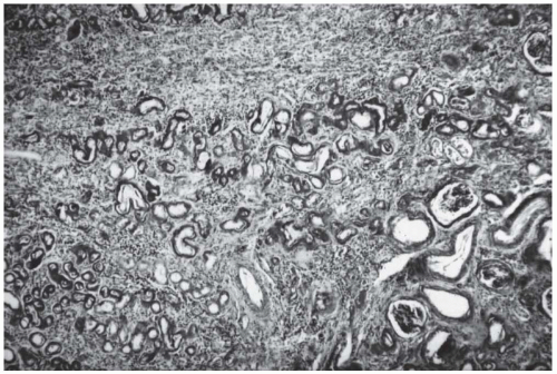
The second shared feature among diseases of the NPHP-MCKD complex pertains to renal histology (Table 15.1A[ii]). The histologic changes are characteristic, but not pathognomonic, for the disease group. The characteristic histologic triad of NPHP-MCKD consists of (1) tubular basement membrane disintegration with irregular thickening as well as attenuation of the tubular basement membrane, (2) interstitial round cell infiltration with marked fibrosis and, (3) later in disease development, tubular atrophy with cyst development, which occurs predominantly at the corticomedullary junction (Fig. 15.2). Cysts seem to be the result rather than the cause of the atrophic process, although this time course could not be corroborated by statistical analysis.1,7 Sometimes, a communication between a cyst and a tubule can be seen. The tubular basement membrane (TBM) is extremely thickened and multilayered. Fibroblasts are noted between the membrane layers. TBM changes and diverticulum formation are most prominent in the distal tubules, where cysts are lined with a single layer of cuboidal or flattened epithelium. In the advanced stage, the picture merges into a diffuse sclerosing tubulointerstitial nephropathy, the characteristic picture of end-stage NPHP-MCKD. The only significant glomerular change in early stages involves periglomerular fibrosis with a splitting and thickening of the Bowman capsule and glomerular obsolescence only in nephrons that
have been destroyed by the tubular alterations. An escape of Tamm-Horsfall (uromodulin) protein from damaged collecting tubules into the interstitium has been demonstrated in about 50% of patients with NPHP-MCKD as a periodic acid-Schiff (PAS)-positive material and by specific immunofluorescence staining with an anti-THP antibody.8 Immunofluorescence does not otherwise contribute to the diagnosis of NPHP-MCKD.


TABLE 15.1 Shared and Distinguishing Features Among Diseases of the NPHP-MCKD Complex


A. Shared Features


(i) Macroscopic pathology:


Corticomedullary cysts


(ii) Microscopic pathology:


Tubuli: basement membrane disruption (thickening and attenuation), distal tubular atrophy and cysts


Interstitium: round cell infiltration, fibrosis


Glomeruli: periglomerular fibrosis only


(iii) Symptoms:


Polyuria, polydipsia, anemia, growth retardation, ESRD


B. Distinguishing Features


NPHP


MCKD


(i) Inheritance:


Autosomal recessive


Autosomal dominant


(ii) Median onset of ESRD:


Juvenile NPHP1: 13 yrs


Infantile NPHP2: 1-3 yrs


Adolescent NPHP3: 19 yrs


NPHP4: 20 yrs


NPHP5: 13 yrs


MCKD 1: 62 yrs


MCKD 2: 32 yrs


(iii) Extrarenal associations:


Retinal degeneration, cerebellar vermis hypoplasia, hepatic fibrosis, cone-shaped epiphyses


Hyperuricemia, gout


ESRD, end stage renal disease; NPHP, nephronophthisis; MCKD, autosomal dominant medullary cystic kidney disease.


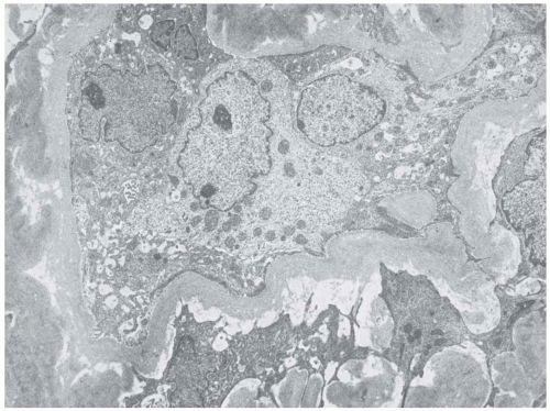
Characteristic changes demonstrated by transmission electron microscopy include thickening, splitting, attenuation, and granular disintegration of the TBM (Fig. 15.3). The transition between these alterations is abrupt.7 Fibroblasts are seen in direct contact with the TBM. At the base of the tubular epithelial cells, a marked increase of microfilaments is seen. The thickening is either homogeneous or has a lamellated, annular, and ringlike appearance. The glomerular basement membrane is normal. Multiple tubular diverticula are seen but the connections between cysts and distal tubular segments are patent.


Clinical Presentation

The third group of features shared among different diseases of the NPHP-MCKD complex involves clinical symptoms (Table 15.1A[iii]). Classical symptoms are polyuria, polydipsia, decreased urinary concentrating ability and, in children, anemia and growth retardation. The insignificance of the symptoms together with the lack of edema, hypertension, and urinary tract infections characteristically leads to a delayed diagnosis and therapy in NPHP-MCKD. In all variants of NPHP-MCKD, terminal renal failure insidiously ensues at characteristic age ranges, necessitating renal replacement therapy (Fig. 15.4). Disease recurrence has never been reported in kidneys transplanted to NPHP patients.9


FEATURES DISTINGUISHING DISEASE ENTITIES OF THE NPHP-MCKD COMPLEX

There are three features that clearly distinguish different disease entities of the NPHP-MCKD complex: (1) the mode of inheritance, (2) the age of onset for ESRD, and (3) the type of extrarenal organ involvement (Table 15.1B).


The Mode of Inheritance

In the NPHP-MCKD complex, the mode of inheritance can be either autosomal recessive or autosomal dominant.
For the recessive forms the term nephronophthisis (NPHP) is used, whereas the designation medullary cystic kidney disease (MCKD) denotes the dominant variants of the complex (Table 15.1B[i]).10,11


FIGURE 15.1 Juvenile nephronophthisis (autopsy case, 13-year-old girl). Note the numerous cysts of varying size in the medulla and at the corticomedullary junction. (Reproduced with permission from Hildebrandt F, Waldherr R, Kutt R, et al. The nephronophthisis complex: clinical and genetic aspects. Clin Invest 1992;70:802.)


The Onset of End-Stage Renal Disease

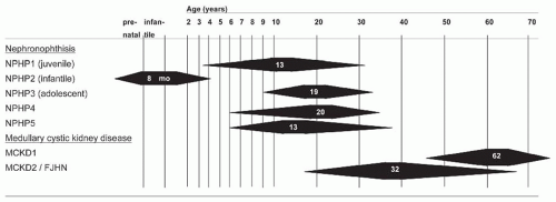
The second distinction pertains to the age of onset of ESRD (Table 15.1B[ii]). In all variants of NPHP-MCKD, ESRD ensues at characteristic age ranges, necessitating renal replacement therapy (Fig. 15.4). In NPHP, chronic renal failure develops within the first 3 decades of life.12,13,14 In a study conducted in 46 children with juvenile nephronophthisis (NPHP1), a serum creatinine value of 6 mg per deciliter was reached at a median age of 13 years (range: 4 to 20 years).12,15 In a study by Waldherr et al.1 ESRD was reached at a median age of 11.5 years. Gretz et al.16 showed that the rate of deterioration of renal function was homogeneous in a study of 29 patients with NPHP1. The median time elapsing between a serum creatinine of 2 and 4 mg per deciliter was 32 months, between 4 and 6 mg per deciliter was 10 months, and between 6 and 8 mg per deciliter was 5 months.16 A high concordance of the development of renal failure was noted in monozygotic twins.17,18 Infantile nephronophthisis (NPHP2) is characterized by an early onset of ESRD between the neonatal period and 3 years of age.14 In adolescent nephronophthisis (NPHP3), terminal renal failure develops at a median age of 19 years, which is 6 years later than in NPHP1.13 The median age of ESRD in patients with NPHP4 and NPHP5 mutations is 20 years19 and 13 years,20 respectively. If renal failure has not developed by the age of 25 years, the diagnosis of recessive NPHP should be questioned and a pedigree analysis should be intensified to exclude dominant MCKD.

In MCKD, terminal renal failure occurs only in adult life. Two different variants are known, MCKD1 and MCKD2, with a median onset of ESRD of 62 years21 and 32 years22 respectively (Fig. 15.4).


Extrarenal Associations

The third distinguishing feature among variants of NPHP-MCKD is represented by the degree to which extrarenal associations occur (Table 15.1B[iii]). Extrarenal disease manifestations have only been described in recessive forms. One exception to this rule is the occurrence
of hyperuricemia and gout in MCKD123 and MCKD2.22 MCKD2 patients with UMOD mutations also may exhibit defects in urine concentrating ability.24 Recently, an extensive study on genotype-phenotype correlations in mutation of NPHP genes has been published.25 NPHP1 can occur in combination with ocular motor apraxia Cogan type,26,27 with retinitis pigmentosa in Senior-Løken syndrome (SLSN),20 with liver fibrosis28 with cone-shaped epiphyses in Mainzer-Saldino syndrome,29 and with coloboma of the optic nerve and cerebellar vermis aplasia in Joubert syndrome type B (JBTSB) (Tables 15.1B[iii] and 15.2).30 Infantile NPHP (type 2) can be associated with situs inversus31 and one case report describes a patient with a nonsense inversin mutation with retinitis pigmentosa.32 NPHP4 patients may have retinitis pigmentosa (SLSN) and Cogan syndrome.33 NPHP5 patients display early onset retinitis pigmentosa (SLSN) in all known cases.20 NPHP6 and NPHP8 patients have SLSN, Joubert syndrome, or Meckel-Gruber syndrome (MKS).25,34,35 NPHP9 is associated with SLSN.36 NPHP10 patients display SLSN and Bardet-Biedl syndrome (BBS)-like phenotypes,37 whereas patients with NPHP11 show JBTS, MKS, and liver fibrosis.38,39 NPHP12 patients exhibit Jeune asphyxiating thoracic dystrophy.40


FIGURE 15.2 Renal histology in juvenile nephronophthisis (NPHP1). Note the characteristic triad, which consists of (1) tubular basement membrane disintegration with thickening as well as attenuation of the tubular basement membrane, (2) interstitial round cell infiltration with marked fibrosis, and later on (3) tubular atrophy and cyst development. (Courtesy of Prof. R. Waldherr, Heidelberg, Germany.)


Epidemiology

NPHP and dominant MCKD seem to be distributed evenly among males and females. NPHP has been reported from virtually all regions of the world.41 Information on the incidence of the disease has been estimated at 9 patients per 8.3 million42 in the United States or 1 in 50,000 live births in Canada.1,43 The condition constitutes the most frequent genetic cause for ESRD in the first 3 decades of life and is a major cause of ESRD in children, accounting for 10% to 25% of these patients.41,44,45 In contrast, in the North American pediatric ESRD population, pooled data indicate a prevalence of less than 5%.46,47

MCKD has initially been reported in the United States.10 Its prevalence in Europe might have been underestimated because recently, kindred have been reported from Cyprus,23 Italy,22,48 France,49 England, Finland,50,51 Belgium, Czech Republic,52 and Germany.53,54 The diagnosis of MCKD may be frequently missed because clinical symptoms and signs are subtle.55


FIGURE 15.3 Thickening, wrinkling, and double layering of tubular basement membranes with intermembranous fibroblasts and dedifferentiation of tubular epithelial cells. An electron micrograph. (Reproduced with permission from Hildebrandt F, Waldherr R, Kutt R, et al. The nephronophthisis complex: clinical and genetic aspects. Clin Invest 1992;70:802.)


FIGURE 15.4 The time course of renal failure in NPHP-MCKD. Range for age of onset of end-stage renal disease is shown as solid triangles. Numbers indicates median age in years. NPHP, nephronophthisis; MCKD, medullary cystic kidney disease; FJHN, familial juvenile hyperuricemic nephropathy.


MOLECULAR GENETICS OF THE NEPHRONOPHTHISIS-MEDULLARY CYSTIC KIDNEY DISEASE COMPLEX

Classification of disease variants of the NPHP-MCKD complex has recently become more definite through the identification of distinct genes for the different variants. The disease complex is genetically very heterogeneous. Aspects of disease nomenclature, known genes, and extrarenal involvement within the NPHP-MCKD complex are summarized in Table 15.2.


Recessive Disease Variants: Nephronophthisis

Within recessive variants of the NPHP-MCKD complex, the different forms are distinguished on the basis of the mutated gene. To date, thirteen distinct genes (NPHP1-12, NPHPL1) have been identified by positional cloning and massively parallel sequencing.


Nephronophthisis Type 1 (Juvenile Nephronophthisis): Clinical Features

The first case of juvenile nephronophthisis was described by Smith and Graham56 in 1945. This report of a sporadic case was followed by the publication of two large kindred with familial disease by Fanconi et al.,57 who introduced the term familial juvenile nephronophthisis. This disease variant is now classified as nephronophthisis type 1 (NPHP1). Since the first description, over 300 cases of NPHP have been published in the literature.2 NPHP1 is the most common variant within the NPHP-MCKD complex. Penetrance of recessive mutations is 100% by adolescence.

In juvenile NPHP, the symptoms of polyuria, polydipsia, decreased urinary concentrating ability, and secondary enuresis are the earliest presenting symptoms in over 80% of cases41 and occur at around 4 to 6 years of age. Pallor, weakness, and generalized pruritus are also common. Anemia51 and, in children, growth retardation occur later and are pronounced. In juvenile NPHP, children usually start to drink regularly at nighttime around age 6 years. This characteristic symptom should actively be sought when taking the patient’s history. The mild nature of symptoms, together with a frequent lack of edema, hypertension, and urinary tract infections, characteristically leads to delayed diagnosis and therapy in NPHP-MCKD. Due to the late detection of symptoms, there is a small but definite risk of sudden death from fluid and electrolyte imbalance. For NPHP1, definite molecular genetic diagnosis is possible (see the text that follows). Disease recurrence has never been reported in kidneys transplanted to NPHP patients.9


Nephronophthisis Type 1: Molecular Genetics

Because little was known about the pathogenesis of NPHP, a positional cloning approach was used for gene identification. By total genome search for linkage, a gene locus for juvenile NPHP1 was mapped to human chromosome 2q12-q13.58,59 The critical genetic region was subsequently cloned in yeast artificial chromosome (YAC) and P1-derived artificial chromosome (PAC) contigs, which led to the identification of the NPHP1 gene, defects in which are responsible for NPHP1.60,61 About 66% of children with juvenile NPHP harbor large (250 kb) homozygous deletions of the NPHP1 gene, whereas some carry point mutations in combination with heterozygous deletions.62,63,64 Through gene identification, a molecular genetic diagnosis in NPHP1 has become possible (see the text that follows).64,65,66,67

The NPHP1 gene spans 83 kb, consists of 20 exons, and encodes an mRNA of 4.5 kb. It is flanked by two large (330 kb) inverted duplications. In addition, a second sequence of 45 kb, which is located between the centromeric inverted duplication and the NPHP1 gene, is repeated directly within the telomeric inverted duplication. In several NPHP1 families, the deletion break points have been localized to the 45 kb direct repeats using pulsed field gel electrophoresis.63 Chromosomal misalignment followed by unequal crossing over or the formation of a loop structure on a single chromosome has been suggested as a potential cause for these deletions. In addition, there is a high degree of further rearrangements known to occur in this region of chromosome 2.63 Furthermore, an unusual maternal deletion in a child with NPHP1 molecularly characterized, showing that the centromeric break point occurred within a long interspersed nuclear element-1 (LINE1).68 The NPHP1 gene is a novel gene, which is not related to any known gene families. Expression studies in humans and mice revealed a broad tissue expression pattern. In addition, in situ hybridization studies of whole mount mouse embryos showed ubiquitous but weak Nphp1 expression at all embryonic stages between days 7.5 and 11.5 postconception.69 In the adult mice, there was also a strong expression in testes.


Nephronophthisis Type 2

A second gene locus (NPHP2) for recessive NPHP has been localized to chromosome 9q31.1 in a large Bedouin pedigree by homozygosity mapping (Table 15.2).14 This disease variant is termed infantile nephronophthisis (NPHP2) due to its prenatal, perinatal, or infantile onset. The clinical course and histology in this disease are quite different from other forms of NPHP.70 The inv mouse model, in which a disruption of the protein inversin led to a consistent reversal of the left-right body axis,71 was noted to have cystic kidney disease.72,73 These observations led to the identification of inversin (INVS) as the gene mutated in NPHP2 with and without situs inversus.31 INVS encodes a 1,062 amino acid protein containing 16 ankyrin repeats, a nuclear localization signal, and an IQ calmodulin domain.74 Yeast two-hybrid and coimmunoprecipitation experiments have confirmed the interaction between inversin and calmodulin. By a knockdown of inversin expression in zebra fish, a polycystic kidney disease (PKD)-like phenotype in addition to a randomization of heart looping was observed.31
Inversin localizes to primary cilia, mitotic spindles, and centrosomes74 and is intimately associated with the microtubule cytoskeleton.75 INVS/NPHP2 mutations remain a rare cause of NPHP, accounting for <1% of cases.


TABLE 15.2 Disease Variants, Gene Loci, and Extrarenal Involvement in the NPHP-MCKD Complex of Diseases


Disease


OMIM#


Inheritance


Gene


Chromosome


Gene (Product)


Extrarenal Organ Involvement


NPHP1 (juvenile)


256100 (607100)


AR


NPHP1


2q12-q13


NPHP1 (nephrocystin-1)


RP, OMA, Coloboma, CVA (JBTS)


NPHP2 (infantile)


602088


AR


NPHP2


9q31.1-q31


INVS/NPHP2 (inversin/nephrocystin-2)


RP, situs inversus


NPHP3 (adolescent)


604387


AR


NPHP3


3q22.1-q22


NPHP3 (nephrocystin-3)


RP, LF


NPHP4


607215


AR


NPHP4


1p36.31


NPHP4 (nephroretinin/nephrocystin-4)


RP, OMA


NPHP5


609237


AR


NPHP5


3q21.13q13.33


IQCB1/NPHP5 (IQ calmodulin-binding motif containing protein-1/nephrocystin-5)


Early onset RP


NPHP6


610142


AR


CEP290


12q21.32


CEP290 (centrosomal protein 290)


RP


NPHP7


611498


AR


GLIS2


16p13.3


GLIS2 (GLI-similar 2)


RP


NPHP8


610937


AR


RPGRIL1


16q12.2


RPGRIP1-like


RP


NPHP9


613824


AR


NEK8


17q11.2


NIMA-related kinase


RP


NPHP10


613524


AR


SDCCAG8


1q43


SDCCAG8 (serologically defined colon cancer antigen 8)


LF


NPHP11


613550


AR


TMEM67


8q22.1


TMEM (transmembrane protein 216)


NPHPL1


613159


AR


XPNPEP3


22q13.2


XPNPEP3 (X-prolyl aminopeptidase 3)


NPHP12


613820


AR


TTC21B


2q24.3


TTC21B (tetratricopeptide repeat domain 21B)


Joubert syndrome type 1


213300


AR


INPP5E


9q34.3


INPP5E (inositol polyphosphate-5-phosphatase) RP, CVA


Joubert syndrome type 2


608091


AR


TMEM216


11p12.2


TMEM216 (transmembrane protein 216)


RP, Coloboma, CVA


Joubert syndrome type 3


608894


AR


AHI1


6q23.3


AHI1 (jouberin)


RP, CVA, cortical polymicrogyria


Joubert syndrome type 4


182290609583


AR


NPHP1


17p11.22q13


NPHP1


CVA


Joubert syndrome type 8


612291


AR


ARL13B


3q11.1


ARL13B (ADP-ribosylation factor-like 13B)


Joubert syndrome type 9


612285


AR


CC2D2A


4p15.32


CC2D2A (coiled coil and C2 domain containing 2A)


Meckel-Gruber syndrome type 1


249000


AR


MKS1


17q22


MKS1


MCKD1


174000


AD


MCKD1


1q21


?


Gout, hyperuricemia


MCKD2


191845


AD


UMOD


16p12.3


UMOD (uromodulin)


Gout, hyperuricemia


AD, autosomal dominant; MCKD, autosomal dominant medullary cystic kidney disease; AR, autosomal recessive; CORS, cerebello-oculorenal syndrome; CVA, cerebellar vermis aplasia; NPHP, nephronophthisis; FJHN, familial juvenile hyperuricemic nephropathy; JBTS1, Joubert syndrome 1; OMA, ocular motor apraxia; OMIM#, Online Mendelian Inheritance in Man (http://omim.org/); RP, retinitis pigmentosa; SMS, Smith-Magenis syndrome; UMOD, uromodulin


Nephronophthisis Type 3

A third locus (NPHP3) for NPHP was mapped to chromosome 3q22.1 in a large Venezuelan kindred by a total genome search for linkage by applying the strategy of homozygosity mapping (Table 15.2).13,76,77 This disease variant was termed adolescent nephronophthisis (NPHP3) because the onset of ESRD occurs 6 years later than in juvenile NPHP1, with a median onset of terminal renal failure occurring at age 19 years (Fig. 15.4).

Identification of the gene NPHP3, which causes adolescent NPHP, was carried out in the same Venezuelan kindred.78 The novel NPHP3 protein interacts with nephrocystin-1. NPHP3 mutations were found in patients with isolated MPHP and in families with NPHP and hepatic fibrosis or tapetoretinal degeneration. Murine Nphp3 was shown to be expressed in the embryonic node, kidney tubules, retina, respiratory epithelium, liver, biliary tract, and neural tissues. A homozygous missense mutation in Nphp3 was identified as the underlying defect in the polycystic kidney disease (pcy) mouse phenotype.78


Nephronophthisis Type 4

By a total genome search for linkage, a fourth gene locus (NPHP4) was localized to chromosome 1p36,79 including a family with SLSN. The respective gene (NPHP4) was subsequently identified as causing NPHP type 4 and SLSN type 4.19,33

Only gold members can continue reading. Log In or Register to continue

Stay updated, free articles. Join our Telegram channel

May 29, 2016 | Posted by in NEPHROLOGY | Comments Off on Nephronophthisis—Medullary Cystic Kidney Disease

Full access? Get Clinical Tree

Get Clinical Tree app for offline access