Isolated Renal Tubular Disorders: Mechanisms and Clinical Expression


Isolated Renal Tubular Disorders: Mechanisms and Clinical Expression


Thomas D. DuBose Jr.

Amanda K. Goode


Renal control of acid and base reabsorption and excretion is controlled by the proximal and distal tubules (see Chapter 6). Thus, it is likely that abnormalities in those mechanisms responsible for these processes will result in clinically significant disorders. The transporters for carbohydrates, amino acids, and ions are polarized to either the apical or basolateral membrane, and the clinical abnormalities of fluid, electrolyte, and acid-base balance regulated by these transporters either have a genetic basis or are the result of acquired abnormalities of specific transport proteins. In this chapter, we review the major tubular transport defects of carbohydrates, amino acids, and some of the ions (H+, K+, Na+). We describe the clinical features of these disorders as they appear in humans and summarize some of the insights learned from experimental models.


RENAL TUBULAR ACIDOSIS

Renal tubular acidosis (RTA) is a clinical syndrome characterized, when fully expressed, by a chronic hyperchloremic nongap metabolic acidosis, which results from a defect in urinary acidification.1 This defect can be localized to the nephron segment responsible for the pathophysiologic expression and may be inherited or, more commonly, acquired by specific diseases or by drug or toxin effects. An incomplete form of distal RTA may be exhibited in family members of patients with genetic abnormalities of specific transporters in this nephron segment. Examples of specific defects involved in the pathogenesis of proximal and classical distal RTA include acquired or inherited abnormalities of the basolateral electrogenic Na+/HCO3 symporter, the apical Na+/H+ exchanger, NHE-3, or the enzyme carbonic anhydrase II in the proximal tubule, and the H+-ATPase and the HCO3/Cl exchanger in the distal tubule. Proximal RTA (type 2 RTA) and classical distal RTA (type 1 RTA) are both associated with chronic hypokalemia. In contrast, a more generalized abnormality in the distal nephron is associated with hyperkalemia. The diagnosis of “complete” RTA requires spontaneous nongap metabolic acidosis in association with either a less than maximal urine pH or low ammonium excretion. Incomplete RTA is seen without spontaneous metabolic acidosis, but with evidence of the inability to acidify urine maximally in response to an exogenous acid load. Most inherited forms cause growth retardation or short terminal stature. Both inherited and acquired forms of proximal and classical distal RTA are accompanied by hypokalemia, often sufficiently severe enough to cause periodic paralysis or even seizures.


DIFFERENTIAL DIAGNOSIS OF NONGAP (HYPERCHLOREMIC) METABOLIC ACIDOSIS

A nongap metabolic acidosis is recognized by a low plasma bicarbonate concentration (or total [CO2]), low blood pH, and a normal anion gap (8 to 10 mEq per L). A compensatory decrease in PCO2 is typical, indicating the presence of a pure, or simple, nongap metabolic acidosis. The differential diagnosis of a nongap metabolic acidosis includes both nonrenal and renal causes. The differential diagnosis of nongap acidosis is displayed in Table 18.1. The evaluation of a nongap or hyperchloremic metabolic acidosis requires one to appreciate the role of the kidney in the acid-base balance, and to determine whether the kidney is responding appropriately to the prevailing acidosis (Table 18.2), which is to increase ammonium (NH4+) production and excretion adaptively. In contrast, ammonium production and excretion are impaired with chronic renal insufficiency, hyperkalemia, and with all forms of nongap acidosis of renal origin, including all examples of renal tubular acidosis.


Loss of HCO3 from the Gastrointestinal Tract versus Renal Tubular Acidosis

Diarrhea is a common cause of hyperchloremic metabolic acidosis. Diarrheal stools contain a large amount of HCO3 and HCO3 decomposed by reaction with organic acids.2 The HCO3 loss and the ensuing volume depletion cause hyperchloremic metabolic acidosis. Hypokalemia develops due to direct K+ loss in the stool and increased renal K+ secretion (in the cortical collecting tubule) due to a secondary increase
in elaboration of renin and aldosterone in response to volume depletion. Both hypokalemia and nonrenal metabolic acidosis increase renal NH4+ synthesis, which increases urinary buffering capacity and urinary pH. The presence of NH4+ in the urine can be used clinically to distinguish hyperchloremic metabolic acidosis due to diarrhea from renal tubular acidosis (Table 18.2). In the latter, NH4+ excretion is invariably low. Urine pH, although a time-honored method to distinguish these disorders, is less reliable because urinary ammonium excretion is augmented in chronic metabolic acidosis due to diarrhea, causing urine pH to increase over time.


TABLE 18.1 Differential Diagnosis of Nongap Acidosis


Extra-Renal Causes


Diarrhea or other GI losses of bicarbonate (e.g., tube drainage)


Posttreatment of ketoacidosis (dilutional) (occasional: initial DKA)


Renal Causes Not Due to Renal Tubular Acidosis


Ureteral diversion (e.g., ileal loop, ureterosigmoidostomy)


Progressive chronic kidney disease


Toluene ingestion (excretion of hippurate)


Drugs


With associated hypokalemia


Carbonic anhydrase inhibitors (acetazolamide and topiramate)


Amphotericin B


With associated hyperkalemia


Amiloride


Triamterene


Spironolactone


Trimethoprim


With normal potassium


CaCl2, MgSO4


Cholestyramine


Exogenous acid loads (NH4Cl, acidic amino acids, total parenteral nutrition, sulfur)


Posthypocapnic state


Renal Tubular Acidosis


Low [K+]p


Type 1 (classical distal) RTA


Type 2 (proximal) RTA


Type 3 (mixed proximal and distal) RTA (carbonic anhydrase II deficiency)


High [K+]p


Type 4 (generalized distal RTA)


Hypoaldosteronism (hyporeninemic and isolated)


Aldosterone resistance


Voltage defect in collecting duct


DKA, diabetic ketoacidosis; GI, gastrointestinal; RTA, renal tubular acidosis.


TABLE 18.2 Diagnostic Criteria for Causes of Nongap Acidosis


Nonrenal Etiology


Nongap acidosis expect:


Increase in NH4+ excretion


Negative urine anion gap


Acid urine pH (<5.5)—exceptions


Renal Etiology


Nongap acidosis expect:


Inability to increase NH4+ excretion


Positive urine anion gap


Urine pH typically >5.5 but variable in type 4


Other Causes of Nongap Metabolic Acidosis

In addition to diarrhea and RTA there are other less common causes of hyperchloremic metabolic acidosis (Table 18.1). External pancreatic and biliary diversion may lead to the loss of HCO3-rich fluid and result in hyperchloremic metabolic acidosis. The excretion of sodium salts of ketones during the recovery phase of ketoacidosis represents the loss of potential HCO3 and may result in hyperchloremic metabolic acidosis. Ureteral diversion is commonly associated with hyperchloremic metabolic acidosis because the ileum and colon are both endowed with an apical Cl/ HCO3 exchanger. When chloride from urine comes into contact with the gut, chloride is absorbed in exchange for bicarbonate leading to excretion of bicarbonate, absorption of chloride, and hyperchloremic metabolic acidosis.3 The degree of acidosis is magnified by stasis and prolonged contact of urine with the HCO3/Cl exchanger in the pouch. Because of bicarbonate secretion, potassium secretion is also stimulated and leads to hypokalemia. Finally, the administration of acid or acid equivalent (arginine HCl, lysine HCl, or NH4Cl) or medications such as cholestyramine, calcium chloride, and magnesium sulfate are associated with hyperchloremic metabolic acidosis.4 Dilutional acidosis occurs in conjunction with rapid infusion of isotonic saline. In these latter examples, the serum potassium is usually normal.

Progressive renal failure is associated with metabolic acidosis. Hyperchloremic metabolic acidosis is commonly seen when the glomerular filtration rate (GFR) is between 20 to 50 mL per min.5 As renal failure progresses to a GFR of less than 10 to 15 mL per min, the acidosis converts to the typical high anion gap acidosis of “uremic” acidosis. The
principle defect in advanced renal failure is impaired ammoniagenesis and ammonium excretion.5 The latter is a result of impaired medullary ammonium transport and trapping of NH3/NH4+ in the outer and inner medulla.

In summary, the defect in renal acidification in RTA may be manifest by one of three clinical syndromes: (1) an acid urine pH and low urine anion gap (UAG) during metabolic acidosis or frank bicarbonaturia and hypokalemia during NaHCO3 therapy (as in proximal RTA); (2) an inappropriately alkaline urine pH, hypokalemia, and a positive urine anion gap (classical distal RTA); or (3) hyperkalemia and a positive urine anion gap but variable pH with aldosterone deficiency, aldosterone resistance, or a “voltage” defect in the collecting tubule (generalized defect in distal nephron) (Table 18.2).


Clinical Laboratory Evaluation

Because the measurement of urinary NH4+ concentration may be problematic for the routine hospital clinical pathology laboratory, it is helpful to estimate the urine ammonium concentration by considering the electrolytes present in urine. Because NH4+ is a cation, its presence in urine, especially when in large amounts as expected in nonrenal forms of hyperchloremic metabolic acidosis, should be denoted by an increase in urinary anions (Cl) in excess of the usual cations (Na+ + K+). The urine anion gap (UAG) is calculated on a “spot” urine sample as follows (Table 18.3)6:


TABLE 18.3 Clinical Application of Urine Anion Gap to Approximate Urine Ammonium Excretion


1. Spot urine electrolytes: [Na+, K+, Cl]u in a patient with hyperchloremic metabolic acidosis


2. Calculate urine anion gap:


UAG = (Na + K)u – Clu


3. Interpretation:


(Na + K)u > Clu: NH4+ low (ammonium excretion impaired)


Clu > (Na + K)u: NH4+ adequate (nonrenal hyperchloremic acidosis)


4. Pitfalls: Unusual anions in the urine (perform urine osmolar gap)


Ketones


Toluene


TABLE 18.4 Urine Osmolar Gap to Approximate Urine Ammonium Concentration


Urine Osmolar Gap = Measured Urine Osmolality — Calculated Urine Osmolality


Urine = Uosm 0.5[2 (Na+ + K+)u + urea/2.8 + glucose/18] [NH4+]


Interpretation: Urine ammonium = 75 mEq/L or > anticipated in acidosis with normal renal tubule function


NH4+ is assumed to be present in the urine if the sum of the major cations (Na+ + K+) is less than the concentration of the major anion (Cl). Therefore, a negative urine anion gap denotes the presence of ammonium in the urine and signals a “normal” renal response to acidosis of nonrenal origin (i.e., diarrhea). Hyperchloremic metabolic acidosis of renal origin (i.e., RTA) is supported by the presence of a positive urine anion gap. A positive urine anion gap confirms a deficiency of NH4+, and obtains when the sum of the major cations (Na+ + K+) in the urine exceeds the major urinary anion (Cl). A positive urine anion gap, therefore, denotes an “abnormal” renal response to acidosis and is consistent with a defect in net acid secretion. The presence of urinary anions other than chloride can invalidate the UAG. Examples of urinary anions which invalidate this shorthand method of estimating urinary ammonium concentrations include drug anions, ketones, and toxins such as toluene. If these constituents are suspected, urinary NH4+ may be estimated reliably by measuring urine osmolality (Uosm); the concentrations of Na+ + K+, urine urea, and glucose (Table 18.4); and calculating the urine anion gap. Urine ammonium (UNH4+) is calculated as:


The fractional excretion of sodium may also be helpful to differentiate hyperchloremic metabolic acidosis due to diarrhea from RTA. The fractional excretion of sodium is typically low (<1%—2%) in patients with diarrhea compared to RTA (2%-3%).


PROXIMAL RENAL TUBULAR ACIDOSIS


Role of the Proximal Tubule in Bicarbonate Reabsorption and Urinary Acid Excretion

The kidney employs two fundamental mechanisms to maintain acid-base homeostasis: bicarbonate absorption and H+ secretion. Of the 4,000 mEq of HCO3 filtered by the kidney
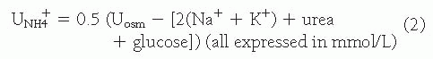
each day, 80% to 90% is reabsorbed in the proximal tubule. Effective HCO3 absorption in the proximal tubule is mediated by H+ secretion. Even though the distal tubule is responsible for the secretion of 50 to 80 mEq of H+ and final acidification of the urine, the vast majority of H+ secretion obviously occurs in conjunction with HCO3+ reclamation in the proximal tubule.

Bicarbonate absorption is dependent on H+ secretion across the apical membrane of the proximal tubule in exchange for Na+ entry into the cell via the Na+/H+ exchanger (NHE-3; see Chapter 6).7,8 A low intracellular Na+ concentration is maintained by the active extrusion of Na+ across the basolateral membrane via the Na+,K+-ATPase. The enzyme carbonic anhydrase present in the cytoplasm (type II) and on the apical and basolateral membrane (type IV) is critical to accelerate the reaction as indicated here:


The active secretion of H+ into HCO3-rich glomerular filtrate by the NHE-3 results in the formation of H2CO3. Luminal carbonic anhydrase (type IV) facilitates the conversion of H2CO3 to CO2 and H2O. CO2 freely diffuses through the luminal membrane and, under the influence of cytoplasmic carbonic anhydrase (type II), forms H2CO3 that dissociates rapidly to H+ and HCO3, which are transported, respectively, across the apical and basolateral membranes. Bicarbonate exits the cell via the electrogenic Na+-3 HCO3 symporter (NBCe1; see Chapter 6). The negative cell potential is the primary driving force for this transport process. In addition to H+ secretion via the Na+/H+ exchanger, an apical H+-ATPase is also responsible for a small but significant fraction of bicarbonate reclamation in the proximal tubule.


Other Proximal Tubular Functions

In addition to its role in H+ secretion and HCO3 absorption, the proximal tubule is the primary site for glucose, amino acid, phosphate, and organic anion reclamation. Each of these solutes is transported across the apical membrane via an Na+-cotransport process. Sodium enters the apical membrane down its electrochemical gradient. Low intracellular Na+ concentrations and the negative intracellular potential are maintained via the basolateral Na+,K+ ATPase. Once inside the cell, these solutes are either metabolized or diffuse passively across the basolateral membrane. Citrate is reabsorbed in the proximal tubule in parallel with Na+– via the NaDC-1 (Na dicarboxylate cotransporter-1).9 The metabolism of citrate within the cell leads to the generation of HC O3. The presence of citrate in tubular fluid and urine has been shown to be protective in the prevention of calcium oxalate stones and nephrocalcinosis. In the presence of all forms of metabolic acidosis except proximal RTA, citrate is preferentially reabsorbed resulting in hypocitraturia and predisposing patients to nephrolithiasis. In proximal RTA, because this Na+-coupled transport system is impaired, urinary citrate remains high, even with metabolic acidosis, and nephrolithiasis rarely, if ever, occurs.


Generalized and Isolated Proximal Tubular Transport Defects

Renal tubular acidosis involving the proximal tubule can be divided into two major categories: generalized disorders of proximal tubule reabsorption and isolated abnormalities in renal acidification (Table 18.5). Those potential abnormalities that have been documented to date include, in order of frequency, genetic defects in the basolateral Na+-3HCO3
symporter or NBCe1, the enzymes carbonic anhydrase type II or IV, and the Na+/H+ exchanger (NHE-3) (Fig. 18.1).


TABLE 18.5 Etiology of Proximal Renal Tubular Acidosis (Type 2) with or without Fanconi Syndrome


1.Primary disorders


Inherited—isolated pure bicarbonate wasting


Autosomal recessive: Mutations of NBCe1/ SLC4A4 (several examples associated with ocular abnormalities)


Autosomal dominant: Mutation of NHE-3 with short stature (defect not determined)


Familial disorders associated with proximal RTA


Cystinosis


Tyrosinemia


Hereditary fructose intolerance


Galactosemia


Glycogen storage disease (type 1)


Wilson disease


Lowe syndrome


2. Acquired disorders


Multiple myeloma, amyloidosis, light chain nephropathy


Chemotherapeutic agents


Ifosfamide


Carbonic anhydrase inhibitors


Topiramate


Acetazolamide


Sulfamylon


Heavy metals


Lead, copper, cadmium, mercury


Renal transplantation


Paroxysmal nocturnal hemoglobinuria


3. Mixed proximal and distal RTA (type 3 RTA)


Carbonic anhydrase II deficiency: osteopetrosis and ocular abnormalities (Guibaud-Vainsel syndrome)


RTA, renal tubular acidosis.


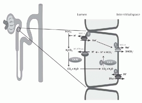
FIGURE 18.1 Pathogenesis of proximal renal tubular acidosis. Model of bicarbonate reabsorption in the proximal tubule showing described inherited defects of genes encoding proximal transport proteins that cause proximal renal tubular acidosis. 1, defect of basolateral Na+-HCO3 cotransporter; 2, defect of carbonic anhydrase type 2; 3, defect of Na+/H+ exchanger; 4, defect of Na+,K+ ATPase. See text for detail.


Generalized Proximal Tubular Dysfunction

Generalized proximal tubular dysfunction is the more common of the two types of defects, and is appreciated by the co-occurrence of renal tubular acidosis (type 2 RTA), glycosuria, aminoaciduria, phosphaturia, and hypercitraturia. This constellation of symptoms is referred to collectively as “Fanconi” syndrome, which can be either hereditary or acquired. Excessive urinary excretion of glucose occurs, although plasma glucose concentration is normal, and is usually less than 10 g per day. A generalized aminoaciduria also occurs, but because it usually does not result in deficiencies, supplementation is not needed. Sodium and potassium losses occur, which may be massive, resulting in severe secondary hyperaldosteronism and even metabolic alkalosis.

The onset of Fanconi syndrome following an exposure to a triggering agent can vary widely, from minutes (as in the case of patients with hereditary fructose intolerance exposed to fructose10), to a few days (in galactosemic patients exposed to galactose11,12), to years (following exposure to cadmium13). Patients with hereditary fructose intolerance lack the enzyme fructose-1-aldolase, which results in sequestration of intracellular phosphate and is associated with ATP depletion.

Pathophysiologic Mechanisms of Fanconi Syndrome. Most studies suggest that the generalized defect and the defect in transcellular HCO3 absorption are due to depletion of intracellular ATP, with inhibition of the Na+,K+-ATPase (Fig. 18.1, number 4). Disruption of active HCO3, amino acid, and solute absorption in the proximal tubule due to ATP depletion and inhibition of Na+,K+ATPase has also been observed in an experimental model of cystinosis.14 Generalized dysfunction of the proximal tubule could occur through three possible mechanisms: (1) an increase in paracellular permeability resulting in backleak of all reabsorbed solutes into the lumen; (2) a generalized defect in proximal tubule absorption, such as ATP depletion; and (3) a defect in basolateral Na+,K+-ATPase activity. The second mechanism is the most widely accepted to date.

Regarding paracellular permeability, enhanced efflux causes increased urinary electrolyte and solute excretion, as shown in studies using the maleic acid model (see below). Both mechanisms have been demonstrated in some studies using the maleic acid model of proximal renal tubule acidosis.15,16 A generalized defect in proximal tubule absorption may be related to the brush-border membrane, such as an abnormality in the sodium-binding domain of the multiple heterogeneous carriers. Alternatively, there may be an abnormality in the way the different carriers are moved to the brush-border membrane. Finally, defects in basolateral Na+,K+-ATPase activity can result in abnormal energy generation, as Na+,K+-ATPase fuels transport in the proximal nephron. Hereditary fructose intolerance, galactosemia, and cadmium poisoning may result in Fanconi syndrome by reducing Na+,K+-ATPase activity. Thus, Fanconi syndrome may be best described as a defect in energy generation in the proximal tubule, with the most important cause being deficient Na+,K+-ATPase activity in the basolateral membrane, which decreases sodium-coupled reabsorption due to alterations in the sodium gradient across the luminal membrane. Most of the solutes lost in Fanconi syndrome are those
coupled to apical sodium reabsorption. Fanconi syndrome is also associated with distal nephron dysfunction in some patients. The mechanisms involved are not known, but the evidence suggests that there may be a defect in Na+,K+-ATPase throughout the nephron.

Experimental Models of Fanconi Syndrome. Maleic acid is a toxin, relatively specific for the proximal tubule, which results in the best characterized model of Fanconi syndrome. Animals (such as rodents) injected with maleic acid develop proximal RTA and exhibit symptoms similar to those seen in humans with Fanconi syndrome, including decreased activity and expression of Na+,K+-ATPase16 and bicarbonaturia. Maleic acid given to rats also impairs vitamin D conversion.17

Because ifosfamide can cause Fanconi syndrome in human patients, it has been used to induce Fanconi syndrome in animals as well. However, the use of this agent in animals is mainly to determine if other agents given with ifosfamide can block the development of Fanconi syndrome without affecting ifosfamide’s antitumor activity.18,19,20,21,22 Heavy metals can also induce Fanconi syndrome in animals and humans. Cadmium, uranium, lead, and mercury all induce Fanconi syndrome in animals, although because cadmium-induced Fanconi syndrome reverses after cadmium administration ceases and because uranium and mercury affect GFR (which is unaffected in people with the syndrome), the study of heavy metal-induced Fanconi syndrome may not necessarily provide accurate information about mechanisms underlying the human disorder.

Dent Disease. Dent disease is a disorder of the proximal renal tubule caused by an X-linked genetic mutation in the CLCN5 gene. Symptoms include proteinuria, nephrocalcinosis, hypercalciuria, and slow progression of renal failure. Because it can also include phosphaturia, aminoaciduria, glycosuria, and rickets, it may be considered a form of Fanconi syndrome. Treatment for patients with Dent disease usually consists of vitamin D to manage rickets and recommendations to reduce hypercalciuria (e.g., thiazide diuretics and citrate supplementation) in order to limit or prevent progression of nephrocalcinosis.23


Isolated Proximal Tubule Bicarbonate Transport Abnormalities

Isolated abnormalities of proximal tubular renal acidification in the absence of Fanconi syndrome are less common, but may be associated with depolarization abnormalities or genetic mutations. One model, the infusion of L-lysine in dogs, results in marked bicarbonaturia due to inhibition of HC O3 absorption.24 The presence of luminal L-lysine has been shown to depolarize proximal tubular cells, which could alkalinize the cell by decreasing HCO3 extrusion across the basolateral membrane. Mice, in which the gene encoding the renal Na+/H+ exchanger (NHE-3) has been knocked out, have metabolic acidosis with proximal RTA25 (Fig. 18.1, number 3).

Sly et al.26 have described a group of patients with inherited carbonic anhydrase II deficiency (Fig. 18.1, number 2). These patients develop osteopetrosis, cerebral calcification, and combined proximal and distal RTA.26 This observation is not unexpected considering the role carbonic anhydrase plays in HCO3 reclamation.


Clinical Features of Proximal RTA

Patients with proximal renal tubular acidosis commonly present with hyperchloremic metabolic acidosis, an acid urine pH (pH<5) (when systemic acidosis prevails), and minimal HCO3 excretion. As bicarbonate is administered to correct the metabolic acidosis, bicarbonaturia occurs and the fractional excretion of HCO3 often exceeds 10% to 15% (bicarbonate wasting). This response to alkali therapy, the ensuing increase in potassium excretion in response to the bicarbonate leak into the distal tubule, and the difficulty with which the plasma bicarbonate is corrected are unique features of proximal RTA. Sebastian et al. demonstrated that the level of K+ excretion correlates directly with HCO3 excretion.27 Decreased NaCl absorption associated with proximal tubular dysfunction enhances K+ excretion by increasing delivery of Na+ to the distal nephron and the increase in aldosterone elaboration in response to volume depletion.

The most common causes of proximal RTA in children are acquired either from the administration and toxicity of ifosfamide or from cystinosis.28,29 Most children present with Fanconi syndrome, but proximal RTA can be limited to an isolated impairment in proximal bicarbonate reabsorption. In contrast, the most common cause in adults is from multiple myeloma or light chain disease. The proximal tubular toxicity related to increased excretion of monoclonal immunoglobulin light chains in patients with multiple myeloma appear to induce a unique biochemical toxicity because of resistance to degradation by lysosomal proteases in proximal tubular cells.30,31 Accumulation of the variable domain fragments is presumably responsible for the impairment in tubular function. Other causes of proximal RTA that lead to isolated bicarbonate wasting include acetazolamide or the administration of any carbonic anhydrase inhibitor. The most common offending agent currently is topiramate, which is a potent carbonic anhydrase inhibitor.

The majority of cases of proximal RTA are associated with generalized proximal tubule dysfunction (Fanconi syndrome) so that glycosuria, aminoaciduria, proteinuria, hyperphosphaturia, hypophosphatemia, hyperuricosuria, hypouricemia, and hypercitraturia are observed commonly. Table 18.5 lists several familial disorders that may be associated with proximal RTA that result when the abnormal product of metabolism impacts proximal tubule function. Hypercalciuria, a common feature of metabolic acidosis, is absent in
proximal RTA, presumably as a result of enhanced calcium absorption in the distal nephron in response to increased bicarbonate delivery. Rickets, a frequent manifestation of the Fanconi syndrome, is a result of phosphate wasting, not proximal RTA or acidosis. Of particular concern in children with proximal RTA is growth retardation, a direct consequence of acidosis. Because growth retardation will correct with alkali therapy, this complication becomes one of the major indications for correction of the serum bicarbonate concentration.

Isolated proximal RTA without features of the Fanconi syndrome can occur rarely in an autosomal recessive disorder affecting the gene SLC4A4 that encodes for the sodium bicarbonate cotransporter. In addition, a defect in the gene that encodes the Na+/H+ exchanger (NHE-3) on the apical membrane has been described in a single family as an autosomal dominant disease.


Management

The primary therapeutic objective in the management of patients with proximal renal tubular acidosis is to maintain a near normal serum HCO3 concentration and arterial pH. The bicarbonaturia associated with this disorder, which amplifies potassium excretion, requires administration of a mixture of sodium and potassium salts (e.g., K-Shohl’s solution) (Table 18.6). A feature of proximal renal tubular acidosis is the large amount of HCO3 required to correct the acidosis, which, in turn, aggravates renal potassium excretion. As an adjunct, the administration of thiazide diuretics has been used to decrease GFR from chronic volume depletion. Sequelae of proximal RTA vary according to cause (generalized vs. isolated). Nevertheless, in children with isolated proximal RTA, stunted growth is normalized by correction of the acidosis. Additionally, the manifestations of isolated proximal RTA in children tend to improve with age, but alkali therapy is usually necessary throughout life.


TABLE 18.6 Treatment of Proximal Renal Tubular Acidosis


Large amounts of alkali required to correct acidosis:


10-20 mEq/kg/day of alkali (typically enhances urinary K loss)


Preparations that include potassium:


Potassium Shohl’s Solution (K-Shohl’s:


Polycitra-LC: Citric acid 334 mg, sodium


citrate 500 mg, and potassium citrate 550 mg


per 5 mL [480 mL] [alcohol free, sugar-free])


Thiazides (may be helpful)


DISTAL RENAL TUBULAR ACIDOSIS


Mechanism and Regulation of Distal Acidification (see also Chapter 6)

The role of the distal nephron in maintaining acid-base homeostasis occurs through HCO3 absorption and net acid secretion. As discussed previously, the proximal tubule absorbs approximately 90% of the filtered HCO3 load, with the distal nephron absorbing the remaining 10%. In addition, the distal nephron is responsible for secreting daily approximately 50 to 80 mEq of hydrogen ions, which matches daily net acid production from metabolism. Thus, excretion of net acid stoichiometrically replaces the bicarbonate lost in extracellular buffering of those acids gained from metabolism of dietary protein. Proton secretion in the distal nephron generates large pH gradients between blood and the lumen. The kidney utilizes an elaborate buffering system to avoid unsustainably large transepithelial H+ concentration gradients and the ensuing tubular toxicity associated with the expected local acidity which would be necessary to secrete 50 to 80 mEq of H+ per day without buffering. This buffering system is divided into two components: (1) ammonium and (2) titratable acids. Titratable acids in urine include phosphate, creatinine, and other miscellaneous buffers. Total net acid excretion (NAE) is represented by the sum of titratable acid (TA) and ammonium (NH4+) excretion (minus minimal HCO3 excretion, if any). Therefore, to maintain acid-base balance, net acid excretion must approximate net acid production.


The Pathophysiologic Basis of Classical Distal Renal Tubular Acidosis


Anatomic and Physiologic Segregation of the Collecting Duct

The collecting duct can be divided into three functional segments: the cortical collecting tubule (CCT), the outer medullary collecting tubule (OMCT), and the inner medullary collecting duct (IMCD). The CCT is a low capacity H+ secretory segment where the rate of H+ secretion is modulated by aldosterone, Na+ and K+ absorption, and systemic acid-base balance. The CCT has the capacity for both H+ and HCO3 secretion.32,33,34,35 The former function is accomplished by type A intercalated cells, and the latter by type B intercalated cells (Fig. 18.2). The OMCT, in contrast, has a high capacity for H+ secretion, which is regulated by systemic acid-base homeostasis, the serum K+ concentration, and aldosterone.36 Finally, the IMCD is a low capacity proton secretory system. In this segment ammonium transport is regulated by acid-base homeostasis and the serum K+ concentration.

In each segment of the distal nephron, HCO3 absorption is mediated by apical membrane H+ secretion.37,38 Because of the negative cell potential, H+ secretion must occur by an active transport.39,40,41 Two ATP-dependent proton pumps, the H+-ATPase and the H+,K+ATPase, have been identified
in the distal nephron and together are responsible for H+ secretion42 (Fig. 18.2). Immunohistochemical studies have localized the H+-ATPase to the apical membrane of acid secreting cells (type A intercalated cells) in the CCT and OMCT. Both the gastric and colonic H+,K+-ATPase subunits are expressed in intercalated cells of the cortical collecting duct and outer medullary collecting duct.42 These two isoforms of H+,K+-ATPase have been designated as the HK1 (“gastric”) and HKα2 (“colonic”) subunits. HKα1 is identical to the H+,K+-ATPase in gastric parietal cells, whereas HKα2 is homologous to the H+,K+-ATPase in distal colon. Several studies have demonstrated that HKα2 mRNA and protein (but not HKα1) are dramatically upregulated by chronic hypokalemia and chronic acidosis.43 Furthermore, increased H+,K+-ATPase activity in the outer and inner medullary collecting duct results in enhanced HCO3 absorption.44


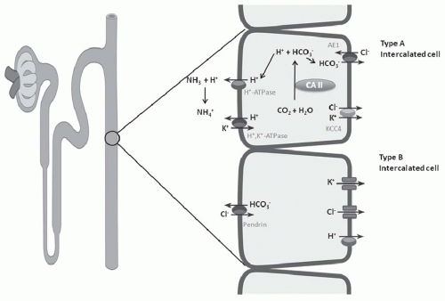
FIGURE 18.2 Type A and B intercalated cells of collecting duct. See text for detail.

Apical proton secretion generates HCO3 intracellularly, which then exits the cell via the Cl/HCO3 exchanger (AE-1) present on the basolateral membrane (encoded by the gene SLC4A1).45 Thus, these three transporters, the apical H+-ATPase and H+,K+-ATPase, and the basolateral HCO3 exchanger could be involved, if defective, in the development of an acidification defect in the distal nephron (Fig. 18.3). The reader is referred to Chapter 6 for additional detail. Examples of genetic and acquired abnormalities of the H+-ATPase and AE-1 have been described and are discussed later.

Additionally, defective net H+ secretion could occur by the insertion of a “leak” pathway for H+ into the collecting duct (Fig. 18.3). This abnormality, also referred to as a “gradient lesion,” occurs most commonly with amphotericin B nephrotoxicity. Whether this latter abnormality accounts for acidification defects in other forms of inherited or acquired distal RTA has been described in case reports, but has not been established clearly.


Ammonium Production and Excretion

Although ammonium is secreted in several segments of the nephron, the majority of ammonium secretion occurs in the proximal tubule and is regulated by acid-base homeostasis (Fig. 18.4). Ammonium transport involves both ammonia (NH3) diffusion and ammonium (NH4+) transport. NH4+ secretion into the proximal tubule lumen occurs via the apical membrane Na+/H+ exchanger (NHE-3) through substitution of NH4+ for H+. Ammonium secretion is augmented dramatically by systemic metabolic acidosis. At physiologic pH, α ketoglutarate, a major metabolic product of ammoniagenesis, is converted to HCO3 ions, which are transported across the basolateral membrane to the extracellular fluid (ECF). This end product of ammoniagenesis therefore represents “new bicarbonate” when returned to systemic circulation via the renal vein. As mentioned previously, and
discussed in detail in Chapter 6, “new bicarbonate” restores the HCO3 lost in the ECF from buffering the acid products of metabolism.


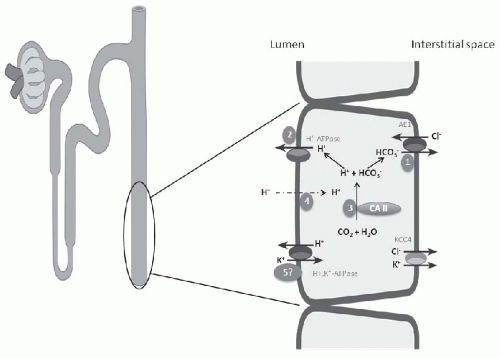
FIGURE 18.3 Pathogenesis of distal renal tubular acidosis (RTA). Model of type A intercalated cell in medullary collecting duct showing described inherited defects encoding distal transport proteins that cause classical distal RTA. 1, defect of basolateral HCO3/Cl exchanger; 2, defect of specific subunits of H+-ATPase; 3, carbonic anhydrase II deficiency; 4, backleak of H+ or gradient lesion (amphotericin B and presumed rare inherited abnormalities); 5, abnormality of H+,K+-ATPase (not verified, but may explain endemic [Northeastern Thailand] distal RTA with severe hypokalemia).

After ammonium enters the proximal tubule lumen, an elaborate system exists to generate high medullary interstitial concentrations of ammonium46 (Fig. 18.5). First, the HCO3 concentration and pH of tubular fluid increases progressively along the thin descending limb of the loop of Henle as a result of water abstraction.47 This alkaline environment favors NH3 diffusion out of the tubule lumen. In addition, direct uptake of NH4+ is accomplished via the apical Na+-2Cl-K+ cotransporter (through competition for the K+ site) in the medullary thick ascending limb of the loop of Henle (TALH).46 Ammonium absorption at this site is stimulated by acidosis and hypokalemia and is impaired by hyperkalemia.48,49,50,51,52 NH3 is capable of reentering the proximal straight tubule from the interstitium.46 Active absorption of NH4+ in the TALH allows for trapping of NH4+ in the medullary countercurrent multiplication system.53 The end result of this system is a medullary-to-cortical concentration gradient for ammonium with medullary concentrations exceeding cortical concentrations severalfold. This corticomedullary ammonium gradient is augmented by metabolic acidosis.50 Ammonium is trapped in the medullary collecting duct by a combination of NH3 diffusion from the interstitium and active H+ secretion by the medullary collecting duct (H+-ATPase and the H+-K+-ATPase).50,54 This process generates high concentrations of ammonium in the final urine. Because NH4+ uptake by the TALH is accomplished by the Na+-K+-2Cl cotransporter, competition between K+ and NH4+ helps explain the association between hyperkalemia and metabolic acidosis.55 Additional detail on ammonia/ammonium transporters and their regulation is provided in Chapter 6.


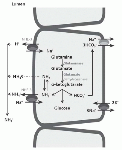
FIGURE 18.4 Ammoniagenesis and transport of ammonia/ ammonium in the proximal tubule.


Regulation of Distal Acidification

Apical proton secretion and basolateral HCO3 transport together and regulate net HCO3 absorption in the distal nephron. The responsible transporters include the
electrogenic H+-ATPase, the electroneutral H+,K+-ATPase on the apical membrane, and the HCO3/Cl exchanger on the basolateral membrane (Fig. 18.6). Alteration in the negative transepithelial potential difference, which is dependent on the rate of Na+ absorption, has a significant secondary impact on proton secretion by the electrogenic H+-ATPase. Thus, a decline in Na+ delivery or Na+ acidity through either impairment of epithelial Na+ channel (ENaC) function or through absence of mineralocorticoid will secondarily impair H+ secretion. A defect in H+ secretion in the CCT in response to a decline in Na+-transport dependent transepithelial voltage has been termed a “voltage defect.” Mineralocorticoids have been demonstrated to be a potent determinant of proton secretion. In the CCT, mineralocorticoids stimulate Na+ absorption (ENaC) increasing the lumen negative transepithelial potential, which stimulates electrogenic proton secretion secondarily.56 This early effect of aldosterone on ENaC is reinforced after several hours to upregulate the basolateral Na+,K+-ATPase as well. Taken together, mineralocorticoid increases the negative transepithelial potential, thus enhancing Na+ absorption. Mineralocorticoids have also been shown to stimulate the H+-ATPase in the cortical, outer, and inner medullary collecting tubules in the absence of Na+.40,57 Thus, in summary, both mineralocorticoids and Na+ absorption in the CCT have important regulatory effects on net H+ secretion in the collecting duct.


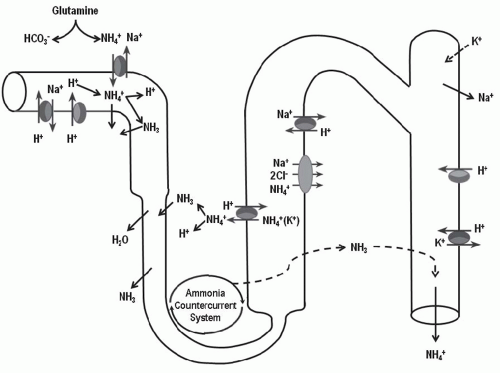
FIGURE 18.5 Summary of ammonia/ammonium transport pathways in the nephron. Possible defective ammoniagenesis and/or ammonium transport associated with distal renal tubular acidosis, discussed in text in detail.

Potassium homeostasis also plays a significant role in the regulation of renal acidification. Clearance studies have suggested that potassium deficiency stimulates distal proton secretion. It has now been established that this regulatory response occurs, at least in part, through upregulation of the H+,K+-ATPase. Potassium status can also affect renal acidification indirectly. First, potassium is an important determinant of aldosterone, and as discussed previously, aldosterone is an important determinant of H+ secretion. Potassium also affects ammonium synthesis and excretion.52 Chronic hypokalemia stimulates ammonium production while hyperkalemia suppresses ammoniagenesis.48 Alterations in ammonium production may also affect the medullary interstitial gradient and buffer availability. Hyperkalemia impairs ammonium absorption in the thick ascending limb, also decreasing medullary concentrations of total ammonia and secretion of NH3 into the medullary collecting duct.48,49,58


Pathogenesis of Distal Renal Tubular Acidosis

Classical Hypokalemic Distal Renal Tubular Acidosis. The mechanisms involved in the pathogenesis of hypokalemic distal RTA (DRTA) are not yet completely resolved.
The occurrence of hypokalemia demonstrates that generalized CCT dysfunction or aldosterone deficiency is not causative. Initially the cause of classical hypokalemic DRTA was considered to be a “gradient lesion.” Alternatively, a defect in proton secretion is the most widely accepted explanation for the inability to maximally acidify the urine,59 in the majority of forms of classical DRTA (Fig. 18.3). The most notable exception of this mechanism is the defect induced by amphotericin B nephrotoxicity, for which insertion of a leak pathway in the apical membrane of the distal tubule by the antibiotic is causative (Fig. 18.3, number 4). An important feature in the determination of the pathogenesis of the acidification defect in these patients is the response of the urine PCO2 to NaHCO3 infusion. Infusion of NaHCO3 to produce a high HCO3 excretion rate results normally in distal nephron hydrogen secretion and the generation of a high CO2 tension in the urine. The magnitude of the urinary PCO2 (referred to as the urine-minus-blood PCO2, or U-B PCO2) is quantitatively related to distal nephron hydrogen ion secretion.38,60 A decrease in the rate of hydrogen ion secretion as a result of a defect of one of the H+ transporters on the apical membrane (H+– or H+,K+-ATPase) or the basolateral HCO3/Cl exchanger will lead to a low U-B PCO2. In contrast, a backleak of H+, as occurs with a “gradient” defect, has been shown in experimental models to be associated with a normal U-B PCO2. In patients with classical hypokalemic DRTA, the U-B PCO2 is usually subnormal, except in amphotericin B-induced DRTA.60,61,62 This finding supports the view that most patients with DRTA have a “rate” or “pump” defect (Fig. 18.3, number 1).


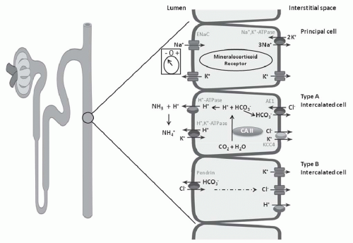
FIGURE 18.6 Three cell types in the collecting tubule: principal cell, and type A and type B intercalated cell. Mechanisms of transport discussed in text.

H+-Secretory Defects. The rate of proton secretion could be affected by an abnormality in a specific transporter or mechanism involved in proton secretion. These include the apical H+-ATPase or H+-K+-ATPase, and the basolateral Cl/HCO3 exchanger, AE-1 (Fig. 18.3, number 2). Impairment of the H+-ATPase in classical DRTA has been documented in both acquired and inherited disorders. Acquired defects of H+-ATPase have been demonstrated in renal biopsy specimens of patients with Sjögren syndrome with evidence of classical hypokalemic DRTA. These biopsy specimens revealed trapping of this transporter in intracellular compartments and an absence of H+-ATPase protein in the apical membrane of type A cells. To further underscore the importance of H+-ATPase in classical RTA, several
investigators have described autosomal dominant mutations in the ATP6V1B1 gene encoding the B-subunit of the H+-ATPase in the kidney and cochlea, and these mutations are associated with sensorineural deafness. Another form of classical DRTA, inherited as an autosomal recessive defect, is associated with normal hearing and has been shown recently to be a mutation in the ATP6V0A4 gene that encodes for the A-subunit of this transporter.63,64,65

Only gold members can continue reading. Log In or Register to continue

Stay updated, free articles. Join our Telegram channel

May 29, 2016 | Posted by in NEPHROLOGY | Comments Off on Isolated Renal Tubular Disorders: Mechanisms and Clinical Expression

Full access? Get Clinical Tree

Get Clinical Tree app for offline access