Nephrolithiasis


Nephrolithiasis


Keith A. Hruska

Anne M. Beck


In the United States, the prevalence of kidney stones has risen over the past 30 years.1 By 70 years of age, 11% of men and 5.6% of women will have a symptomatic stone.1 Nephrolithiasis is a costly malady to society. Estimates in the 1970s exceeded $5 billion annually in the United States.2 More current estimates exceed $30 billion. The incidence of nephrolithiasis in the United States is highest in the Southeast,3,4 with peak incidence occurring in the late summer months. In addition, sedentary, white collar workers are more likely to form stones than are active, blue collar laborers.5,6 The rising prevalence of renal calculi highlights the importance of environmental factors, such as diet, in their formation. Fructose consumption, which increased since the introduction of high fructose corn syrup, is associated with stone formation.7 Increased rates of hypertension and obesity are linked to nephrolithiasis.8 Stones are distinctly less common in African Americans, American Indians, and people of Asian descent. The age and sex distribution of patients referred for evaluation of nephrolithiasis shows a 2:1 ratio of male to female patients and a maximum incidence in the 30- to 50-year-old group (excluding patients with cystinuria and infection stones).

The natural history of stone disease is characterized by recurrence. Although many studies of the natural history and incidence of stones have been biased by referral practices and differences in definition of recurrence, there is clear evidence in the literature of the recurrent nature of stone disease.

A common dilemma faced by the clinician is whether or not to evaluate and treat the first-time stone former. The risk of recurrence after an initial episode has been estimated to be about 50% by 5 years, with almost two thirds of patients having a recurrence by 9 years.6,9 Even when nephrolithiasis is evaluated and treated appropriately, the incidence of recurrence in first-time stone formers is not significantly different from that in treated patients with a history of multiple stone episodes.10 In fact, these patients most likely represent recurrent stone formers in the initial stage of their disease. These and other data demonstrating Randall’s plaque11,12 suggest that such patients should be evaluated and treated in the same manner as patients with recurrent nephrolithiasis.

Patients who have undergone shockwave lithotripsy represent another group at risk for stone recurrence. Stone fragmentation often leads to regrowth of stone material, because residual fragments may act as a nidus for ongoing crystal deposition and stone formation. Medical therapy aimed at correcting underlying urinary abnormalities in these patients may prevent or limit stone growth and recurrence.13

Despite the almost inevitable risk of nephrolithiasis recurring if it is left untreated, diagnostic evaluation and selective treatment of metabolic abnormalities that decrease the incidence of new stone formation and can induce complete remission are not routinely practiced.14 Calcium, uric acid, cystine, and struvite stones differ from one another in terms of pathogenesis and treatment. Therefore, each stone type is described separately. The clinical manifestations by which the stones present are not related to the composition of the stone, and this should be kept in mind.


CLINICAL MANIFESTATIONS

The classic presentation of nephrolithiasis is acute renal colic manifesting as colicky flank pain radiating to the groin. As stones descend in the ureter, the pain may localize to the abdominal area overlying the stone and radiate to the gonad. Peritoneal signs are generally absent. Stones at the ureterovesical junction may cause lower quadrant pain that radiates to the urethral tip, urinary urgency, frequency, and dysuria resembling bacterial cystitis. Exam often reveals patients in distress, unable to find a comfortable position, with tenderness in the costovertebral angle or lower quadrant. Gross or microscopic hematuria is present 90% of the time, but the absence of hematuria does not preclude the diagnosis of nephrolithiasis. Owing to the shared splanchnic innervation of the renal capsule and intestines, hydronephrosis and distension of the renal capsule may produce nausea and vomiting. Thus, renal colic may mimic acute abdominal or pelvic conditions.

The best means of confirming the diagnosis of a urinary stone is unenhanced computed tomography (noncontrast helical CT scan) of the abdomen and pelvis. The sensitivity approaches 96% with a specificity of 100%.15 Positive and
negative predictive values are 100% and 91%, respectively. Negative CT scans often detect other abnormalities including appendicitis, pelvic inflammatory disease, diverticulitis, abdominal aortic aneurysm, and bladder cancer. Plain abdominal radiography assesses whether the stone is radiopaque, which is the case 75% to 90% of the time. Ultrasonography has a high specificity but a much lower sensitivity than CT. Ultrasonography is appropriate as the initial imaging test in pregnancy and in pediatrics and in patients who should avoid radiation. Intravenous pyelography has been replaced by helical CT as the preferred imaging test.16 Secondary signs of urinary tract obstruction such as ureteral dilatation, hydronephrosis, and perinephric stranding, are variably seen depending on the duration of pain prior to imaging and the sign itself.17

Management of acute renal colic involves a decision whether urgent intervention is required or not. The presence of an obstructed infected upper urinary tract, renal deterioration, intractable pain or vomiting, anuria, or obstruction of a solitary or transplanted kidney are all indications for urgent intervention. Intervention is carried out by urology, and is beyond the scope of this chapter. Pain management has traditionally included narcotics, but stimulation of dependency and long-term effects make nonsteroidal anti-inflammatory drugs (NSAIDs) attractive alternatives. When urgent intervention is not selected, the interval between acute colic and elective intervention for failure of stone passage is an important topic. Observation for up to 4 weeks is considered generally reasonable.18


CALCIUM STONES


Classification of Calcium Nephrolithiasis by Urinary Chemistries

Biochemical and physical disturbances that contribute to the formation of calcium stones are quite varied, based on two surveys from the mid-1990s.19,20 Several disturbances have the potential to create the environment conducive to renal stone formation. Several investigators utilize the presence of such disturbances as the basis for diagnostic categorization of nephrolithiasis.19,20,21 Earlier studies based on ambulatory evaluations of patients with nephrolithiasis reported 10 metabolic etiologies composing four types of hypercalciuria, hyperuricosuria, hyperoxaluria, renal tubular acidosis (RTA), uric acid stones, and infection stones, and an 11% incidence of finding no metabolic abnormalities.22 Now more than 15 etiologic categories of nephrolithiasis have been described (Table 20.1). A single diagnosis is found in the minority of patients whereas approximately 60% have more than one diagnosis. The finding of no metabolic abnormality can be reduced to the range of 2% to 4% of patients with care and repeated measures. Hypercalciuric nephrolithiasis accounts for about 60% of the patients. Hyperuricosuria associated with calcium nephrolithiasis can be subdivided into hyperuricosuric calcium nephrolithiasis and patients with gouty diathesis. Hyperoxaluric calcium nephrolithiasis, which occurs in about 8% of patients with recurrent stones, has been subdivided into enteric, primary, and dietary variants. Hypocitraturic calcium nephrolithiasis, which affects about 30% of patients in its idiopathic variant, is also associated with incomplete RTA and the chronic diarrheal syndrome. Hypomagnesiuric calcium nephrolithiasis, infection stones, and cystinuria are uncommon, accounting for 7%, 6%, and 1% of patients, respectively. The acquired problem of low urinary volume, less than 1 L per day according to Levy and colleagues19 and less than 1.5 L per day according to Seltzer and Hruska,20 is the single most common abnormality.

The descriptions of clinical subtypes that follow represent the minimal diagnostic criteria used to establish the presence of the entities listed in Table 20.1, according to Hruska and Seltzer.


Absorptive Hypercalciuria Type I

Diagnostic criteria include: calcium nephrolithiasis, normocalcemia, normophosphatemia, hypercalciuria (>200 mg per day) on a calcium-restricted diet, normal fasting urinary calcium (<0.11 mg per dL glomerular filtrate [dL GF]), exaggerated calciuric response to an oral calcium load (>0.20 mg urinary calcium per mg urinary creatinine), and normal to suppressed serum parathyroid hormone (PTH) function.23,24,25,26,27,28,29,30


Absorptive Hypercalciuria Type II

Criteria are the same as for type I, except for normal urinary calcium (<200 mg per day) on the restricted diet.23,24,25,26,27,28,29,31


Absorptive Hypercalciuria Type III (Renal Phosphaturia)

Diagnostic criteria are characterized as similar to type I, except for persistent hypophosphatemia (2.5 mg per dL or less).32


Sodium-Linked Phosphate Transporter

Low serum phosphate concentrations due to a decrease in renal phosphate reabsorption occur in some patients with renal calcium stones and/or bone demineralization. Two different heterozygous mutations in the sodium-linked phosphate transport protein encoded by the NPT2a gene have been associated with this disorder.33 Subsequent studies have shown that although genetic variants of NPT2a are not rare, they do not seem to be associated with clinically significant renal phosphate or calcium handling anomalies in a large cohort of hypercalciuric stone-forming pedigrees.34


Renal Hypercalciuria

Diagnostic criteria include: calcium nephrolithiasis, normocalcemia, normophosphatemia, hypercalciuria on the restricted diet, elevated fasting urinary calcium (>0.11 mg per dL GF), and elevated serum parathyroid hormone (PTH).35


Primary Hyperparathyroidism (Resorptive Hypercalciuria)

Criteria for diagnosis include: nephrolithiasis, hypercalcemia, hypercalciuria, and high serum PTH with surgical confirmation of abnormal parathyroid tissue.23,24,25,26,28,36,37


TABLE 20.1 Urinary Chemistries in Evaluation of Nephrolithiasisa


Seltzer and Hruska80 Jewish Hospital (n = 587)


Levy et al.79 University of Texas Southwestern (n = 1270)


Category


Sole occurrence(%)


Combined occurrence(%)


Sole occurrence(%)


Combined occurrence(%)


Hypercalciuria


14


51


Male (>250 mg/24 hr)


8


34


Female (>225 mg/24 hr)


6


17


Absorptive hypercalciuria


37


6.1


23.1


Fasting hypercalciuria


14


4.3


13.9


Renal hypercalciuria


1


0.3


1.3


Renal phosphaturia


2


2.1


7.6


Primary hyperparathyroidism


1


0.8


1.3


Hyperuricosuria


8


42


Male (>0.75 g/24 hr)


6


30


Female (>0.70 g/24 hr)


2


12


Hyperuricosuric calcium nephrolithiasis


8.3


27.6


Gouty diathesis


3.1


6.9


Hypocitraturia


9


34


Male (<250 mg/24 hr)


5


18


Female (<300 mg/24 hr)


4


16


Complete distal RTA


0.08


0.16


Incomplete distal RTA


0.0


1.1


Chronic diarrheal syndrome


0.2


1.8


Idiopathic


3.5


24.4


Hyperoxaluria (>40 mg/24 hr)


8


34


Enteric hyperoxaluria


0.2


1.4


Primary hyperoxaluria


0.0


0.4


Dietary hyperoxaluria


0.4


5.7


Hypomagnesuria (<5 mEq/24 hr)


5


26


0.3


6.5


(<50 mg/24 hr)


Low urinary volumes (<1500 mL/24 hr)


26


61


1.7


13.5


(<1,000 mL/24 hr)


No diagnosis/difficult to classify


2


4.0


aThe category definitions in the table refer to Jewish Hospital, Washington University, St. Louis. Criteria for the University of Texas Southwestern data are provided in the text.


RTA, renal tubular acidosis.


Fasting Hypercalciuria and Elevated Fasting Urinary Calcium

Calcium nephrolithiasis and hypercalciuria on a restricted diet can be categorized into a resorptive form because of fasting hypercalciuria. Fasting hypercalciuria is further characterized by normal to suppressed parathyroid function, eliminating renal calciuria, normocalcemia, and normophosphatemia (>2.0 mg per dL).


Hyperuricosuric Calcium Nephrolithiasis

The diagnostic criteria for hyperuricosuric calcium nephrolithiasis (HUCN) include: calcium nephrolithiasis, hyperuricosuria
(>700 mg per day for females; >750 mg per day for males), and frequently a low urinary pH of ≤5.5.38,39,40


Gouty Diathesis

Criteria include uric acid or calcium nephrolithiasis and low urinary pH (<5.5) in the absence of excessive gastrointestinal alkali losses41,42,43 or animal protein excess. Hyperuricemia, hypertriglyceridemia, and a history of gouty arthritis may be present.


Hyperoxaluric Calcium Nephrolithiasis

Criteria include calcium nephrolithiasis and hyperoxaluria (>44 mg per day). The three forms of hyperoxaluric calcium nephrolithiasis are:

1. Enteric hyperoxaluria, defined as the presence of ileal disease (Crohn disease, ulcerative colitis, jejunoileal bypass, or intestinal resection), or fat malabsorption with hyperoxaluria on the random and restricted diets.44,45,46,47

2. Primary hyperoxaluria, consisting of marked hyperoxaluria (>80 mg per day) without evidence of bowel disease, high oxalate diet, low calcium diet, treatment with calcium-binding agents, enhanced oxalate absorption, or high doses of vitamin C.48

3. Dietary hyperoxaluria, marked by high oxalate diet, hyperoxaluria on a random diet, and normal urinary oxalate excretion on the restricted diet.47,49,50

Enteric hyperoxaluria is typically associated with hypocitraturia due to intestinal loss of HCO3, low urinary volume, and low normal urinary calcium excretion.


Hypocitraturic Calcium Nephrolithiasis

Diagnostic criteria include calcium nephrolithiasis and hypocitraturia (<320 mg per day),39 that compose:

1. Distal RTA, which is characterized by systemic metabolic acidosis or defective urinary acidification following an ammonium chloride load and urinary pH above 6.5. The acidosis is a hypokalemic, hyperchloremic nonanion gap metabolic acidosis.50 In the complete form, metabolic acidosis is present before an ammonium chloride load, whereas in the incomplete form, urinary acidification following an ammonium chloride load is impaired despite normal serum electrolytes before the load.

2. Chronic diarrheal syndrome, which is defined as chronic diarrhea with excessive alkali loss from various gastrointestinal disorders (e.g., gastric resection, ileal disease, Crohn disease, and ulcerative colitis).51

3. Idiopathic hypocitraturia of unknown etiology.


Hypomagnesiuric Calcium Nephrolithiasis

Criteria for diagnosis include: nephrolithiasis, hypomagnesuria (<50 mg per day) on the random diet, and absence of a diarrheal disorder.52


Infection Stones

Criteria for diagnosis include struvite or carbonate-apatite nephrolithiasis.53


Cystinuria

Diagnosis is based on cystine nephrolithiasis and urinary cystine level higher than 200 mg per day.54


Low Urine Volume

Diagnosis is based on calcium or uric acid nephrolithiasis and urine volume less than 1 L per day.55


No Metabolic Abnormality

This is attributed to calcium nephrolithiasis and a normal biochemical evaluation.56


Difficult to Classify

Those that fall into this category are generally nephrolithiasis with recognized stone risk factors.57 A definitive diagnosis cannot be made due to borderline or inconsistent laboratory values or to the absence of critical data (e.g., stone analysis or roentgenographic visualization).


HYPERCALCIURIC NEPHROLITHIASIS

The majority of patients with calcium nephrolithiasis exhibit hypercalciuria (Table 20.1) and have idiopathic hypercalciuria, which is a term used to describe recurrent nephrolithiasis associated with hypercalciuria, and is probably a distinct entity. However, most of the data suggest that multiple metabolic abnormalities besides excess calcium excretion contribute to nephrolithiasis associated with hypercalciuria. Furthermore, there is a much higher incidence of idiopathic hypercalciuria than nephrolithiasis in the general population.58 Several estimates place the incidence of idiopathic hypercalciuria at 2% to 4%,59 whereas the incidence of nephrolithiasis is no more than 0.5% to 1.0%. Between 40% and 50% of calcium stone formers excrete excess calcium in their urine, defined as more than 300 mg per 24 hours (men), 250 mg per 24 hours (women) on ≥1,000 mg calcium intake, or 4 mg per kg body weight per 24 hours (either sex). The term idiopathic hypercalciuria applies if the serum calcium level is normal and sarcoidosis, RTA, hyperthyroidism, malignant tumors, rapidly progressive bone disease, immobilization, Paget disease, Cushing disease (or syndrome), and furosemide administration have been excluded. Virtually all normocalcemic hypercalciuria encountered in patients with nephrolithiasis falls under the umbrella of “idiopathic hypercalciuria.”19

Among patients with recurrent calcium stones who have served as control subjects in randomized, controlled trials of interventions, new stones formed in 43% to 80% of subjects within 3 years.14,60,61,62


FIGURE 20.1 Pathogenesis of absorptive hypercalciuria. Intestinal hyperabsorption of calcium leads to an increase in serum calcium and a reduction of parathyroid hormone (PTH). The increase in the filtered load of calcium and the reduced calcium reabsorption produced by loss of PTH activity lead to hypercalciuria.


Idiopathic Hypercalciuria

Idiopathic hypercalciuria is an inherited syndrome. Studies of families of patients with hypercalciuric nephrolithiasis reveal a high incidence of hypercalciuria in first-degree relatives.63 The pattern of inheritance in consecutive generations with high frequency is compatible with an inherited trait with the broad characteristics of autosomal dominant transmission. This pattern of inheritance was demonstrated in large kindred.64 Hypercalciuria also occurs in children at the same rate as in adults.65 Spontaneous hypercalciuria similar to that observed in hypercalciuric nephrolithiasis of humans has been demonstrated in the laboratory rat,66,67 which has become an animal model of the human disorder.

The pathogenesis of idiopathic hypercalciuria involves excessive intestinal calcium absorption and depressed renal tubular calcium reabsorption (Fig. 20.1). The latter is largely due to suppression of PTH68,69 and can be considered as a major factor in preventing hypercalcemia associated with increased intestinal absorption. When placed on low calcium diets, patients with hypercalciuric nephrolithiasis often demonstrate a negative calcium balance.70 This could be due to defective renal tubular calcium reabsorption, but renal hypercalciuria should produce secondary hyperparathyroidism, which is rarely observed.19,20 In addition, considerable evidence indicates that PTH levels are suppressed and that the negative calcium balance stems from excessive skeletal remodeling and bone resorption.68,69 The question of whether depressed renal tubular calcium reabsorption greater than that expected with PTH suppression contributes to the hypercalciuria of nephrolithiasis remains unanswered. It is supported only by data from a few patients in whom secondary hyperparathyroidism has been documented (see “Renal hypercalciuria” in Table 20.1).


FIGURE 20.2 Renal phosphaturia. Hypophosphatemia leads to increased production of 1,25(OH)2D3, intestinal hyperabsorption of calcium, and increased skeletal calcium mobilization. As a result, hypercalciuria develops.

In hypercalciuric calcium oxalate stone formers, the initial site of calcium/phosphate crystal deposition is the basement membrane of the thin limbs of Henle’s loop. There is subsequent extension to the vasa section, then the interstitium and, in the most severe cases, to the papillae. Alternatively, in patients with hyperoxaluria secondary to intestinal bypass and idiopathic calcium phosphate stones, the initial crystals were again calcium/phosphate complex, but these arose within the tubule lumens of terminal collecting ducts. Non-stone formers, when subjected to nephrectomy, had neither plaque nor crystals. Thus, there are different sites of crystallization depending on the metabolic abnormalities leading to stone formation.71,72

Additionally, in patients with idiopathic hypercalciuria, there was evidence for crystal-induced cell injury in areas of dense crystal deposition, whereas in the bypass patients there was not only cell injury, but also cell death.11

The genetically hypercalciuric stone forming rats spontaneously form calcium/phosphate stones unless their diet is augmented with an oxalate precursor.73

In the genetic hypercalciuric stone forming rats, calcium oxalate (but not calcium/phosphate) stones induce marked proliferation of the urothelium resulting in sequestration of stones.67

Thus, rats and humans appear protected against calcium oxalate stone formation unless a nucleation site, such as the calcium/phosphate crystal, is present.


Absorptive Hypercalciuria

Increased intestinal absorption of calcium is a uniform finding in patients with hypercalciuric nephrolithiasis20 (Fig. 20.1). At issue is whether increased absorption is the primary defect or caused secondarily in the idiopathic hypercalciuric syndrome (Figs. 20.1,20.2,20.3,20.4). All forms of hypercalciuric nephrolithiasis are associated with increased intestinal calcium absorption. Those associated with intestinal calcium hyperabsorption on a secondary basis—renal hypercalciuria, primary hyperparathyroidism, and renal phosphaturia—are relatively uncommon forms of hypercalciuric nephrolithiasis. Furthermore, fasting hypercalciuria appears to be the expression of increased skeletal remodeling and intestinal calcium hyperabsorption together. All of this indirectly suggests that a specific problem producing intestinal calcium hyperabsorption is a major, if not the basic, underlying defect in idiopathic nephrolithiasis.


FIGURE 20.3 The pathogenesis of renal hypercalciuria. Defective renal calcium reabsorption leads to hypocalcemia and a stimulation of parathyroid hormone (PTH) secretion. The latter increases the production of 1,25(OH)2D3, stimulating calcium absorption and leading to hypercalciuria. The hypercalciuria in turn compensates for the reduction in serum calcium but compensation never completely restores normal calcium, and elevated PTH values are required in this syndrome.


Intestinal Calcium Absorption

Net calcium absorption is the difference between the mucosal absorptive rate and the secretion of calcium into biliary, duodenal, and pancreatic fluids. Although calcium absorption rates may be measured using oral radiolabeled calcium, only overall balance studies in which fecal losses are measured can quantitate net calcium absorption. The mucosal to serosal absorptive rate is higher in patients with hypercalciuric nephrolithiasis than in healthy individuals26,36,74,75,76,77,78,79,80,81 (Table 20.2), but overlap is extensive. In six studies, individuals with no signs of hypercalciuric nephrolithiasis absorbed an average of 27% to 52% of an oral dose of radioactive calcium, whereas those with hypercalciuric nephrolithiasis absorbed 22% to 80%. If one chooses only the six studies incorporating normal control subjects, the more efficient calcium absorption by hypercalciuric nephrolithiasis subjects is particularly evident. Increased mucosal-to-blood transport of calcium, but not magnesium, has also been demonstrated directly by in vivo jejunal perfusion in hypercalciuric nephrolithiasis.29 At normal calcium intakes, <1,500 mg per day, calcium absorption in the duodenum and proximal jejunum is an active process mediated by a mucosal membrane calcium pump (ECaC)82,83 and efficient cytosolic calcium binding proteins (calbindin),84 both transcriptionally regulated by calcitriol. At high calcium intakes, passive transport mechanisms in the more distal small bowel and colon may account for most of calcium absorption as the proximal active calcitriol-regulated mechanisms are suppressed.

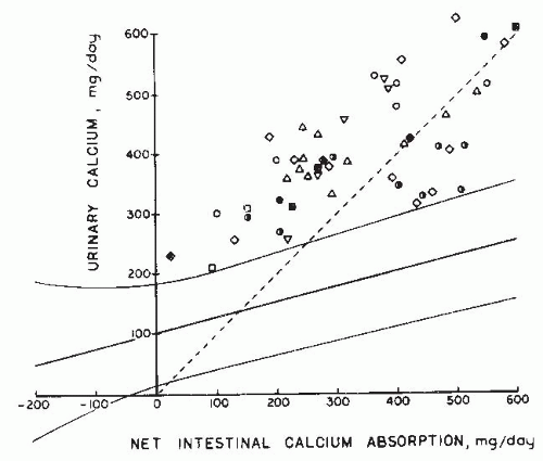
In normal individuals, urine calcium excretion rises slowly with net absorption85 (Fig. 20.5), and calcium balance is usually positive when the absorption rate exceeds 200 mg per 24 hours. At all levels of net absorption, urinary calcium excretion was higher in hypercalciuric than in normal subjects, so much so that none of the patient data fell within the 95% confidence band derived from studies of normal individuals (Fig. 20.6). For example, in the range of 200 to 300 mg of net calcium absorption, not one of 38 normal subjects excreted as much as 300 mg of calcium in the urine, whereas 16 hypercalciuric patients did (compare Figs. 20.5 and 20.6). In other words, hypercalciuric nephrolithiasis subjects excreted in the urine an abnormally high percentage of the calcium they absorbed from the intestine. This is compatible with suppression of renal tubular calcium transport rates by low levels of PTH. Net absorption rates exceeded 200 mg per 24 hours in 55 normal subjects (Fig. 20.5). Urine calcium excretion was less than net absorption; that is, calcium balance was positive in 48 subjects. If a generous margin for error (50 mg per 24 hours) is allowed in the balance data, none of the 55 normal individuals were in negative calcium balance. Among 37 hypercalciuric patients with a calcium absorption rate above 200 mg per 24 hours, however, calcium excretion exceeded net absorption in 23 patients by more than 50 mg per 24 hours (Fig. 20.6). In other words, negative calcium balance was frequent in idiopathic hypercalciuria subjects but not in normal individuals. This is compatible with either reduced tubular reabsorption, which should produce elevated levels of PTH, or with excessive bone resorption. The latter appears most likely.


FIGURE 20.4 Pathogenesis of hypercalciuria and primary hyperparathyroidism. Excess secretion of parathyroid hormone (PTH) stimulates bone remodeling and elevations of 1,25(OH)2D3 and calcium absorption. The resultant increase in serum calcium leads to an increase in the filtered load of calcium.The latter overwhelms the stimulation of renal tubular calcium transport by PTH, and hypercalciuria results.


Renal Tubular Calcium Reabsorption

Two systematic studies86,87 have evaluated overall renal fractional calcium reabsorption (Table 20.3). In both, the filtered load of calcium was calculated from inulin clearance or creatinine clearance and ultrafilterable serum calcium concentration. The fraction of the filtered calcium load excreted was calculated for several clearance periods in normal and hypercalciuric nephrolithiasis subjects. Fractional calcium excretion was clearly high in the hypercalciuric nephrolithiasis subjects. The effects of hydrochlorothiazide and acetazolamide on the renal tubular handling of sodium, magnesium, and calcium suggested to the authors a generalized defect in proximal tubular reabsorption.86,87,88 These studies did not examine the role of suppressed or elevated PTH levels in the subjects with hypercalciuric nephrolithiasis. The general finding of a tendency for PTH levels to be
suppressed and the rarity of secondary hyperparathyroidism call for a reexamination of the issue regarding renal tubular calcium fluxes.


TABLE 20.2 Intestinal Calcium Absorption in Normal Subjects and Patients with Idiopathic Hypercalciuria


Dietary calcium absorbed (%)a


Reference (no.)


Method


Calcium intake (mg/24 hr)


Normal subjects


Idiopathic hypercalciuria


Caniggia et al.124


Fecal 45Ca


Free dietb


None studied


22.0 (1)


Birge et al.123


47Ca, PO/IV


800


52.2 ± 13.2 (6)


58.5 ± 8.6 (4)


Wills125


47Ca, PO/IV


400


49.0 ± 10.0 (4)


76.0 ± 17.0 (5)


Pak126


Fecal 47Ca


400


45.6 ± 9.0 (29)


69.7 ± 7.0 (9)


58.1 ± 13.0 (11)c


Pak et al.86


Fecal 47Ca


400


50.0 ± 7.0 (20)


71.0 ± 7.0 (22)d


50.0 ± 17.0 (2)e


Ehrig et al.126


47Ca/45Ca, PO/IV


462-952


None studied


47.8 ± 11.0 (22)f


37.6 ± 11.0 (22)g


Kaplan94


Fecal 47Ca


400


48.0 ± 8.0 (11)


80.0 ± 9.0 (211)d


73.0 ± 7.0 (3)e


Shen128


47Ca/45Ca, PO/IV


Free dietb


27.0 ± 9.0 (14)


40.0 ± 9.0 (15)


Barilla129


Fecal 47Ca


400


None studied


69.5 ± 6.4 (10)d


70.1 ± 10.4 (8)e


Zerwekh and Pak130


Fecal 47Ca


400


None studied


69.0 ± 7.0 (11)d


68.0 ± 9.0 (10)d


a Values are means ± standard deviations; numbers in parentheses represent numbers of patients studied.

b Usual diet but not measured.

c Eleven patients listed as having normocalcemic primary hyperparathyroidism may be considered hypercalciuric.

d Absorptive idiopathic hypercalciuria.

e Renal idiopathic hypercalciuria.

f Prior to therapy.

g Three to 16 months after administration of hydrochlorothiazide.


Ca, calcium; PO, orally; IV, intravenously.


A rare syndrome, X-linked hypercalciuric nephrolithiasis (XLHN), or Dent disease, is characterized by recurrent calcium nephrolithiasis and has been found to be due to mutations in a proximal tubular intracellular vesicle chloride transport protein, CLCN5.89,90,91,92,93 Two other types of hypercalciuric nephrolithiasis, which map to the same defective gene on the X chromosome (Xp11.22) as Dent disease, X-linked recessive nephrolithiasis and recessive hypophosphatemic rickets, are associated with inactivating mutations in CLCN-5.89

The CLCN-5 gene is a member of a family of genes that encode voltage-gated chloride channels.90 CLCN-5 is found in the kidney tubules and in bone cells. All mutations in the CLCN-5 gene found to date have been functional, with loss of function manifested as a lowered conductance of the mutated channel. CLCN-5 is distributed in the human kidney in the proximal tubule, in the thick ascending limb of the loop of Henle, and in the α-type intercalated cells of the collecting duct.90 These sites are where calcium is resorbed from the filtrate. CLCN-5 knockout animals are hypercalciuric and proteinuric.91

CLCN-5 colocalizes with the vacuolar H+-ATPase in proximal tubular cells and a-type intercalated cells. CLCN-5 mutations are associated with modifications in the polarity and expression of H1-ATPase, but not ultrastructural alterations in proximal tubular cells.93 The variability in diseases
with the CLCN-5 mutations involves impaired solute reabsorption by the proximal tubule and range from kaliuresis, glycosuria, phosphaturia, and/or hypouricemia. Constant findings of Dent disease include hematuria and low molecular weight proteinuria. Dent disease progresses to renal failure due to nephrocalcinosis and tubulointerstitial nephritis.
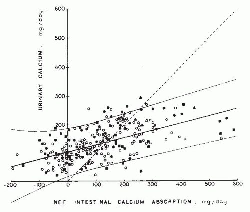


FIGURE 20.5 Urinary calcium excretion as a function of net intestinal calcium absorption. Data are derived from 6-day balance studies on 195 normal adults. Each symbol represents individual subjects from different sources: circles, Knapp238; squares, Lafferty and Pearson239; diamonds, Liberman et al.240; and triangles, Edwards and Hodgkinson.241 Open figures represent women, and solid figures represent men. Solid lines represent mean and 2 standard deviations. The dotted line is the line of identity; points above the line reflect negative calcium balance. (From Coe FL, Favus MJ. Nephrolithiasis. In: Brenner BM, Rector FC, eds. The Kidney, 2nd ed. Philadelphia: WB Saunders; 1991, with permission.)


FIGURE 20.6 Urinary calcium excretion as a function of net intestinal calcium absorption from 6-day balance studies performed on 51 patients with idiopathic hypercalciuria reported as follows: open square, Henneman et al.243; open squares with dot in center, Jackson and Lancaster244; open triangles, Harrison245; open circles with dot in center, Dent et al.246; open inverted triangles, Parfitt et al.247; closed diamonds, Edwards and Hodgkinson241; open diamonds, Liberman et al.240; and half-darkened circles, Lemann.248 Solid lines represent mean and 2 standard deviations derived from balance studies from 195 normal adults, shown in Figure 26.10. The dotted line is the line of identity, with positive calcium balance below the line. (From Coe FL, Favus MJ. Nephrolithiasis. In: Brenner BM, Rector FC, eds. The Kidney, 2nd ed. Philadelphia: WB Saunders; 1991, with permission.)

Thiazide diuretics reduce hypercalciuria in patients with CLCN-5 mutations, but thiazides can make these patients become hypokalemic. The beneficial effect must be weighed against the potential side effect profile.92 Langman finds little reduction in stone formation even when urinary calcium is lowered to normal in such individuals.94


TABLE 20.3 Fraction of Filtered Calcium Excreted in the Urine by Normal and Hypercalciuric Subjectsa


Normal subjects


Hypercalciuric subjectsb


Edwards and Hodgkinson137


0.94% (7)


2.94% (14), P < 0.001


Peacock and Nordin135


1.27% (5)


4.25% (9), P < 0.01


aNumber of subjects studied are shown in parentheses next to fractional excretion values.

b Urine calcium >300 mg/24 hr (men) or 250 mg/24 hr (women).


Another Voltage-Gated Chloride Channel

Bartter syndrome is a disease arising from one of three possible genes in the thick ascending limb that bear mutations in the Na+-K+-2Cl- gene NKCC2, in the K+ channel ROMK, or in the chloride channel CLCNKB. Each of these mutations produces a phenotype that includes hypercalciuria and kidney stone formation with or without nephrocalcinosis. A missense mutation in the CLCNKB gene leads to disease of intrafamilial heterogeneity of urinary calcium levels. Some family members have Bartter syndrome with frank hypercalciuria, but others have hypocalciuria and a clinical phenotype of Gitelman syndrome.95


Hypomagnesemia/Hypercalciuria

Familial hypomagnesemia with hypercalciuria and nephrocalcinosis (FHHNC) is an autosomal recessive tubular disorder that is frequently associated with progressive renal failure. The primary defect is related to impaired tubular reabsorption of magnesium and calcium in the thick ascending loop of Henle. Mutations in PCLN-1, which encodes the renal tight junction protein paracillin-1, were identified as the underlying genetic defects. Affected patients usually present in childhood or adolescence with symptomatic hypocalcemia.96,97,98 Recurrent nephrolithiasis and nephrocalcinosis are also seen and progression to renal insufficiency and an acidification defect are common. The problem with acidification has been attributed to defective ammonia transfer to the deep nephrons and impaired medullary hydrogen ion secretion due to nephrocalcinosis.99 Treatment with magnesium salts and thiazides seems to have no effect on the progression of the disease.

A second form of primary hypomagnesemia, Gitelman syndrome, is associated with hypocalciuria. It is due to mutations in the gene encoding the thiazide-sensitive sodiumchloride-cotransporter. Because thiazides are used to treat hypercalciuric nephrolithiasis, it is important to know that they can mimic the syndrome of hypomagnesemia including hypokalemia induced by magnesuria.100


Skeletal Remodeling

The high frequency of negative calcium balance in patients with idiopathic hypercalciuria on low calcium diets was the first indication that exaggerated bone resorption characterized the syndrome. Additional evidence for elevated skeletal remodeling in hypercalciuric nephrolithiasis has accrued. Several investigators69,101 have documented reduced vertebral bone density in hypercalciuric nephrolithiasis by both CT and dual energy X-ray absorptiometry. Patients who exhibit fasting hypercalciuria tend to have a greater reduction in trabecular bone density than do other hypercalciuric patients, but there is significant overlap, and patients with absorptive hypercalciuria and normal fasting calcium excretion exhibit a high prevalence of reduced bone mineral density. Increased rates of skeletal remodeling with resorption favored over formation are supported by the findings of increased osteocalcin secretion and increased urinary hydroxyproline levels in patients with fasting hypercalciuria.69 The pathogenesis of exaggerated bone remodeling rates may be due to elevations in 1α,25-dihydroxycholecalciferol (1α,25[OH]2D3) levels, or due to elevations in bone cytokine activity such as prostaglandin activity102 and interleukin-1 activity.69 The result of this exaggerated skeletal remodeling is an increase in calcium release to the systemic circulation and suppression of PTH secretion.103 One possibility is that exaggerated skeletal remodeling is a component of the syndrome of idiopathic hypercalciuria. Activation of skeletal remodeling in the hypercalciuric patient results in increased skeletal remodeling, leading to the loss of a quantum of the skeleton before counterregulatory influences decrease remodeling rates, removing the component of fasting hypercalciuria from the hypercalciuric syndrome. Such a scenario is sufficient to explain the clinical picture, as we currently understand it. Greater clarification of the roles of fasting hypercalciuria, and of bone remodeling, and their pathogenesis is required in patients with hypercalciuric nephrolithiasis. The role of skeletal remodeling in nephrolithiasis was further clarified by the recent discovery of a mutation in the type 2a, sodium-dependent phosphate cotransporter gene found in the proximal tubule and osteoclasts.33


Fasting Hypercalciuria

Except for negative calcium balance, either primary intestinal calcium absorption or a primary renal calcium leak could produce the findings summarized in Tables 20.2 and 20.3 and in Figures 20.5 and 20.6. Primary intestinal overabsorption increases postprandial serum calcium levels
above normal and increases the filtered load of calcium (Fig. 20.1). PTH secretion is reduced by the hypercalcemia, and suppression of PTH secretion would reduce calcium reabsorption because PTH stimulates renal tubular calcium reabsorption. In contrast, a renal tubular transport defect (Fig. 20.2) leading to hypercalciuria would produce secondary hyperparathyroidism. PTH, in turn, would stimulate the production of 1α,25(OH)2D3 and produce intestinal calcium hyperabsorption. Hyperabsorption would elevate postprandial serum calcium levels, raising the filtered calcium load and decreasing the magnitude of secondary hyperparathyroidism. The only way of distinguishing one mechanism from the other is by testing specific predictions that differ in the two forms of hypercalciuria. Clinically, PTH levels are the most clear-cut basis of distinction. Fasting hypercalciuria is not a means of detecting a renal calcium leak because it can be and is caused by exaggerated bone remodeling.

Absorptive hypercalciuria is associated with low or normal fasting immunoreactive PTH (iPTH) levels. The absorptive hypercalciuria hypothesis predicts a spectrum of fasting PTH values, but it forbids the combination of elevated fasting urinary calcium-creatinine ratio, and normal-to-suppressed iPTH levels. Normal PTH levels are typically observed in patients with fasting hypercalciuria and hypercalciuric nephrolithiasis. The renal model requires elevated fasting urinary calcium-creatinine ratios and a high serum iPTH level. This is seen uncommonly (Table 20.1).

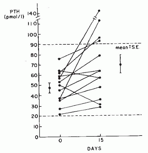
On the other hand, evidence exists for suppressed PTH levels in patients with hypercalciuric nephrolithiasis who exhibit fasting hypercalciuria103 (Fig. 20.7). When fasting hypercalciuric subjects are treated with sulindac, an NSAID agent, their urinary calcium excretion is decreased but, more importantly, their PTH levels are increased. This suggests that a bone resorptive process is releasing calcium to the circulation and suppressing PTH. Inhibition of bone resorption by sulindac results in an increase in PTH levels, suggesting that the levels are suppressed in patients with fasting hypercalciuria.103

Past studies attempting to detect low or normal PTH levels26,35,79,104,105 suffer from difficulties with the radioimmunoassay for PTH. More recent double-antibody techniques that enable the measurement of intact hormone and the detection of low circulating PTH levels circumvent these problems and support the finding of low or normal PTH levels in patients with hypercalciuric nephrolithiasis.


Pathogenesis of Absorptive Hypercalciuria

A potential explanation for the pathogenetic process identified in absorptive hypercalciuria is abnormally elevated 1,25(OH)2D3 levels. Patients with idiopathic hypercalciuria tend to exhibit elevations in 1,25(OH)2D3 levels.32,36,70,79,106 The frequency of high 1,25(OH)2D3 levels in idiopathic hypercalciuria is controversial, but it appears to range from 30% to 40%. Kaplan36 demonstrates that fractional calcium absorption correlates with the serum concentration of 1,25(OH)2D3. Two thirds of the patients in this study did not have elevated 1,25(OH)2D3 levels.


FIGURE 20.7 Changes in serum immunoreactive parathyroid hormone (PTH) in patients with fasting hypercalciuria after 15 days of diclofenac treatment. Dotted lines indicate the normal range of PTH. (From Filipponi P, Mannarelli C, Pacifici R, et al. Evidence for a prostaglandin-mediated bone resorption mechanism in subjects with fasting hypercalciuria. Calcif Tissue Int. 1988;43:61, with permission.)

On the other hand, evidence exists that shows that increased intestinal absorption of calcium may be primary and independent of vitamin D. Several studies indicated that the hypophosphatemia observed in idiopathic hypercalciuria is not sufficient to stimulate 1,25(OH)2D3 levels.107,108 Breslau109 used ketoconazole, an imidazole antimycotic agent110 capable of reducing serum 1,25(OH)2D3 levels by 40%, in normal subjects and in patients with primary hyperparathyroidism after 1 week of therapy.111 Ketoconazole was used as a probe to investigate the pathogenetic importance of 1,25(OH)2D3 in patients with absorptive hypercalciuria. Twelve of 19 patients responded to ketoconazole with a reduction in serum 1,25(OH)2D3 levels, intestinal calcium absorption, and 24-hour urinary calcium excretion. In the responding patients, intestinal calcium absorption was directly correlated with serum 1,25(OH)2D3 levels and 24-hour urinary calcium excretion. In seven nonresponders, a reduction in 1,25(OH)2D3 produced no change in intestinal calcium absorption or 24-hour urinary calcium excretion. The authors conclude that absorptive hypercalciuria is a heterogeneous disorder composed of both vitamin D-dependent and vitamin D-independent subsets.109 The vitamin D-dependent subsets incorporate patients with elevated 1,25(OH)2D3 levels, patients with abnormally responsive vitamin D receptors, and patients with allelic variations
in the vitamin D receptor that have been incriminated in causing osteoporosis.112 Animal studies in the genetically hypercalciuric rat support the possibility that an abnormal vitamin D receptor could contribute to the pathogenesis of absorptive hypercalciuria.113


TABLE 20.4 Types of Renal Stones Formed and Frequency of Occurrencea


Only gold members can continue reading. Log In or Register to continue

Stay updated, free articles. Join our Telegram channel

May 29, 2016 | Posted by in NEPHROLOGY | Comments Off on Nephrolithiasis

Full access? Get Clinical Tree

Get Clinical Tree app for offline access


CaOx and Cap


CaOx


Cap


Uric acid


Cystine


Struvite


Number of stones


Nordin and Hodgkinson1


46.0


14.7


8.0


2.9


3.3


25.1


243


Lagergen2


44.2


15.1


7.6


3.6


1.1


28.1


460