Fig. 6.1
The port placement for a medial-to-lateral laparoscopic sig-moidectomy/left colectomy. Optional port5-mm
The laparoscopic sigmoid colectomy is facilitated by a dedicated cameraperson (2nd assistant), as this allows the assistant to help with two instruments. This is important for adequate retraction of a floppy sigmoid colon and during splenic flexure mobilization. At the start of the operation, the surgeon stands at the right side of the patient and uses the two right-sided ports for dissection, and the camera assistant stands to the left of the surgeon. The assistant is positioned on the left side of the patient, using the two left-sided ports to retract the sigmoid colon. Two monitors are helpful at this point, placed on both sides of the patient near the knees, as the assistant and surgeon should view separate monitors (Fig. 6.2).
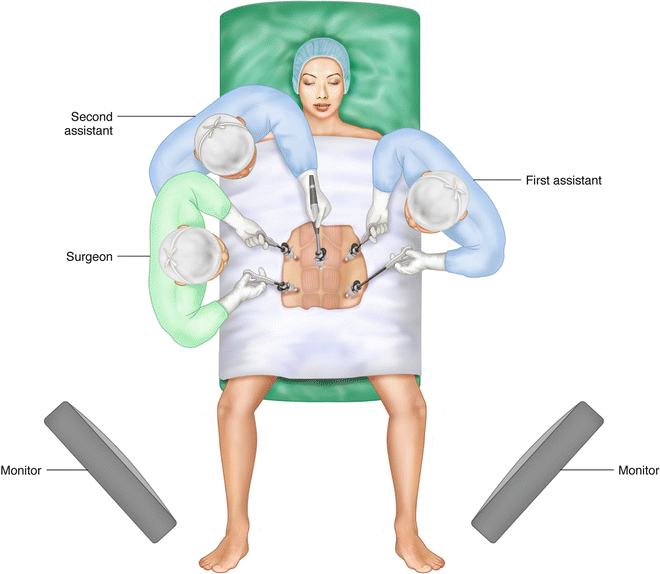
Fig. 6.2
Position of the monitors and surgical team for vascular ligation, medial and lateral bowel mobilization, and anastomosis
For splenic flexure mobilization, a monitor is moved to the patient’s left shoulder. The surgeon moves in between the legs and uses the two left-sided ports for dissection. The assistant moves to the patient’s right side, using the two right-sided ports to assist (Fig. 6.3).
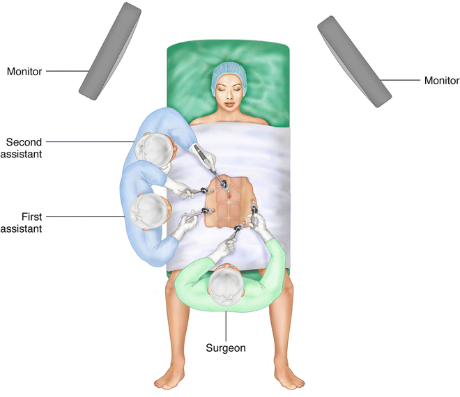
Fig. 6.3
Ports on the patient’s left abdomen should be in the same position as Fig. 6.1. It’s a little off in this illustration
It is important to remember that different port placements are used by different surgeons, and what is described above is only one option of many. A minimally invasive sigmoid colectomy by two operators using 2 or 3 working ports has been described, and port placement should eventually be tailored by each surgeon to meet one’s needs. However, one should never limit the number of ports to the point that basic surgical principles such as tissue triangulation are sacrificed. An additional 5-mm trocar presents minimal morbidity to patients and could be of tremendous benefit during a difficult laparoscopic colectomy. The port placement described above remains the most common configuration used by the author.
Operative Steps
The following are the general operative steps in a medial-to-lateral laparoscopic sigmoid colectomy:
Isolation and division of the inferior mesenteric artery
Isolation and division of the inferior mesenteric vein, ligated together or separately from the artery
Separation of the sigmoid colon and mesentery from the retroperitoneal fascia in a medial-to-lateral direction
Dissection of the lateral attachments of the sigmoid and descending colon
Splenic flexure mobilization (when necessary)
Division of the bowel proximally and distally
Anastomosis
Vascular Isolation and Division
It is common for the distal small bowel loops to drape into the pelvis and obstruct the exposure of the sigmoid mesentery, and retraction of these loops out of the pelvis can at times be difficult. Gravity is used to help with this cause, and thus, positioning of the patient is very important. Initially, the patient is placed in a steep Trendelenburg position and tilted (air-planed) with the left side up. The omentum is lifted over the transverse colon, and the small bowel loops are swept into the right upper quadrant away from the mesosigmoid. It is important that the sacral promontory is visualized and can be palpated with a laparoscopic instrument at this point. The terminal ileum not uncommonly is adhered to the right pelvis, making superior retraction of the terminal ileum impossible. In this case, the ileum must be freed from the right pelvis as the initial step.
The sigmoid colon is then retracted by the assistant in two areas. The sigmoid mesentery is retracted in a ventral direction and toward the left. Especially in a redundant “pelvic” sigmoid colon, traction must also be in a superior direction (pulling the sigmoid colon out of the pelvis). The surgeon feels for the sacral promontory using a laparoscopic instrument as this should be the initial location where the peritoneum overlying the sigmoid mesentery is incised. Access to the proper avascular plane posterior to the inferior mesenteric vessels is easiest at the sacral promontory. While adequate mesenteric traction is provided by the assistant, a wide mesenteric window is created around the origin of the inferior mesenteric artery (IMA) toward the inferior mesenteric vein (IMV) (Fig. 6.4). The right and left hypogastric nerves are adhered to the inferior mesenteric vessels and must be bluntly swept in a dorsal direction and preserved in order to avoid autonomic nerve injury that causes retrograde ejaculation. The dissection must stay anterior to the iliac vessels and these autonomic nerves and posterior to the inferior mesenteric vessels. Inability to find the correct avascular plane is usually due to inadequate ventral traction on the vessels. Creating a mesenteric window that is wide enough will allow for better traction on the vessels and avoiding “tunneling,” which results in limited visualization.
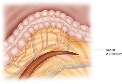
Fig. 6.4
The peritoneal incision of the sigmoid mesentery commences at the sacral promontory. This is extended in a cephalad direction around the origin of the inferior mesenteric artery toward the inferior mesenteric vein
The line of the fusion of the sigmoid mesentery and retroperitoneal fascia is identified underneath the inferior mesenteric vessels, and the retroperitoneal fascia is bluntly swept dorsally. It is with this step that the left ureter and left gonadal vein should be identified and preserved (Fig. 6.5). It is easy to dissect in a plane that is too deep, i.e., deep to the ureter and gonadal vessels. Only after identification of the left ureter should the IMA be divided. The origin of the IMA is cleaned off and skeletonized as the preaortic superior hypogastric plexus is further swept away from the origin of the IMA and preserved (both left and right branches). A mesenteric window is created lateral to the IMV, isolating the inferior mesenteric vessels. The IMA and IMV are then divided using a vessel-sealing energy device, clips, or a laparoscopic vascular stapler (Figs. 6.6 and 6.7). It is common practice to leave a 1–2 cm stump on the IMA so it can be grasped and sealed in the case it bleeds. Vascular calcification leads to failure of energy devices, and thus in cases of long-standing diabetes or cardiovascular disease, the use of a vascular stapler or clips is preferred. The IMV is usually not calcified and can be readily divided with an energy device.
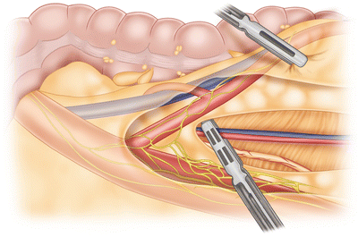
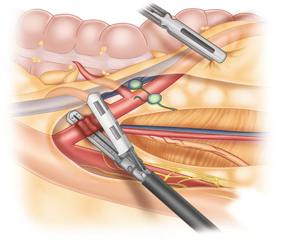
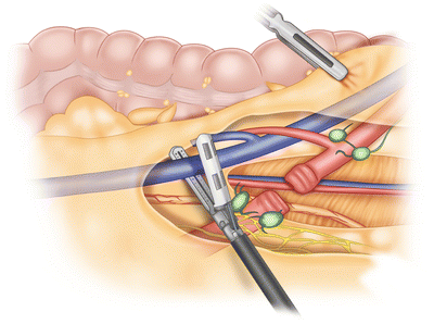
Fig. 6.5
Dissection of the inferior mesenteric artery, with preservation of the hypogastric nerves and left ureter and gonadal vessels
Fig. 6.6
The division of the inferior mesenteric artery
Fig. 6.7


Division of the inferior mesenteric vein
Related posts:


Stay updated, free articles. Join our Telegram channel
Full access? Get Clinical Tree
