Infections and Infertility

Chapter 33 Infections and Infertility


INTRODUCTION


The connection between a woman’s infertility and her husband’s history of gonorrhea was first reported in the late 1800s. This eventually led to the discovery of gonorrhea-related pelvic inflammatory disease (PID) as an important cause of infertility in young women. At the beginning of the 20th century, only 6% of women with a history of PID ever conceived. Surgery was the mainstay of therapy. With the advent of antibiotics, this picture has improved dramatically so that medical treatment options are available to help preserve fertility.


Infertility is one of the most disturbing consequences of infection of the female genital tract. Although the causes of infertility are multiple, 30% to 40% of cases are due to pelvic adhesive disease secondary to acute or chronic PID. Prevention, therefore, is the best healthcare policy in this field. Failing that, early detection and aggressive treatment are imperative.


Sexually transmitted diseases (STDs) and pelvic infections are associated with a great deal of socioeconomic and cultural impact in addition to substantial morbidity. In the United States alone, 12 million cases of STDs are acquired each year, with one fourth of these occurring in teenagers.


The specific pathogens involved are geographically dependent, in part as a reflection of available local healthcare and preventive services. For example, in India 15% of cases of secondary amenorrhea are related to tuberculosis. The World Health Organization estimates that, throughout the world, over 300 million episodes of curable STDs occur annually. In many parts of the developing world, a large percentage of the population is infected with one or more STDs, many of which have become antibiotic resistant.


In this chapter, we discuss the wide range of pathogens that cause impaired fertility in both men and women around the world. We also discuss the anatomic location and pathologic effects of the infections. With this anatomic approach, we hope to guide the clinician to appropriate differentials and diagnostic options as they evaluate their patients for infectious causes of infertility. Finally, we review the implications for fertility of acquired immunodeficiency syndrome (AIDS).


EPIDEMIOLOGY AND PATHOPHYSIOLOGY


Several pathogens associated with STDs are known to cause infertility (Table 33-1) or other syndromes (Table 33-2). In Western society the most common of these pathogens are Neisseria gonorrhoeae and Chlamydia trachomatis. However, other pathogens may influence fertility as well.


Table 33-1 Sexually Transmitted Diseases


Table 33-2 Syndromes Associated with Curable Sexually Transmitted Diseases


Cervical cancer


Gonorrhea


There are an estimated 600,000 new cases of gonorrhea in the United States alone each year. In the United States, 75% of reported cases of gonorrhea occur in men and women between ages 15 and 29. The highest rates of infection are found in women age 15 to 19 and men age 20 to 24.


The main site of infection in women is the endocervix. Patients can be asymptomatic, although more frequently symptoms occur, ranging from increased vaginal discharge to bleeding between periods, dyspareunia, and dysuria.


Neisseria gonorrhoeae is a gram-negative coccus that grows in pairs, hence the term diplococcus. N. gonorrhoeae mainly infects columnar or cuboidal cells and not the squamous epithelium of a postpubertal girl. After attaching to the mucosal epithelium, it penetrates through the epithelial cells and into the submucosal tissues. Neutrophils respond and cause sloughing of the epithelium, submucosal microabscesses, and formation of pus. If left untreated, neutrophils are eventually replaced by macrophages and lymphocytes.


Gonorrhea usually spreads along contiguous mucosal surfaces, producing a superficial infection. This is in contrast to staphylococci or streptococci, which tend to penetrate more deeply. Gonorrhea can then extend through the cervical os into the uterine cavity, producing endometritis. It can then spread to the fallopian tubes, causing an acute inflammatory reaction and, if not treated early enough, chronic inflammation, damage to the cilia, and ultimately scarring.


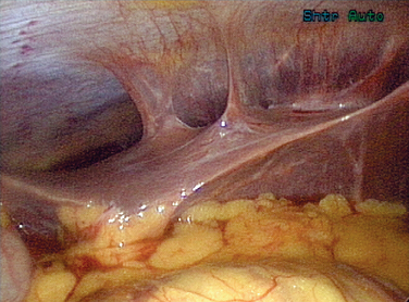
Gonorrhea can also pass beyond the fimbraie of the fallopian tubes and travel up the paracolic gutters and produce an infection around the liver. This is perihepatitis, or Fitz-Hugh–Curtis syndrome (Fig. 33-1). In unusual cases, gonorrhea can produce a disseminated disease with skin rash, infectious arthritis, and rarely endocarditis.


Treatment of a gonococcal infection depends on the location of the infection. If limited to the cervical region, the Centers for Disease Control and Prevention (CDC) recommends treatment with fluoroquinolones, ceftriaxone, or azithromycin (Table 33-3).


Table 33-3 Sexually Transmitted Disease Treatment Guidelines for Adults


Chancroid Azithromycin 1 g po × 1 or ceftriaxone 250 mg IM in a single dose or ciprofloxacin 500 mg po BID × 3 d or erythromycin base 500 mg po TID for 7 d
Herpes simplex virus — primary Acyclovir 400 mg po TID for 7–10 d or acyclovir 200 mg po five times a day for 7–10 d or famciclovir 250 mg po TID for 7–10 d or valacyclovir 1 g po BID for 7–10 d
Herpes simplex virus recurrences
Herpes simplex virus — suppression Acyclovir 400 mg PO BID or famciclovir 250 mg po BID or valacyclovir 500 mg QD or Valacyclovir 1 g po QD
Syphilis — primary and secondary Benzathine penicillin G 2.4 million units IM × 1
Early latent syphilis Benzathine penicillin G 2.4 million units IM × 1
Late latent syphilis Benzathine penicillin G 7.2 million units total, administered as three doses of 2.4 million units IM at 1-week intervals
Neurosyphilis Aqueous crystalline penicillin G 18–24 million units per day, administered as 3–4 million units q4h for 10–14 d
Chlamydia cervicitis Azithromycin 1 g po × 1 or doxycycline 100 mg po BID for 7 d
Uncomplicated gonococcal infections of the cervix, urethra, and rectum Ceftriaxone 125 mg IM × 1 or cefixime 400 mg po × 1 or ciprofloxacin* 500 mg po × 1 or ofloxacin* 400 mg po × 1 or levofloxacin* 250 mg po × 1 plus treatment for chlamydia if not ruled out
Bacterial vaginosis Metronidazole 500 mg po BID × 7 d or metronidazole gel 0.75% one full applicator intravaginally qd × 5 d or clindamycin cream 2% one full applicator intravaginally qhs × 7 d
Trichomoniasis Metronidazole 2 g po × 1 or tiridazole 2 g po × 1
Human papillomavirus — external genital warts Patient-applied: podofilox 0.5% solution or gel BID × 3 d then none for 4 d, repeated up to 4 cycles prn or imiquimod 5% cream qhs 3 × week for up to 16 wk
Human papillomavirus — external genital warts Provider-applied: cryotherapy q 1–2 wk or podophyllin resin 10%–25% in tincture of benzoin q wk or trichloroacetic acid q wk or surgical removal

* Should not be used in men who have sex with men or in those with a history of recent foreign travel or partners’ travel or infection acquired in California or Hawaii.1


Adapted from CDC Sexually Transmitted Diseases Treatment Guidelines 2006. MMWR 55:1–95, 2006.


Fluoroquinolone resistance in gonococci is becoming common in the Far East and Hawaii. Recently, it has begun emerging as a significant problem in some populations in the mainland United States. In men having sex with men, the prevalence of fluoroquinolone resistance has reached nearly 5%, prompting the CDC in 2004 to recommend that ofloxacin is not the drug of choice in this population.1 The resistance will continue to spread to other populations, and we will undoubtedly see further changes in the recommendations for the general population.


Treatment of gonococcal infections involving the upper genital tract is discussed under Pelvic Inflammatory Disease in this chapter.


Chlamydia


There are an estimated 3 million new cases of chlamydia in the United States each year. As with most sexually transmitted infections, these cases are more common among younger women. According to the CDC 2002 Sexually Transmitted Disease Surveillance Chlamydia Supplement, more than 5% of 15- to 24-year-old women screened at family planning clinics will be found to have chlamydia. For those young women age 15 to 24 entering the National Job Training Program in 2002, the chlamydia prevalence was approximately 10%. For adolescents entering juvenile detention facilities, the prevalence of chlamydia is higher than 16%.


The National Longitudinal Study of Adolescent Health (Wave III), using ligase chain reaction assays on first-void urine specimens, found chlamydia prevalence of 4% among young adults. The prevalence was greatest among minority women, including African Americans, Native Americans, and Latinos.2 The rate for young African American women was 6 times greater than for white women. From 1987 through 2002, the reported rate of chlamydial infections among all U.S. women increased nearly sixfold, from approximately 78 to 455 cases per 100,000 women. The etiology of this dramatic increase is likely multifactorial and includes increased screening, the use of the more sensitive nucleic acid amplification test (NAT), and better reporting, in addition to an increased infection rate.2


Chlamydia trachomatis is the pathologic agent of chlamydial cervical and upper genital tract infections, and certain serotypes also cause an ulcerative sexually transmitted infection termed lymphogranuloma venereum.


Chlamydia cervicitis is clinically indistinguishable from gonococcal cervicitis, with symptoms of vaginal discharge, intermenstrual bleeding, dysuria, and dyspareunia. In contrast to gonorrhea, which is usually symptomatic, chlamydia is often carried asymptomatically, and permanent damage to fertility can occur without notice. Chlamydia can ascend from the cervix to the endometrium and continue to progress into the fallopian tubes, producing PID.


Chlamydia can be detected using nucleic acid amplification testing of a cervical swab sample. NAT can also be used to detect chlamydia in first-void urine. This is an effective screening tool, with sensitivities comparable to those seen with cervical swabs and samples much easier to obtain.


Chlamydia can also be detected with serology. This is not useful for acute cervical or upper genital tract infections, but is the only way to diagnose certain serotypes if the diagnosis of lymphogranuloma venereum is considered. The serologic studies are also useful in determining prior exposure of patients presenting with infertility. By the time a patient presents with infertility, NAT results of a cervical specimen will not likely be positive for chlamydia. Positive serology for chlamydia in this setting helps to direct an infertility workup.


Cervical and upper genital tract chlamydial infections are treated with doxycycline, azithromycin, or erythromycin. The patient and all partners must be treated simultaneously. Treatment options are listed in Table 33-3.


Syphilis


Syphilis was referred to as the great pox in the 1500s and affected a vast number of people in Europe. Although the incidence of syphilis saw a major decline from 1970 through 1990 in the United States, there has been a rise recently in the numbers of cases, especially among homosexual and bisexual men. In 2002, there were 6862 cases of primary and secondary syphilis reported to the CDC. There were also 412 cases of congenital syphilis.


Syphilis is caused by the spirochete Treponema pallidum. This bacterium is extremely fragile, and the infection is almost exclusively transmitted by sexual contact with an infected person or from an infected pregnant woman to her unborn child. In rare circumstances, it can be passed through broken skin on other parts of the body.


The course of syphilis is described as being in one of four stages: primary, secondary, latent, and tertiary (or late) syphilis. Primary syphilis begins as a painless ulcer (i.e., chancre) that develops approximately 3 weeks after exposure on the exposed area. This lesion can be difficult to detect if it is on the vaginal wall or cervix.


Secondary syphilis presents on average at 6 weeks after exposure and is manifest by a diffuse skin rash that classically involves the palms and soles, verrucous-appearing genital lesions, or alopecia. This stage, if untreated, is followed by a long asymptomatic, or latent, stage.


Latent syphilis refers to untreated infections that are no longer symptomatic or contagious. In many untreated patients, no further signs or symptoms of the disease will ever occur.


Tertiary syphilis will eventually develop in approximately one third of untreated patients who have had secondary syphilis. Systemic spread of the bacteria can lead to damage of almost any other part of the body, most notably the brain, nervous system, bones, joints, heart, and eyes. This stage can last for decades, and can eventually lead to progressive neurologic dysfunction, cardiovascular disease, bone destruction, blindness, and death.


The effect of syphilis on pregnancy has been well documented. Syphilis clearly produces miscarriages, prematurity, stillbirths, neonatal deaths, and congenital disease in the infant. The effects of syphilis on fertility are not clear, but a study out of Senegal did find that women age 40 or older with past or active syphilis were significantly more likely to have no history of gestation than women without evidence of syphilis infection.3


The diagnosis of syphilis is made using one of two screening nontreponemal blood tests, known as the rapid plasma reagin (RPR) and the Venereal Disease Research Laboratory (VDRL) tests. These tests are measured in titers that can be followed serially to evaluate treatment response. Both the RPR and VDRL usually become positive within 2 weeks of developing the chancre. If positive, one of two available confirmatory treponemal tests is required, either the fluorescent treponemal antibody absorption (FTA-ABS) test or the microhemagglutination assay for Treponema pallidum (MHA-TP) test. These will remain positive lifelong regardless of treatment status.


Although syphilis may have devastating long-term consequences if not treated, this disease is relatively easy to treat once diagnosed. Penicillin is the mainstay of therapy (see Table 33-3).


Trichomonas


Trichomoniasis is a common type of vaginitis that is sexually transmitted and causes approximately 5 million genital infections in the United States each year. Although it is clearly a sexually transmitted infection, it is not a reportable disease; thus, data on its prevalence is an estimate and likely an underestimate.


Trichomoniasis is caused by the single-celled motile protozoa, Trichomonas vaginalis. Infected men rarely have symptoms. In women, trichomonads cause superficial infection of the vagina and ectocervix, resulting in a foamy yellow vaginal discharge, vulvovaginal irritation, dysuria, dyspareunia or, in many cases, no symptoms at all. Although trichomonads may be cultured from the endocervix, they do not cause a clinical cervicitis.


Several studies have found that women with T. vaginalis infections had an increased risk of tubal infertility (up to 1.9-fold), and that this risk increased sixfold with more than four episodes of T. vaginalis infection.46 These organisms have been found in the abdominal cavity of women with acute salpingitis, and there is speculation as to whether or not the motile trichomonads are able to carry bacteria or viruses to the upper genital tract.7


Diagnosis is made most frequently using a wet mount prep of the vaginal discharge and visualizing the motile protozoa. Culture of the trichomonad is the gold standard test but is not widely used in clinical practice. A rapid test utilizing an immunochromatographic strip that can be performed rapidly in a clinical office is being evaluated for clinical use, with reported sensitivities near that of culture.


Treatment is with metronidazole 2 grams orally in a single dose. Tinidazole has also been approved in the United States for treatment of trichomoniasis in a 2-gram single oral dose, but is more costly (see Table 33-3).


Human Papillomavirus


Human papillomavirus (HPV) is one of the most common causes of sexually transmitted infections in the world. According to the CDC, there are more cases of genital HPV infections in the United States than any other STD, with at least 20 million people currently infected. At least 5 million new cases of sexually transmitted HPV infections are reported annually.


Human papilloma viruses are small double-stranded circular DNA viruses. Although more than 100 different types of HPV exist, only 30 types are sexually transmitted. Many men and women with HPV infections have no symptoms. Symptomatic HPV infections manifest in women as genital warts, abnormal results on Papanicolau (Pap) smears, and both premalignant and malignant lesions of the cervix, vagina, vulva, and rectum.


There are high-risk and low-risk types of HPV. Low-risk HPV types 6 and 11 are the more common causes of visible external genital warts, although any of the types that infect humans can cause warts. These lesions are often asymptomatic, although they can be painful or pruritic. The lesions can grow large enough to interfere with coitus. Perianal warts can be seen even if a patient does not have anal sex, but intra-anal warts are only seen with receptive anal intercourse. Vaginal and cervical verrucous warts can also be seen.


Nucleic acid amplification testing is widely available for use with Pap smear specimens to determine the presence of high-risk HPV types. However, the external genital warts are not routinely typed. High-risk HPV types 16, 18, 31, 33, and 35 have all been associated with neoplasia, particularly of the cervix. For this reason, the presence of high-risk HPV types warrants further evaluation with colposcopy and cervical biopsies if indicated.


Although HPV has not been shown to directly affect a woman’s fertility, lesions large enough to affect coitus are certainly problematic. The treatments required for localized preneoplastic or neoplastic lesions (i.e., cryotherapy, loop electrosurgical excision; see Table 33-3) can rarely cause scarring or affect the production of cervical mucosa, both of which could hinder the passage of sperm, thus affecting fertility. Invasive cervical neoplasias can require hysterectomy or other significant pelvic surgery.


Treatment does not eradicate the virus; thus prevention becomes especially important for this sexually transmitted infection. Recent advances have shown that a three-dose HPV-16 vaccine prevented the development of persistent HPV-16 infection and HPV-16-related cervical neoplasia in college age women for the first 17 months after vaccination.8 This finding will certainly lead to further vaccine developments for all of the major high-risk HPV types.

Only gold members can continue reading. Log In or Register to continue

Stay updated, free articles. Join our Telegram channel

Aug 27, 2016 | Posted by in UROLOGY | Comments Off on Infections and Infertility

Full access? Get Clinical Tree

Get Clinical Tree app for offline access