The standard plain x-ray of abdomen are supine AP and erect AP views. If the patient cannot stand, a lateral decubitus view may be taken. In the plain film of the abdomen, identify and look for the following:
Screening:
1. The lateral border of psoas muscle may be hazy in retroperitoneal abnormalities.
2. Sub-diaphragmatic areas may contain gas due to rupture of an abdominal viscous (Figure 58). An air–fluid level can be appreciated in case of an abscess.

Figure 58 There is air under the diaphragm (arrows), suggesting a leak from an abdominal viscus. The patient had obstipation, vomiting, fever, and abdominal tenderness with rigidity
3. The diaphragm may also be raised due to absorption collapse of the lung or from push from the abdomen as with enlarged liver (liver abscess), or gross splenomegaly. It may also be seen with weakness of the diaphragmatic muscle (eventration of the diaphragm).

Figure 59 Eventration of the diaphragm on the left, with raised right hemidiaphragm in patient with gross hepatomegaly on the right top. On the bottom left to right, plain x-ray abdomen showing bilateral kidney stones with two kidney stones on CT in the middle and kidney and gall stones on the right
4. Renal outline may be enlarged in hydronephrosis.
5. Ureteric and urinary bladder areas may demonstrate calcific areas due to stones. Calcification is seen with stones in the gallbladder and urinary tract. Phleboliths, fecoliths, mesenteric lymph nodes, blood vessels, adrenal glands (TB), uterus (fibroids), liver, spleen, and pancreas may also demonstrate calcific areas.
7. Dilatation and fluid levels may be seen in obstruction of the large and small bowel (Figure 60). In toxic dilatation of the colon, as in ulcerative colitis, Crohn’s disease, or severe diarrhea, the transverse colonic diameter is more than 6 cm (Figure 68).

Figure 60 Multiple air–fluid levels on plain x-ray abdomen on the left. With dilated small bowel loops in the supine film are diagnostic of intestinal obstruction, in this patient small bowel obstruction. On the right, supine view with dilated bowel and fluid level shown by arrows
8. Spine and sacroiliac joints may also demonstrate abnormalities on a plain x-ray of the abdomen.

Figure 61 On the left, a barium meal shows a typical peptic ulcer crater on lateral view. On the right, a barium meal shows a thinned stomach with reduced capacity. This is seen in infiltrating cancer called linitis plastica
Malignancy in stomach is best diagnosed on endoscopy (see Figures 91, 103, and 107 in the endoscopy section. Also see page 97 in Volume I). Features suggesting on barium study are given as follows:
1. Irregular filling defect.
2. Irregular edges.
3. Location at the antrum or greater curvature is more commonly malignant.
4. The mucosal folds do not reach the edge of the ulcer in malignancy.
5. Linitis plastica (infiltrating adenocarcinoma in Figure 61).
Carcinoma of the colon may present with an obstructing mass with shouldering sign (overhanging edges) and irregular pattern (Figure 62 and page 121, Volume I). The diagnosis is established on colonoscopy and biopsy (Figure 101).

Figure 62 Barium enema showing classical mass and filling defect with shouldering (arrow) and over-hanging edges. An apple core appearance is also seen when there is annular constriction. This was carcinoma of the colon
Ulcerative colitis is characterized by uninterrupted inflammation and ulceration of the colon. These continuous lesions especially involve the rectum. The ulcers are usually shallow, with granularity of the wall, loss of haustrations, and hosepipe-like colon on barium study, in later stages (Figure 63). The colon may be narrowed and shortened. Pseudopolyps are swollen mucosa between areas of ulcerations that project into the lumen (Figure 63). Also see endoscopic features in Figure 99 in the endoscopy section and details on page 108, Volume I.

Figure 63 Barium enema in advanced ulcerative colitis with hosepipe-like large bowel and total loss of haustration in pancolitis on the left. In the middle, showing granularity (small black arrows), pseudopolyps (white arrows), and extensive ulceration (black large arrows). On the right, CT showing colon from the mid descending down to the rectum moderately thickened (inflamed) in a patient with chronic bloody diarrhea, characteristic of ulcerative colitis
Crohn’s disease is associated with transmural inflammation, fibrosis, narrowing, producing skip lesions (normal intervening bowel). Sinus tracts, micro-perforations, and fistula may be seen. It classically involves the terminal ileum, but may involve any part of the gut. About 20 percent have disease limited to the colon and one-third have perianal disease (skin tags, fissure, abscess, and fistula). Fatigue, chronic diarrhea, crampy abdominal pain, weight loss, and fever, with or without overt bleeding (uncommon), are the hallmarks of Crohn’s disease. The major signs are stricture and mucosal lesions. Strictures are of variable length and are responsible for the string sign (Figure 64). Fine ulcerations produce a cobblestone appearance. Rose thorn ulcers are characteristic. Thickening of the bowel wall and inflammation produce displacement of the bowel. Malabsorption may be present. Extraintestinal manifestations include eye, skin, joint involvement. Also see endoscopic features in Figure 88 in the endoscopy section and page 108, Volume I for details.
Lymph node biopsy and MBTB PCR may be necessary to make a definitive diagnosis.

Figure 64 CT abdomen with IV and oral contrast, showing terminal ileal concentric mural thickening and narrowing (arrows) and multiple small abdominal lymph nodes. Differential diagnosis could be Crohn’s disease, tuberculosis terminal ileitis, and lymphoma
Esophageal varices are best seen on endoscopy (Figure 87) when they can be counted and graded according to the size in relation to the lumen of the esophagus. They are characteristically seen in chronic liver disease with portal hypertension. Any other cause is rare. A barium esophagogram with varices is shown in Figure 65 and page 66, Volume I for details.
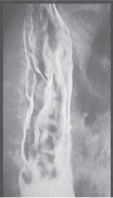
Figure 65 Barium swallow showing variable worm-like filling defects classical for esophageal varices. Esophageal varices are best evaluated by endoscopy where they can be numbered and classified according to the size and position (see Figure 101). Signs of threatening bleeding can be identified and therapeutic intervention (band ligation or sclerotherapy) offered at the same time
Short bowel syndrome usually occurs when less than 120 cm of small bowel remains functional. It may be associated with GI operations (cancer, mesenteric vascular disease, inflammatory bowel disease, bariatric surgery, strangulated hernia, bowel injury, volvulus, and radiation or may be congenital) (Figure 66).

Figure 66 Congenital short bowel syndrome. R = rectum. A = ascending colon. D = descending colon. Short bowel as seen by upper GI contrast study with rapid transit
Complications and problems associated with short bowel syndrome include:
• Watery diarrhea due to loss of small intestinal surface and bacterial over growth and electrolyte disturbance with metabolic acidosis.
• May require parenteral nutrition, which may be associated with sepsis, liver disease, gallbladder disease, and nephrolithiasis.
•
Related posts:


Stay updated, free articles. Join our Telegram channel

Full access? Get Clinical Tree


