Fig. 14.1
Fluoroscopic image of a “rendezvous” transgastric assisted ERCP. Arrows denote the guide wire being passed from a cholangiogram catheter, down the common bile duct, and into the duodenoscope
Percutaneous. Percutaneous management of choledocholithiasis is traditionally performed by interventional radiology via a transhepatic approach. This approach is indicated for patients in whom ERCP has failed and/or are poor operative candidates. To perform this procedure, confirmation of choledocholithiasis is performed via a percutaneous transhepatic cholangiogram. Once confirmed an external biliary catheter is placed. The tract is then dilated over 2–6 weeks by placement of progressively larger catheters until a 16 French size is reached. Once dilation is complete, the CBD stones are then extracted. The success rate for this procedure is reported between 75 and 96 %. The associated morbidity and mortality rate of this procedure are 10 % and 1 %, respectively. Major complications include bleeding, duct injury, bile leakage, and cholangitis [30, 31].
14.3.2 Postoperative Patients
For patients who present with symptomatic biliary colic or cholecystitis, management is the same. Laparoscopic cholecystectomy should be performed with timing based on acuity of presentation, clinical exam, and patient preference. For the patient status post-RYGB or DS, preoperative or intraoperative evaluation of the CBD is recommended. Routine inspection of limbs and potential spaces for occult internal hernia should also be considered at the time of cholecystectomy.
For patients with choledocholithiasis, management options remain the same in patients who have undergone gastric specific operations such as SG, AGB, or VBG . For patients who have undergone AGB, the band may require deflation prior to ERCP to accommodate the endoscope. Previously described surgical management options remain the same for patients who have undergone RYGB or DS. Endoscopic management, however, is technically challenging secondary to anatomic constraints. Accessing the duodenum is difficult via standard endoscopic approaches secondary to the length of the alimentary limb. As such, novel techniques have been developed and employed to facilitate duct clearance via ERCP. The remainder of this chapter focuses on endoscopic CBD management options specific to this patient population.
14.3.2.1 Management of Choledocholithiasis in RYGB and DS Patients
Surgically Assisted ERCP
Transgastric Access
Surgically assisted ERCP, generally performed laparoscopically, involves the creation of a transluminal access point to permit subsequent endoscopic access to the ampulla. For RYGB patients, the remnant stomach is the preferred location because it is generally easily accessed, is defunctionalized (creating a low risk of significant postoperative leak ), and places the side-viewing duodenoscope in a standard position for ERCP. This is the most widely reported method in the literature [32–45].
Access to the body of the remnant stomach varies by author with some preferring more proximal access and some preferring more distal access [32, 34, 37, 38, 45]. It is important, however, not to place access too close to the pylorus as this can make endoscopic navigation and endoscope stability more difficult. Several methods of access have been described, including the creation of a gastrotomy with direct placement of an ethylene oxide gas-sterilized endoscope through the abdominal wall. Alternatively, a 15 mm laparoscopic trocar can be placed through the abdominal wall and into the remnant gastrotomy [37, 45]. A non-sterile endoscope can subsequently be passed into the stomach through a sterile ultrasound probe cover placed onto the trocar itself.
Standard ERCP methods are then used to clear the common bile duct. One notable difference is the endoscopist’s position; because the patient is supine for laparoscopy, the endoscopist’s position is reversed making cannulation more challenging. In this circumstance a “rendezvous” technique, as described earlier, can greatly facilitate ERCP (Fig. 14.1) [46]. At the conclusion of the procedure the gastrotomy can be closed with sutures, resected with a stapler, or converted into a gastrostomy. Gastrostomy formation permits repeat transabdominal access to the remnant stomach without the need for additional surgery. This is the preferred method when additional endoscopic biliary interventions are anticipated.
Transgastric ERCP has a high technical success rate. A recent literature review of 113 patients undergoing transgastric ERCP noted technical success in 112 (98.8 %) with a complication rate (7.2) similar to ERCP alone [45]. The sole failure was due to an impacted stone in the ampulla [34]. While there is added morbidity (3.6 %) from laparoscopic access to the remnant stomach ( leak, wound infection), many of these patients require an additional surgical procedure (most notably cholecystectomy) that can be conducted simultaneously under a single anesthetic setting. Lysis of adhesions and reduction of internal hernias are also common interventions performed [40, 47]. While this method is quite beneficial, it can require significant coordination between the surgeon, the endoscopist, and the ancillary staff. Adequate room setup to optimize both laparoscopic and endoscopic interventions is important (Fig. 14.2).
Fig. 14.2
Recommended room setup for transgastric assisted ERCP (E = endoscopist, S = surgeon)
Transjejunal Access
In lieu of a gastrotomy, the small bowel can be accessed in the biliopancreatic limb in both RYGB and DS patients to permit retrograde access to the ampulla [47, 48]. For DS patients, this is the preferred route of access for endoscopic CBD access. Access to the BP limb means that endoscopic visualization of the ampulla will be retrograde, and ERCP can be conducted with either a side-viewing or forward-viewing scope. At the conclusion of the procedure, the enterotomy can be closed with sutures, turned into a stapled entero-enterostomy, or converted into a large-caliber jejunostomy. There are only case reports of this technique in the literature, which have all been successful [47, 48]. No case series are described to report the technical success rate or complications.
Per-Oral ERCP
In patients with RYGB anatomy, direct access to the ampulla with a duodenoscope is an extreme technical challenge. This is due to a combination of factors including the distance that needs to be traversed to reach the ampulla due to proximal gastric division (40 cm esophagus, 5–10 cm gastric pouch, 100–150 cm alimentary limb, 50–80 cm biliopancreatic limb) as well as the fact that safely navigating the small bowel with side-viewing duodenoscope is challenging even over short distances.
To address these issues, multiple alternative means of accessing the ampulla with forward-viewing endoscopes have been described. Even when successful in reaching the ampulla, such methods of bile duct clearance are hampered by several technical factors; the ampulla is approached from the distal duodenum at an upward angle, there is no channel elevator to facilitate cannulation, and traditional accessories for ERCP (sphincterotomes, balloons, stents) may be too short for the scope or angled inappropriately to permit easy bile duct access or be too wide to fit through the accessory channel. Some of these limitations, however, can be overcome with sufficient technical prowess. The literature supports several methods of per-oral access to the biliary tree in bariatric patients.
ERCP with a Standard Duodenoscope
As noted above, patients with gastric specific operations can undergo ERCP via standard methods. For RYGB patients, a 33 % technical success rate with this method has been reported [49]. Because of this low success rate, this method is rarely utilized and should be considered only in patients with very short bypass limbs and no other viable options for clearing the duct.
ERCP via Push Enteroscopy
ERCP utilizing a push enteroscope or a pediatric colonoscope has also been described. Navigation with these types of scopes is time consuming and requires frequent loop reduction maneuvers, external pressure to prevent loop formation, and changing patient position. Most series describing this method unfortunately include both bariatric and nonbariatric patients [50, 51]. When considering just bariatric patients, the reported success rate is only 45 % [51].
ERCP via Balloon-Assisted Enteroscopy
Balloon-assisted endoscopic methods utilize high-volume, low-pressure balloons and overtubes to permit small bowel stabilization on the endoscope. Single-balloon techniques have one balloon attached to an overtube while double-balloon methods have two balloons (one on an overtube, one on the endoscope insertion tube). These methods have a proven track record of deep intubation of the small bowel for a variety of endoscopic interventions. They have gained favor in the bariatric population for their ability to navigate the long limbs of the RYGB anatomy to access the bile ducts without the need for surgery. Technical success rates in reaching the ampulla with balloon-assisted methods range from 55 to 100 % with an 83–100 % chance of biliary orifice cannulation once there [52–60]. Therapeutic success of 77–100 % is reported if the biliary orifice is able to be cannulated [52–60]. When taken together, these methods are overall of low risk, but have a higher technical failure rate than the surgical and hybrid methods described above. They are still not universally available and require a skilled endoscopist.
ERCP via Spiral Enteroscopy
Percutaneous Access to the Biliary Tree
Any method of percutaneous access to the gastrointestinal tract can theoretically be used to access the biliary tree. This includes percutaneous gastrostomy tubes, jejunostomy tubes, cholecystostomy tubes, and transhepatic tubes. Following track maturation and upsizing, flexible endoscopes can be used to access the biliary tract (directly or indirectly) to manage disease processes.
Novel Therapies on the Horizon
Gastro-Gastric Fistula Formation
The exploitation of a preexisting gastro-gastric fistula (GGF) to permit per-oral endoscopic interventions in the excluded portions of the RYGB foregut has been described. Due to the close proximity of the gastric pouch and the proximal remnant stomach, some authors have proposed the intentional endoscopic creation of a GGF as a means of accessing the excluded stomach. Under fluoroscopic or endoscopic ultrasound guidance, needle access is obtained from the pouch into the remnant stomach and a guide wire is passed. Over-the-wire balloon dilation and/or enteral stent placement then follows. Endoscopic interventions (include ERC) can then be conducted via this GGF tract. The advantage of this method is that a standard duodenoscope can be used to reach the ampulla without the need for surgical incisions or a gastrostomy and the full array of ERCP accessories are at the disposal of the endoscopist. Obvious disadvantages include the creation of an acute perforation of the pouch and remnant stomach (with the inherent risk of leak), the possibility of long GGF persistence following the intervention, and stent removal (with the risks of marginal ulcer formation, weight regain, and recrudescence of diabetes due to the presence of food within the stomach and duodenum). Novel methods of perforation and fistula closure, including over-the-scope clips and endoscopic suturing devices, may negate some of the risks of intentional GGF creation and may ultimately make this method a viable endoscopic option.
Percutaneous Cholelithectomy
Percutaneous transhepatic access and cholangioscopy have an established track record for CBD clearance in the RYGB patient (as described above). More recently, it has been recognized that in some patients with a gallbladder in situ a percutaneous cholecystostomy tube can both decompress the acutely obstructed CBD and permit an access route for complete removal of all gallstones. Following cholecystostomy tube placement, wire access is obtained through the CBD via the cystic duct. The cystic duct can be dilated to permit larger instruments to be passed into the CBD to clear the duct and perform a sphincteroplasty. Subsequently, standard choledochoscopic methods can be used to remove gallstones from the gall bladder itself, negating the need for subsequent cholecystectomy. This method requires several favorable factors including the presence of a gallbladder, favorable cystic duct anatomy, and size-appropriate gallstones. Multiple interventions are required but this method may be beneficial in poor operative candidates.
Device-Assisted Endoscopy
Several investigators have described case reports of transprosthetic endoscopy to permit immediate access and therapy via the remnant stomach. Wire access to the remnant stomach is first obtained via double balloon-assisted PEG method or via trans-abdominal ultrasound [63]. The tract is subsequently dilated to 20 mm, and T-anchors and/or stents are placed to secure the tract (Fig. 14.3). ERCP via the stent is then performed using a standard duodenoscope and accessories. The stent is then removed and converted to a gastrostomy tube at the conclusion of the procedure.
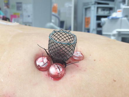
Fig. 14.3
Gastrointestinal T anchors (white buttons) and percutaneous fully covered stent placement into remnant stomach to permit subsequent transprosthetic-ERCP
Endoscopic Ultrasound-Assisted Biliary Access
Utilizing EUS methods, Weilert and colleagues gained wire access from the gastric pouch into the left lateral segment biliary tree. A wire was able to be advanced across the ampulla in six consecutive patients which permitted successful biliary intervention in all six, four using an antegrade transgastric, transhepatic method and two using a retrograde DBE technique that was able to successfully rendezvous with their antegrade-placed wire [64].
14.4 Choosing the Route of CBD Clearance
Because of the variety of options available, no single method of CBD access following RYGB is considered the gold standard. Table 14.1 provides an overview of the five major methods of bile duct clearance. The bariatric surgeon, in conjunction with the endoscopist, must determine which method has the highest chance of success and the lowest risk of morbidity for the individual patient. Factors to consider include the following:
Table 14.1
Overview of the five main methods of CBD clearance in RYGB and DS patients
Method | Pros | Cons | Pitfalls | Ideal patient |
|---|---|---|---|---|
Double-balloon ERCP | • Non-operative | • Repeat intervention difficult • Technically challenging • High expertise required • Time consuming | • Long limb lengths • Internal hernia • DS patient | • Known limb length
Related posts: Nutritional Complications and Emergencies
Medical Malpractice in the Twenty-First Century Nutritional Complications and Emergencies
Medical Malpractice in the Twenty-First Century
 Enteric Leaks after Gastric Bypass: Prevention and Management Enteric Leaks after Gastric Bypass: Prevention and Management
 Gastrointestinal Obstruction in the Bypass Patient Gastrointestinal Obstruction in the Bypass Patient
 Internal Hernias: Prevention, Diagnosis, and Management Internal Hernias: Prevention, Diagnosis, and Management
 Enteric Leaks After Sleeve Gastrectomy: Prevention and Management Enteric Leaks After Sleeve Gastrectomy: Prevention and Management
Stay updated, free articles. Join our Telegram channel
Full access? Get Clinical Tree
 Get Clinical Tree app for offline access

|