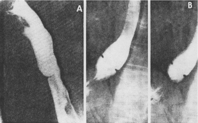
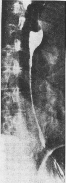
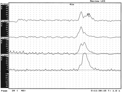
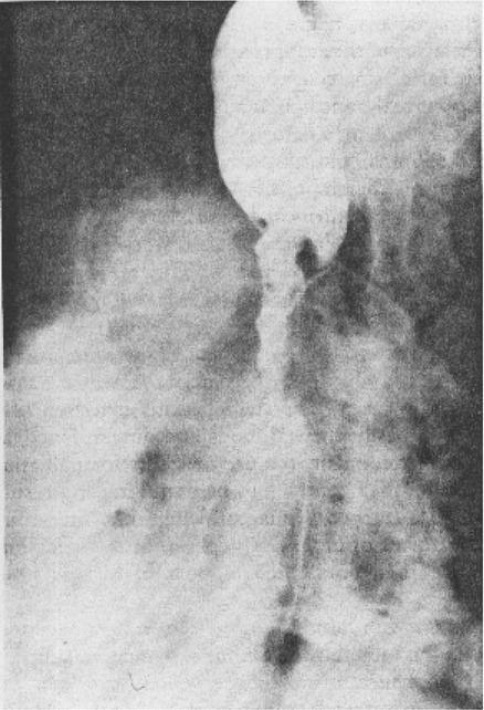
Esophageal Disorders


Esophageal Disorders


MICHAEL E. GROHER


ROLE OF THE SPEECH-LANGUAGE PATHOLOGIST


It is not the role of the speech-language pathologist (SLP) to diagnose and treat dysphagia of esophageal origin. In most cases this is done by the gastroenterologist. However, because of the interdependency of the oral, pharyngeal, and esophageal stages of swallowing it is important for the speech pathologist to be aware of how esophageal-based dysphagia might affect other compartments involved in swallowing. It also is important for the SLP to be aware of the types of swallowing problems that should be referred to the gastroenterologist and what types of treatments they might recommend. Sometimes treatments (e.g., medications) might affect other parts of the swallowing chain, and patients might need certain instructions about taking their medications reinforced by other health care providers. One example is patients who take medication to control their gastroesophageal reflux disease (GERD). On careful questioning, it is revealed that they only take their medicine when they perceive they might be eating a meal that would cause an increase in reflux events. In most cases patients should be taking their medication on a daily basis, so reinforcing this point or reviewing proper dietary restrictions may be needed to avoid an increase in reflux events. In these circumstances knowledge of other professional roles and methods of evaluation and treatment can be beneficial to improve patient compliance and answer any questions patients might have about their dysphagia.


It is becoming more common for the SLP to include a screening of the esophagus in patients who are able to stand during the modified barium swallow study. The screenings are useful because they might detect a disorder that helps explain the patient’s oropharyngeal symptoms. It is the role of the radiologist—rather than the SLP—to document and comment on these abnormalities. If further tests are warranted the SLP might include them in his or her progress note but with the approval of the radiologist.


STRUCTURAL DISORDERS


Esophageal dysphagia can be caused by a change in the ability of the esophagus to fully open during swallowing, resulting in a blockage of bolus passage. A change in the structure of the esophagus may be caused by a luminal stenosis or narrowing or by a luminal deformity such as another structure compressing it, thereby limiting its ability to open.


Esophageal Stenosis


Esophageal stenosis is conceptually the easiest mechanism of dysphagia to understand. When the lumen narrows, solid food may be too large to pass through it. Esophageal stenosis typically causes dysphagia for solid food. In addition, the type of solid material ingested often is important for symptom production. For instance, dysphagia of esophageal origin is more likely when solids are tough or fibrous. Softer, more easily chewed foods are much less likely to cause symptoms of esophageal dysphagia. An exception to this tough food–soft food dichotomy is that many patients also have particular trouble with soft, absorbent foods such as bread or pasta, which swell when mixed with saliva during mastication. Once bolus impaction occurs, the patient may have difficulty with liquids as well, obscuring the characteristic solids-only nature of esophageal stenosis. However, a careful history usually reveals that liquid dysphagia begins with ingestion of solids (see differential diagnosis later in this chapter).


Clinicians often rely too much on the patient’s sensation of where food is sticking. The common wisdom that patients accurately localize symptoms to the site of obstruction is often inaccurate. In fact, approximately one third of patients with obstructing lesions of the distal esophagus point to the neck as the site of obstruction.1 Conversely, one third of patients with dysphagia localized to the pharynx have an isolated abnormality of the esophagus.2


It is surprising how well some patients fare despite dramatic stenosis. Based on radiographic observations in patients with Schatzki’s rings, it is often stated that patients with luminal diameters of more than 18 to 20 mm are never symptomatic, whereas those with diameters less than 10 to 12 mm are always symptomatic.3 When the radiologist examines the esophagus for suspected stenosis, a radiopaque pill that is 13 mm in diameter is used to detect a stenosis. Between these extremes (20 and 10 mm), symptoms vary both in frequency and severity depending on the presence of associated motor dysfunction and the choice and preparation of food. Stenosis is treated by opening or removing the narrowed segment, depending on the specific cause. This is usually accomplished with Maloney (bougie) dilators or with balloon dilatation.


Common intrinsic structural abnormalities that narrow the esophagus include mucosal rings, benign strictures, and malignant tumors.


Rings and Webs


The esophagus may be narrowed by a band of tissue composed of mucosa and submucosa. By tradition, this type of lesion is called a ring when located at the esophagogastric junction and a web when located elsewhere in the esophagus or hypopharynx.


Although classically described in patients with iron-deficiency anemia (sideropenic dysphagia), the majority of esophageal webs are not associated with iron deficiency. Webs of the pharyngoesophageal segment or cervical esophagus are frequently asymmetric, most often impinging on the esophageal lumen from the anterior wall (Figure 7-1, A). A suspected web at the cervical level also can be seen in Video 7-1 on the Evolve site accompanying this text.


Schatzki’s rings are the most common bandlike constriction of the esophagus. This lesion is typically symmetric and located at the esophagogastric junction (Figure 7-1, B). Asymptomatic Schatzki’s rings are detected in approximately 10% of the population.4 The ring is always noted in the presence of a hiatal hernia. However, most hiatal hernias are not associated with Schatzki’s rings. The etiology of a Schatzki’s ring is unknown. Because they are rarely seen in childhood and generally are first noticed in middle age, it is unlikely that a Schatzki’s ring represents a congenital abnormality. A video image of a Schatzki’s ring can be seen in Video 7-2 as a narrowing of the esophageal lumen in the distal esophagus during swallowing of a thicker bolus.


Webs and rings typically produce dysphagia for solids only. Patients often report that symptoms are intermittent and less likely if they select their food wisely and chew carefully (see the section on differential diagnosis). Conversely, symptoms are more likely if the patient eats away from home or carries on a conversation while eating; in these situations the choice of food is more restricted and proper preparation of food before swallowing is more difficult. The patient often must end the episode by inducing regurgitation. Once the food is dislodged, the patient often can return to the meal without further difficulty.


The extent to which attention to the mechanics of cutting and chewing controls symptoms is limited. When the lumen is severely compromised, the patient may find it impossible to maintain the level of attention required to remain symptom free without avoiding solids entirely. The patient may describe symptoms without any apparent progression in frequency or severity that date back for many years. Progression, when it does occur, usually is slow.


Radiographically, rings and webs appear as thin (2 to 4 mm) bands that form shelflike constrictions anywhere along the esophagus. Although radiologists occasionally refer to thicker lesions as webs or rings, these are probably short strictures or abnormal muscular contractions.


Treatment of webs or rings involves dilatation or rupture of the ring by any one of a variety of esophageal dilator systems. The ring is thin, nonfibrotic, and easy to dilate. Complete, or nearly complete, symptomatic relief can be anticipated. Failure to respond is unusual. Dilatation may provide permanent relief, although a large proportion of patients need periodic redilatation at variable intervals.5


Benign Stricture


Strictures are rarely seen in children, although congenital strictures do occur. The majority of benign esophageal strictures are acquired in adulthood as a consequence of esophagitis. In a circular structure such as the esophagus, edema resulting from ongoing inflammation and fibrosis as part of the healing process occurs at the expense of luminal diameter.


As with webs and rings, dysphagia is generally for solids only. However, dysphagia is progressive, with episodes becoming more frequent and severe over a period of months or years. As luminal narrowing increases, the patient reports trouble swallowing food that previously caused no difficulty. Stenosis occasionally can become so severe that even thick liquids cause dysphagia. Even then, however, dysphagia is virtually always greater for solids than liquids.


Benign strictures are usually secondary to reflux-induced esophagitis, although most patients with gastroesophageal reflux disease (GERD) do not have esophagitis. Esophagitis refers to inflammation of the lining of the esophagus. Esophagitis may vary in severity from microscopic inflammation to mucosal edema to erosion, ulcerations, and stricture. Patients usually describe a history of heartburn or chest pain and may report the frequent use of antacids or other ulcer medications. In some patients the esophagus appears to be relatively insensitive to acid exposure. These individuals never experience significant reflux symptoms despite severe esophagitis and progression to stricture formation. Although most benign esophageal strictures are a result of reflux esophagitis, any source of esophagitis can cause stricture formation (Box 7-1).


Drug-induced or pill esophagitis can be seen in young or elderly patients. Typically, commonly administered medications that are larger in size (tetracycline, potassium, quinidine) become lodged at the level of the aortic arch and dissolve, causing inflammation and stricture. Symptoms of chest pain, odynophagia, heartburn, and dysphagia may be present, usually more acutely in younger patients.6


Radiographically, a benign stricture is seen as a narrowed segment of esophageal lumen that may range from 1 cm to many centimeters long (Figure 7-2). The stricture usually is smooth and gradually tapering, with a symmetric lumen that follows the anticipated path of the normal esophagus. Ongoing inflammation may produce an eroded appearance along its course. A lateral and anteroposterior (AP) video image of a midesophageal stricture caused by GERD can be seen in Video 7-3 on the Evolve site. In the AP view barium flow is interrupted, with barium building up above the stricture. The lateral view shows a long, tapered appearance of a stricture in the esophagus.


Proper management requires both treatment of the underlying inflammation and dilation of the stricture. Treatment of the cause of esophagitis requires accurate diagnosis. Although reflux is the most common cause of esophagitis, other possibilities must be considered, especially in patients with atypical histories, an unusual distribution of inflammation, or failure to respond to reflux treatment.


Dilatation often can be performed by using the same techniques available for a Schatzki’s ring. However, the stricture may be relatively unyielding and require stiffer dilator systems. Effective dilatation usually improves symptoms, although edema from inflammation may result in less-complete symptomatic relief than with a Schatzki’s ring and in relatively rapid restenosis. Frequent dilatations are more often required in benign strictures than with Schatzki’s rings. Even when ongoing inflammation completely ceases, periodic dilatation may be necessary, especially during the first year after initial treatment, when maturation of the fibrotic reaction continues at the expense of luminal diameter.


Malignant Stricture


Although benign tumors may arise from the esophagus, the majority of clinically significant tumors of the esophagus are malignant. In the past, most esophageal malignancies were squamous cell carcinomas, although recent studies suggest a dramatic increase in adenocarcinoma of the distal esophagus. Most esophageal adenocarcinomas appear to arise from Barrett’s esophagus, a premalignant condition in which columnar cells replace the usual squamous epithelium covering the lower end of the esophagus as a result of severe GER.


As with other types of stenotic lesions, dysphagia initially occurs for solids only. However, it usually progresses rapidly, with dysphagia for soft foods and even liquids developing within a few months of the onset of symptoms.


Radiographically, esophageal malignancies appear as strictures of variable length. By the time of presentation, the cancerous tumor or area is usually many centimeters long and involves the entire circumference of the esophageal lumen, producing a stricture. The typical malignant stricture is characterized by its shelflike proximal margins and irregular channel, which may diverge substantially from the anticipated course of the esophageal lumen (Figure 7-3). However, not all esophageal cancers are obviously malignant on barium radiography, and occasional malignant-looking strictures may be benign.6 For this reason, endoscopy with tissue sampling by biopsy with or without cytologic brushing is essential to differentiate benign and malignant strictures.


Curative treatment is primarily surgical, although apparent cures by radiotherapy have been reported. Unfortunately, by the time symptoms develop, the cancer is usually very advanced and incurable. The overall 5-year survival rate for esophageal cancer is only approximately 5%.1 Even among those in whom resection for apparent cure is possible, the 5-year survival rate is only approximately 15%.7 Recent studies suggested that the 5-year survival rate could be doubled with a combination of preoperative radiotherapy and chemotherapy.8 Surprisingly, almost 25% of patients had no evidence of cancer by gross or histologic examination. Among these patients, survival was improved fourfold over rates reported for surgery alone and twofold over those with evidence of residual tumor at surgical resection.


For patients in whom curative resection is not possible, palliative resection often is still feasible and provides good symptomatic relief. In the past, a high perioperative mortality rate of approximately 29% combined with the infrequency of cure made surgery unattractive.9 However, with better nutrition provided by preoperative and perioperative hyperalimentation the risk of palliative surgery has declined.10


Alternative approaches include dilatation, tumor ablation (thermal treatment to destroy tumor obstructing the esophagus) by laser or bipolar electrocautery, and stent placement. Each of these approaches is directed at opening the esophageal lumen to permit eating, in recognition that the major cause of early death in patients with esophageal cancer is malnutrition and aspiration pneumonia.


Dilatation generally provides limited and short-lived relief but is useful in preparing for other forms of therapy. The choice between other modalities depends on specific features of the tumor and local technical expertise and resources. Endoscopic laser therapy and bipolar electrocautery can be used to destroy tumor tissue that blocks the esophageal lumen; this may provide a number of months of relief, allowing continuing oral intake. Treatment can be repeated if obstruction recurs.


An esophageal stent is a tube with a large channel that can be placed through the strictured segment to maintain luminal patency. The stent permits ingestion of a modified diet, concentrating on soft, easily chewed foods and purees. The use of stents for palliation has decreased dramatically since the development of thermal methods of treatment. However, stents continue to be useful in certain situations, especially in the presence of a tracheoesophageal fistula that often complicates the natural history or treatment of esophageal cancer. In this situation, a properly placed stent can maintain the esophageal lumen while covering the opening to the airway. The recent introduction of expandable metal stents has made insertion easier and provides a larger internal luminal diameter, allowing patients to eat a less-restrictive diet.


Although endoscopic treatment with laser, bipolar electrocautery, or stent placement may be highly successful in reestablishing luminal patency, a substantial proportion of patients with esophageal cancer have poor appetites and are unable to gain weight. The early use of endoscopically placed or fluoroscopically guided gastrostomies should be considered in patients who do not eat once the lumen is reestablished or who are scheduled to undergo chemotherapy or radiotherapy, treatments that may produce or exacerbate anorexia. (See Chapter 8 for a discussion of transhiatal esophagectomy.)


Luminal Deformities


Extrinsic Compression


Some degree of luminal deformity caused by extrinsic compression by normal mediastinal structures (the aortic knob, the left mainstem bronchus, and the left atrium of the heart) is normally seen on barium studies and rarely, if ever, causes symptoms. More pronounced compression can occur with mediastinal conditions, such as aortic aneurysm, cardiomegaly, congenital abnormalities of the large mediastinal arteries (e.g., aberrant subclavian artery), enlarged mediastinal lymph nodes, and lung cancer. Video 7-4 on the Evolve site shows a patient with cardiomegaly and reduced bolus flow. The enlarged heart is seen as a large shadow (note heartbeat) in the middle of the video image. The elasticity of the contralateral esophageal wall usually tends to minimize symptoms until compression is far advanced. Dilatation is usually ineffective because the force of dilatation is absorbed by the elastic, uninvolved wall. Effective treatment, when necessary, requires shrinking or removing the mass producing the compression. Unfortunately, this is often not practical in patients in whom compression produces significant symptoms.


Esophageal Diverticulum


Compared with diverticula of the hypopharynx, esophageal diverticula are rare and usually asymptomatic, even when they are relatively large. When symptoms do occur, they include dysphagia for liquids and solids, regurgitation of previously swallowed food back into the mouth, or both. Regurgitation without dysphagia is not uncommon.


Most often, esophageal diverticula are a consequence of obstruction distal to the region of bolus collection. Increased pressure in the esophagus results in bulging at a point of relative weakness. Less commonly, diverticula can result from periesophageal inflammation, which causes traction on the esophageal wall (traction diverticulum). Although most traction diverticula occur in the midesophagus, most midesophageal diverticula, like their distal esophageal counterparts, are caused by pulsion. Video 7-5 shows a diverticulum that fills and causes a momentary obstruction to bolus flow.


Treatment of pulsion-type diverticula is necessary only if a diverticulum is symptomatic. Because they frequently give rise to motor or structural disorders, it is important to look for pulsion-type abnormalities as causes for the development of the diverticulum. It may be difficult to distinguish between the underlying obstructive disorder and the diverticulum as a cause of symptoms. It is appropriate to attempt to treat the underlying cause of increased pressure with dilatation in the case of structural obstruction or with drugs for dysmotility. In some patients symptoms initially believed to be a consequence of the diverticulum improve significantly or resolve entirely with such conservative therapy.


Surgical removal of the diverticulum is required if medical management fails. Surgery limited to diverticulectomy, however, is associated with a high incidence of early anastomotic leakage or late recurrence, probably because it fails to deal with the underlying cause of increased intraesophageal pressure and creates an area of relative esophageal wall weakness. Therefore diverticulectomy should be combined with treatment of the underlying disorder—motor (with a surgical myotomy) or structural (with dilatation).


ESOPHAGEAL MOTILITY DISORDERS


An orderly, progressive peristaltic wave is not uniformly present after every swallow, even in individuals without dysphagia. The dividing line between normal and pathologic degrees of dysmotility is poorly defined. The incidence of abnormal contractions changes with bolus type (it is increased with dry swallows), although not with age.


A variety of schemes have been proposed to classify esophageal dysmotility. In abnormalities of esophageal peristalsis, contraction amplitude may be too high or low, contraction duration prolonged, or the orderly progression of the contractile wave down the length of the esophagus uncoordinated. In abnormalities of lower esophageal sphincter (LES) function, the pressure may be too high or too low and relaxation may be incomplete. Finally, the esophageal body and LES can misbehave separately or together. The individual characteristics of commonly described motility disorders are not necessarily unique. In many ways the separation between entities is somewhat arbitrary.


Disorders of Peristalsis


Motor dysfunction of the body of the esophagus may cause symptoms of dysphagia, chest pain, or regurgitation. Dysphagia is usually for liquids as well as solids, although not necessarily in equal measure. Chest pain may mimic that of cardiac disease and cause considerable concern on the part of both patient and physician. Although pain initiated or exacerbated by swallowing strongly implicates the esophagus as the site of origin, a clear relation to eating is often absent. Similarly, the presence of other symptoms implicating the swallowing mechanism supports the possibility that the esophagus is the cause of chest pain. However, cardiac disease is sufficiently common, especially in older patients, to justify a cardiology evaluation.


Diffuse Esophageal Spasm


Esophageal spasm is a graphic term with an imprecise meaning. The diagnosis of esophageal spasm is used quite freely among physicians, including gastroenterologists. All too often esophageal spasm is diagnosed on the basis of minor degrees of dysmotility seen radiographically (Figure 7-4) or manometrically (Figure 7-5), or even on the basis of consistent symptoms in the absence of radiographic or manometric confirmation. Esophageal spasm constitutes the end of a spectrum of nonspecific esophageal dysmotility. At one end of the range are the abnormal contractions seen occasionally in normal individuals. At the other are repeated high-amplitude, prolonged, simultaneous, or multiphasic contractions or some combination of these in the absence of any normal peristaltic activity (Figures 7-5 and 7-6). Although few would argue against calling the latter spasm, little agreement exists on where less-severe abnormalities of esophageal peristalsis end and spasm begins.


Stay updated, free articles. Join our Telegram channel

Aug 27, 2016 | Posted by in GASTROENTEROLOGY | Comments Off on Esophageal Disorders

Full access? Get Clinical Tree

Get Clinical Tree app for offline access