Esophageal Cancer
Sharline J. Zacur
Brian J. Kaplan
Mr. Gill is a 61-year-old African American man who was in good health until 3 months ago when he began experiencing difficulty swallowing. Initially, his dysphagia was limited to solid foods when he felt a piece of steak get stuck in his throat. During the past two weeks he has had progressive dysphagia to include difficulty swallowing liquids. Mr. Gill has no history of vomiting, odynophagia, cough, or hemoptysis, but he does report a 20-pound weight loss since the beginning of his symptoms. His past medical history includes poorly controlled hypertension and coronary artery disease. His surgical history is significant for a right inguinal hernia repair. His current medications include hydrochlorothiazide, aspirin, and enalapril. He has a 45-pack-year smoking history and currently smokes a pack a day. He reports social alcohol use.
On physical examination, his vital signs are as follows: pulse 86 beats per minute, blood pressure 152/80 mm Hg, respirations 14 breaths per minute, and temperature 98.3°F. Head and neck examination reveals poor dentition and no lymphadenopathy. He is anicteric and no carotid bruits are heard. His cardiovascular and respiratory examinations are unremarkable. Mr. Gill has normal bowel sounds with no abdominal tenderness or masses. Rectal examination is normal with negative occult blood in the stool. His neurologic examination is normal.
What is the differential diagnosis of dysphagia?
View Answer
Dysphagia due to luminal narrowing may be a result of esophagitis, Schatzki ring, benign strictures, benign tumors, or malignant tumors. Patients with Plummer-Vinson syndrome have cervical dysphagia, secondary to a cervical esophageal web, and iron deficiency. Extrinsic compression of the esophagus may be caused by a Zenker’s diverticulum, mediastinal masses, an enlarged thyroid, vertebral osteophytes, an aberrant right subclavian artery, a left atrial enlargement, an aortic aneurysm, or a right-sided aorta. Dysphagia resulting from esophageal dysmotility may be caused by muscle weakness, achalasia, diffuse esophageal spasm, or scleroderma.
What is the difference between mechanical and motor dysphagia?
View Answer
Mechanical dysphagia may result from a large food bolus, intrinsic luminal narrowing, or extrinsic compression. Motor dysphagia occurs because of difficulty initiating swallowing, abnormal peristalsis, or disorders of the smooth and striated esophageal muscles.
What are the early and late manifestations of esophageal cancer?
View Answer
The most common symptoms of esophageal cancer are dysphagia and weight loss.
Dysphagia with solid foods often progresses to include liquids, which indicates a luminal compromise of 60%. Dysphagia may be associated with odynophagia, emesis, aspiration pneumonia, malnutrition, and weight loss. Body weight loss of greater than 2% has been reported to have a significant decrease on 5-year survival (1). Advanced disease may manifest with tracheoesophageal fistulas, vocal cord paralysis secondary to recurrent laryngeal nerve involvement, and severe pain.
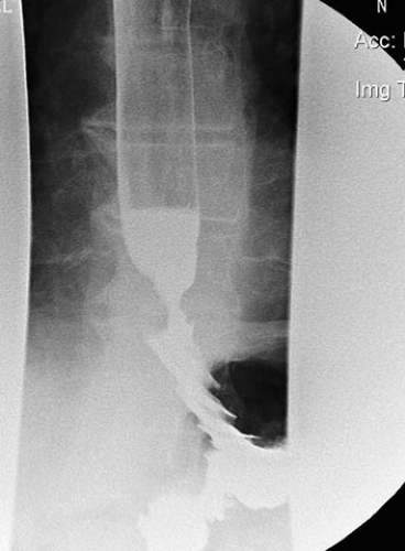
Mr. Gill was seen by his family physician, and the following laboratory tests were obtained: hemoglobin 13.2 g per dL (normal 12 to 15 g per dL), white blood count 8.4 (normal 4.3 to 10), sodium 138 mEq per L (normal 135 to 145 mEq per L), potassium 4.5 mEq per L (normal 3.5 to 5.0 per L), aspartate aminotransferase 10M (normal 10 to 40M), alanine aminotransferase 16M (normal 10 to 40M), alkaline phosphatase 37 IU (normal 21 to 91 IU), bilirubin 0.8 mg per dL (normal 0.3 to 1.0 mg per dL), and albumin 3.5 g per dL (normal 3.5 to 5.5 g per dL). A barium contrast examination revealed a mucosal irregularity with an annular lumen narrowing at the midesophageal level (Fig. 6.1).
What is the diagnostic evaluation of a patient suspected to have esophageal cancer?
View Answer
The patient must undergo esophagoscopy with biopsy of the suspected tumor to confirm the diagnosis of esophageal carcinoma. Direct visualization can also determine location and length of the involved segment. Bronchoscopy can evaluate tracheobronchial invasion in a patient with a history suggestive of advanced disease. The patient may present with progressive pneumonia, chocking, coughing with feedings, or aspiration of bile-stained mucus from the airway. Nishimura et al. determined that the accuracy rate of diagnosing tracheobronchial invasion based on bronchoscopy was 78%; this rate was greater than computed tomography (CT) (58%) but less than bronchoscopic ultrasound (91%) (2).
Mr. Gill was referred to a gastroenterologist who performed an esophagoscopy and biopsy of a nearly obstructing mass seen at 27 cm from the incisors. Pathology of the biopsy specimen revealed squamous cell carcinoma of the esophagus.
Once diagnosed with esophageal cancer, what examinations should be used to stage him?
View Answer
CT is an essential study to evaluate patient anatomy, confirm the presence of distant metastasis, and assess lymph node involvement. CT scan cannot, however, precisely determine the stage of cancer. A CT scan with contrast has a sensitivity of 70% to 80% in detecting hepatic metastasis larger than 2 cm, but smaller liver metastasis may go undetected (3). Invasion of other structures may be difficult to detect on CT and has high interobserver variability. Accuracy rates range from 59% to 82% (4). Furthermore, CT has a sensitivity of 34% to 61% for mediastinal and 50% to 76% for abdominal lymphadenopathy (3).
Esophageal ultrasound (EUS) is capable of showing individual layers of the esophagus by combining endoscopy and high frequency ultrasound. It can identify the extent of tumor wall invasion and especially locate involved lymph nodes that might otherwise be missed by CT scan. Accuracy of EUS in T staging is 84%, and it increases in T3 to T4 carcinomas and with operator experience (3). Fine-needle aspiration biopsy of lymph nodes can be achieved under EUS guidance, providing greater staging information. Positron emission tomography (PET) cannot demonstrate tumor invasion as well as EUS; however, PET can locate distant metastasis undetected by CT scan. The role of PET has yet to be elucidated.
Is there a role for minimally invasive surgery in esophageal tumor staging?
View Answer
Comparable to the use of mediastinoscopy in lung cancer staging, there has been an increase in using mediastinoscopy, thoracoscopy, and laparoscopy to search for and biopsy lymph nodes undetectable by imaging techniques. Laparoscopy is also useful for detecting liver metastasis and omental implants. A review of the literature by Reed and Eloubeidi reported that thoracoscopic accuracy was 93% and laparoscopic accuracy was 94% (3). Luketich et al. reported 32% of patients who underwent CT and EUS had a T stage change after thoracoscopy and laparoscopy (5). Nguyen et al. had similar results, reporting a change in treatment plan for 36% of patients staged with minimally invasive techniques. The specificity of detecting occult metastatic disease was 100% (6). This staging benefit must be compared to the risks of an additional invasive procedure.
Mr. Gill had an EUS, which revealed invasion of the muscularis propria. There were no lymph nodes seen. Thus, his stage is T2, N0. His CT scans of the chest, abdomen, and pelvis showed that they were free of metastasis.
What is the length of the esophagus?
View Answer
The esophagus measures an estimated 25 cm in length, and it begins at the inferior edge of C6, connecting the pharynx to the cardia of the stomach. The cervical portion of the esophagus begins below the cricopharyngeus muscle and spans about 5 cm until T2 or the suprasternal notch. The thoracic esophagus then begins at the thoracic inlet, measures roughly 20 cm, and is further divided into upper, middle, and lower segments. These thoracic portions are located as follows: upper third, T2-5 (suprasternal notch to carina); middle third, T5-8 (carina to inferior pulmonary vein); lower third, T8-12 (inferior pulmonary vein to gastroesophageal junction). Endoscopic examinations report measurements beginning at the incisor teeth and ending about 5 cm beneath the diaphragm. Typically, this measurement is 38 to 40 cm in men and can vary according to height.
What are the layers of the esophagus?
View Answer
The esophagus is only composed of an epithelial layer and a muscular layer. Unlike other gastrointestinal organs, there is no serosa. Squamous epithelium lines the mucosa, and the musculature of the esophagus consists of an outer longitudinal and an inner circular layer. The upper third of the esophagus is composed of striated muscle that allows voluntary movements, such as swallowing; however, the distal two thirds is made up of smooth muscle, which permits involuntary movement.
What is the blood supply to the esophagus?
View Answer
The arterial blood supply is segmental and results in an extensive intramural vascular plexus. The cervical esophagus receives its supply from the inferior thyroid artery. The thoracic esophageal portion is supplied by blood from thoracic branches, some directly off of the aorta. As for the abdominal esophageal segment, the inflow is from both the left gastric and inferior phrenic arteries. Venous drainage parallels the arterial network. The cervical esophagus drains into the inferior thyroid vein, whereas the thoracic region is drained by the bronchial, azygos, or hemiazygos veins. The distal esophagus has venous drainage into the left gastric (coronary) vein.
What is the lymphatic drainage of the esophagus?
View Answer
The lymphatic plexus, located in the submucosa of the esophagus, mainly spreads in a longitudinal fashion. The cervical esophagus drains cephalad into the internal jugular, paratracheal, and deep cervical lymph nodes. However, the middle and lower thoracic segments of the esophagus drain caudad to the subcarinal/pulmonary hilar nodes and to the paraesophageal/celiac nodes, respectively.
Are most esophageal tumors benign or malignant?
View Answer
Most esophageal tumors are malignant because only 0.5% of total esophageal masses are benign. Benign tumors are classified by their location, mucosal or intramural, and consist of leiomyomas, benign polyps, hemangiomas, lipomas, and esophageal duplication cysts. Leiomyomas, as the most common, represent 60% of benign neoplasms. Squamous cell carcinoma accounts for the most common malignant tumor worldwide; however, there is an increase in adenocarcinoma incidences, especially in the United States and Western Europe.
Where are most esophageal malignancies located?
View Answer
Only 15% of esophageal cancers occur in the upper portion of the esophagus compared with 50% in the middle and 35% in the lower segments. The higher percentages seen in the lower segments compared to the upper are, in majority, related to Barrett’s esophagus.
What risk factors does our patient have?
View Answer


In Western countries, esophageal cancer tends to be most common in African American men older than age 50. There is an association between squamous cell carcinoma and tobacco and alcohol usage. An extremely high prevalence of esophageal cancer is also seen in China and Iran. In China, environmental factors such as nitrosamines in the soil and contamination of food by fungi and yeast were found to be carcinogenic. In Iran, the pyrrolysates found in opium and the ingestion of extremely hot teas are thought to contribute to an increase in the incidence of esophageal cancer. Nutritional factors such as malnutrition, vitamin deficiencies, poor dentition, and anemia are also linked to increasing the risk of esophageal cancer. Other risk factors include Plummer-Vinson syndrome, achalasia, lye strictures, irradiation esophagitis, and tylosis (familial keratosis palmaris and plantaris). Gastroesophageal reflux disease (GERD) and Barrett’s esophagus are risk factors for adenocarcinoma of the esophagus, which is more common in whites than African Americans.
Stay updated, free articles. Join our Telegram channel

Full access? Get Clinical Tree


