Endoscopic Ultrasound–Assisted Access to the Pancreatic Duct

Chapter 31 Endoscopic Ultrasound–Assisted Access to the Pancreatic Duct


Endoscopic retrograde pancreatography (ERP) is the method most commonly employed to access the main pancreatic duct (MPD) and is occasionally performed for diagnostic purposes and routinely performed for therapeutic purposes. Historically, percutaneous and surgical approaches were the only options available for patients in whom access could not be achieved via ERP. An emerging alternative is endoscopic ultrasound (EUS)–assisted pancreatic access and drainage. While EUS was introduced nearly 30 years ago as a diagnostic imaging modality, the development of linear instruments allowed for tissue acquisition.1,2 This subsequently led to the development of EUS-guided therapeutic interventions including celiac blockade and neurolysis,35 pancreatic fluid collection drainage,69 cholecystenterostomy10 and delivery of chemotherapy, radioactive seeds, and gene therapy.11,12 The concept of combining ERP with interventional EUS technology led to the first report in 1995 of EUS-guided pancreatography for a patient who required pancreatic duct stone removal following pancreaticoduodenectomy.13 The continued need to develop less invasive alternatives to surgical and interventional radiological therapies has further driven the development of EUS-guided methods for pancreatic intervention.


Video for this chapter can be found online at www.expertconsult.com.


Description of Technique


Equipment Selection


Therapeutic channel linear array echoendoscopes are favored since they allow use of the full spectrum of accessories and placement of large caliber (10 Fr) stents. Smaller caliber diagnostic echoendoscopes may be used for rendezvous wire passage or placement of stents ≤7 Fr in diameter. Duct access may be achieved with any of the currently available 25-, 22-, or 19-gauge fine-needle aspiration (FNA) needles and array of guidewires. It is important to select a guidewire gauge that permits passage through the selected FNA needle. Large caliber (19-gauge) needles are generally preferred, as they allow passage of larger gauge (0.035-in) guidewires that may facilitate traversal of stenotic regions and passage of other accessories and stents. However, the stiffness of 19-gauge needles can hinder access of small caliber ducts. While smaller caliber (22-gauge) needles are more easily advanced into the MPD, their selection demands use of smaller gauge (≤0.021-in) guidewires. These wires are more flexible and may improve duct access and facilitate traversal of obstructed segments, but due to their floppy nature can make subsequent interventions more difficult. In addition to altering the guidewire caliber, one may select either hydrophilic wires or angled tip wires, which may facilitate traversal of narrowed or tortuous segments.


A clear understanding of the procedure goals can help guide equipment selection. For instance, it may be reasonable to use a 22- or even 25-gauge needle if the goal is only to obtain a pancreatogram. Some endosonographers also prefer a smaller gauge needle when simply determining if contrast freely flows into an anastomosed bowel lumen suggesting absence of critical stenosis, thereby potentially obviating the need for therapeutic intervention.


Various accessories may be used to create a fistula between the gut lumen (stomach, duodenum, or jejunum) and pancreatic duct to facilitate passage of other accessories and devices, for dilation of strictures, and for eventual stent placement. A variety of standard biliary and pancreatic catheters and pneumatic dilators or cautery-assisted access may be employed and should be readily available. The techniques of EUS-guided pancreatic interventions are not standardized and there is a lack of comparative trials to clarify the relative value of current devices. Equipment use varies among endoscopists and often requires trial and error between patients and even within the same patient.


Nomenclature


The techniques for EUS-assisted MPD access and drainage may be broadly categorized into the following:


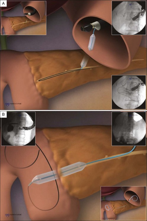
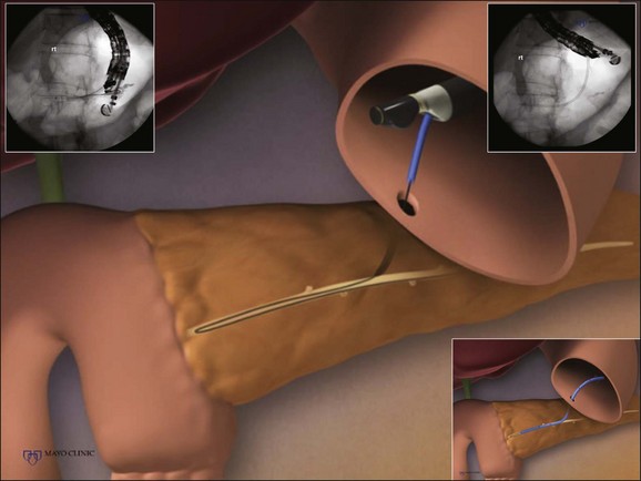
The transpapillary or transanastomotic (following resection) route involves stent placement across the papilla or anastomosis. EUS is initially performed for MPD access and guidewire passage into the small bowel. Thereafter, the stent may be placed with aid of a duodenoscope or long-length forward-viewing endoscope (colonoscopes, push-type and balloon-assisted enteroscopes) inserted following removal of the echoendoscope (retrograde approach), often referred to as a “rendezvous procedure.” The entire procedure including stent placement may also be performed via the echoendoscope (antegrade approach).


The transluminal (or transmural) route may be performed when transpapillary or transanastomotic stenting fails. With this approach, the distal end of the stent is placed within the MPD and the entire exam is performed via EUS (antegrade approach).


Techniques


Transpapillary/Transanastomotic Approach (with Retrograde Stent Placement)


The MPD is localized and accessed under EUS guidance. The optimal point of MPD access varies depending on the site of obstruction and is located anywhere from the gastric cardia to the second portion of the duodenum. A location is selected that provides the least distance between the transducer and MPD in order to facilitate access and subsequent therapy. It is important to exclude intervening structures such as blood vessels, loops of bowel, and undesired ducts.


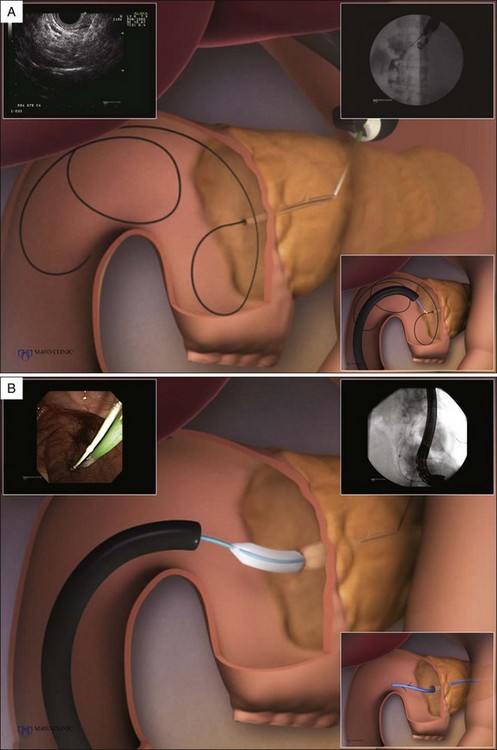
The guidewire is advanced through the FNA needle and antegrade through the site of stenosis, across the papilla/anastomosis, and coiled in the small bowel. Doing so permits subsequent transpapillary or transanastomotic stenting. The guidewire should be sufficiently advanced to form loops within the bowel lumen to reduce the risk of wire dislodgement that may occur either during removal of the echoendoscope or when inserting the duodenoscope. Fluoroscopy is used to verify the echoendoscope position, to perform ductography, and to facilitate guidewire passage.


Following proper guidewire placement, the echoendoscope is then withdrawn, leaving the guidewire in place. The ERP or “rendezvous” portion of the exam is then performed by passing a side- or forward-viewing endoscope to the papilla or site of anastomosis. The end of the guidewire is grasped with a snare or biopsy cable and withdrawn through the channel of the endoscope. A standard duodenoscope is preferred for patients with unaltered gastroduodenal anatomy when interventions are across the papilla. In patients with an afferent jejunal limb or Roux-en-Y anatomy following pancreaticoduodenectomy, an extended forward-viewing instrument such as a colonoscope is often employed. Once the selected instrument is properly positioned and guidewire control is achieved, biliary stent insertion and other interventions may be performed in standard fashion. It is often necessary to work over the internal/external guidewire until initial dilation is performed. After dilating the site of obstruction with a catheter or balloon, a cannula may be passed alongside the indwelling guidewire to allow insertion of a second wire into the pancreatic duct. This allows stent insertion via one guidewire while the other serves as a safety wire in the event of inadvertent loss of duct access.

Stay updated, free articles. Join our Telegram channel

Tags:
Mar 11, 2017 | Posted by in GASTROENTEROLOGY | Comments Off on Endoscopic Ultrasound–Assisted Access to the Pancreatic Duct

Full access? Get Clinical Tree

Get Clinical Tree app for offline access