Essentials of Diagnosis
- Rise of serum creatinine of ≥0.5 mg/dL or 25% above baseline within 48 hours after parenteral contrast administration.
- Majority of cases are nonoliguric.
- Peak in serum creatinine usually occurs within 3–5 days with complete resolution in 7–10 days for most patients.
- Oliguria and need for dialysis are unusual and are primarily seen in patients with diabetes with severe chronic kidney disease.
General Considerations
Contrast-induced nephropathy (CIN) is a common cause of hospital-acquired acute renal failure. Although the incidence of CIN is low, the number of patients receiving intravenous contrast media is enormous and is expected to rise as the population ages and greater numbers of patients will be subjected to diagnostic and therapeutic procedures requiring intravascular contrast.
Observational retrospective studies have demonstrated that patients incurring CIN had significantly higher in-hospital mortality than counterparts who did not develop CIN, particularly so if they went on to need dialysis. Multivariate regression analysis, adjusting for baseline differences in comorbidities, strongly suggests that CIN is, in fact, an independent predictor of mortality.
Various patient and procedural factors influence the likelihood of developing CIN. Among patient-related factors, preexisting chronic kidney disease (CKD) is regarded as the most potent risk factor. Nearly 60% of patients developing CIN have baseline CKD, and the incidence of CIN parallels the severity of prestanding renal impairment. Diabetes mellitus also confers increased risk of developing CIN, but only in patients with CKD. Thus the risk of CIN is greatest in patients with CKD and diabetes, lower in nondiabetic patients with CKD, and lowest in patients without preexisting CKD regardless of their diabetic status. Similarly, patients with CKD and diabetes are at the highest risk of developing oliguric acute renal failure and of requiring dialysis. Patients with CIN who require dialysis have a very high in-hospital mortality.
Volume depletion, congestive heart failure, older age, and hypotension have all been cited as risk factors for CIN but are probably primarily markers for a low glomerular filtration rate (GFR), the true risk factor. Concomitant use of nephrotoxins such as nonsteroidal anti-inflammatory agents also increase the risk for CIN.
Multiple myeloma has traditionally been considered a risk factor for CIN. However, recent studies using modern contrast agents indicate that patients with multiple myeloma are not at a greater risk, provided that they are volume replete at the time of contrast exposure.
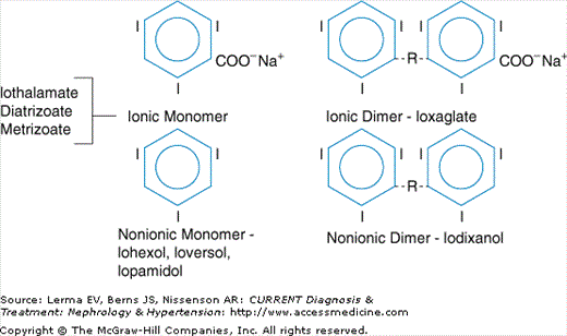
Procedure-related factors will also influence the likelihood of CIN. Most studies, although not all, suggest that exposure to larger volumes of parenteral contrast causes greater predisposition to CIN. In addition, the type of contrast material (specifically its osmolality) influences the development of CIN. Contrast media formulations occur in three types: High-osmolar contrast media (also termed ionic), which have an osmolality of approximately 2000 mOsm/L, low-osmolar contrast media (also termed nonionic), which have an osmolality of 600–900 mOsm/L, and iso-osmolar contrast media (also a nonionic composition), which have an osmolality of 300 mOsm/L (Figure 12–1). Multiple studies in high-risk patients with CKD have demonstrated that low-osmolar contrast media results in less CIN than high-osmolar contrast media, and there is some evidence that iso-osmolar contrast media may be less nephrotoxic than low-osmolar contrast media.
Pathogenesis
Several mechanisms have been proposed to explain the pathogenesis of CIN. Measures aimed to prevent CIN (see Prevention below) are based on these pathogenetic mechanisms. Renal ischemia is currently considered the primary mechanism for CIN. It is known that the outer renal medulla has an extremely low oxygen tension (Po2 10–20 mm Hg), as the result of countercurrent oxygen exchange and removal between vasa rectae and utilization of oxygen by active tubular transport by the ascending loop of Henle. Administration of contrast media has been shown to selectively further reduce oxygen tension in this area of the kidney by a two-pronged mechanism. The first is reduction in renal blood flow, mediated by release of vasoconstrictive compounds such as endothelin and adenosine, an effect that is magnified by blockade of vasodilatory compounds such as nitric oxide and prostaglandins. The second is increased oxygen utilization caused by increased work of active transport in response to an osmotic diuresis induced by contrast media in the renal tubule. A summary of these effects is depicted in Figure 12–2.
Hyperosmolarity may itself be etiologic in the development of CIN. Intratubular hyperosmolality may activate tubuloglomerular feedback or increase intratubular hydrostatic pressure, either of which could reduce glomerular filtration. Hyperosmolality may also increase tubular cell apoptosis.
There is also evidence to suggest that generation of oxygen free radicals contributes to the pathogenesis of CIN. This theory would explain the possible ability of N–acetylcysteine (NAC), a free radical scavenger, and sodium bicarbonate, which inhibits the enzymatic formation of free radicals, to prevent CIN (see Prevention below).


