Fig. 32.1
Bristol Stool Scale (Adapted with permission from Lewis and Heaton, © 1997 Informa Healthcare. Reproduced with permission)
Etiologies of Constipation
Diet affects the size, consistency, and frequency of bowel movements; dietary fiber intake is highly correlated with stool bulk.
Factors associated with constipation are summarized in Table 32.1.
Table 32.1
Factors associated with constipation
Lifestyle
Inadequate fluid intake
Inadequate fiber intake
Inactivity
Laxative abuse
Medications
Opiates
Anticholinergics
Iron
Medical illness
Neurologic
Spinal cord dysfunction/damage
Parkinson’s disease
Multiple sclerosis
Endocrine/metabolic dysfunction
Diabetes mellitus
Hypothyroidism
Electrolyte abnormalities
Uremia
Hypercalcemia
Porphyria
Psychological
Depression
Anorexia
Psychiatric illness
Sexual abuse
Colonic structure/function
Cancer
Crohn’s disease
Irradiation
Endometriosis
Hirschsprung’s disease
Chagas’ disease
Pelvic floor abnormality
Nonrelaxing puborectalis
Anal stenosis
Rectocele/enterocele
Subtypes of Constipation
Patients can be further classified by associated findings such as slow transit (motility disorders), irritable bowel syndrome (IBS), and pelvic floor dysfunction, also described as obstructed defecation syndrome (ODS) or mixed disorders.
Motility disorders can be isolated to the colon (colonic inertia aka slow-transit constipation) or can affect the stomach and small bowel.
Colonic inertia (CI) is frequently associated with constipation since childhood, fewer than 3 BMs per week, and laxative dependence.
IBS-C (constipation subtype) is associated with abdominal pain, irregular bowel habits, and pain relieved by defecation.
ODS refers to a constellation of symptoms such as prolonged repeated straining at bowel movements, sensation of incomplete evacuation, and the need for digital manipulation.
History and Physical Examination
The evaluation of constipation begins with a thorough history.
Query into psychiatric illness and sexual and physical abuse must be performed as they are often associated with defecation difficulties.
Multicompartment pelvic floor symptoms such as urinary dysfunction, pelvic organ prolapse, and sexual dysfunction need to be elicited.
Evaluation for pelvic floor dysfunction includes vaginal, perineal, and rectal examination.
Bulging of the posterior vaginal wall beyond the hiatus is consistent with advanced prolapse and may represent a rectocele, enterocele, or sigmoidocele. *Examination in the standing position with a finger in the rectum and vagina may be performed to elicit the maximal prolapse of the pelvic organs as they descend through the pouch of Douglas and genital hiatus.
A gaping patulous anus may indicate neurological injury, intra-anal intussusception, or full-thickness rectal prolapse.
Digital examination evaluates resting anal tone and squeeze strength and can identify a large rectocele, sphincter defect, or no movement of the pelvic floor muscles.
Valsalva maneuver or simulated defecation on a commode is useful to elicit full-thickness rectal prolapse.
Diagnostic Testing
Laboratory testing with thyroid studies and calcium levels is useful to exclude metabolic etiologies of constipation.
Obstructing colon lesions and inflammatory conditions such as IBD or diverticulitis must be excluded by colonoscopy or GI contrast studies before considering functional etiologies.
Diagnostic Studies to Evaluate Intestinal Transit
The most widely available technique for determining colonic transit uses radiopaque markers and radiographs of the abdomen.
The patient should refrain from all enemas, laxatives, and most medications for 2 days prior to the ingestion of 24 radiopaque markers.
The patient is required to ingest 30 g of fiber daily during the test and must continue to refrain from taking medication and laxatives.
An abdominal radiograph is obtained on the fifth day, and the distribution and number of markers present in the colon is noted. A variation is to obtain radiographs on days 1, 3, and 5.
Eighty percent of normal patients will have passed all the markers by 5 days.
If the markers remain scattered throughout the colon and more than 20 % of the markers remain in the colon, colonic inertia can be diagnosed.
If the markers are found to have accumulated in the rectum, traditional teaching suggests that this is a diagnostic of outlet obstruction constipation (Fig. 32.2 ); however, this is controversial and there may be no correlation between the pattern of marker distribution and type of constipation.
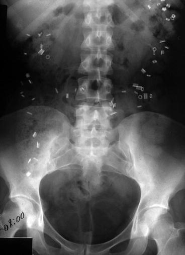
Fig. 32.2
Marker study revealing colonic inertia
In the general population, 95 % of patients will have a transit time of less than 65 h in men and 75 h in women. Patients with normal transit constipation will have a colon transit time that is in the normal range.
Transit times obtained through scintigraphy are generated by following the passage of a radiolabeled meal.
Small-bowel transit time may also be measured with a lactulose hydrogen breath test. The principle of this examination is that hydrogen produced through lactulose fermentation only occurs in the colon. If one records the time from ingestion of lactulose to hydrogen production, small-bowel transit time can be inferred.
Diagnostic Studies to Evaluate Pelvic Floor Dysfunction
Anal manometry evaluates resting and squeeze pressure; often with constipation, patients exhibit internal sphincter hypertonia with poor incremental squeeze pressures.
The volume noted at first sensation can be blunted, i.e., requiring larger volumes to obtain a sensory response, and the maximum tolerated volume can also be blunted.
The presence of the rectal anal inhibitory reflex (RAIR) is useful to exclude Hirschsprung’s disease.
Electromyography aids in the diagnosis of puborectalis syndrome by indicating a paradoxical or nonrelaxing muscle.
Balloon expulsion is an inexpensive method to assess ability to evacuate. Normal studies indicate the ability to evacuate a 50–100 cm3 balloon in less than 1 min.
Defecography is the gold standard to confirm evacuatory dysfunction due to intussusception, rectal prolapse, enterocele, sigmoidocele, rectocele, and perineal descent.
Some centers use a dynamic MRI, but the technique varies. For the best images, complete evacuation of the contrast during the MRI after Valsalva will most likely simulate defecation.
Defecating MRI has advantages over traditional defecography because it involves less radiation and provides multicompartment images. However, defecating in the supine position is not physiologic.
For patients with pelvic floor dysfunction who demonstrate dysnergic defecation, randomized controlled trials show that biofeedback is superior to laxatives, sham treatments, and alternative therapies.
In the setting of rectal intussusception, rectocele, and mucosal prolapse, stapled transanal rectal resection (STARR) can be offered.
STARR employs a double stapled technique
Prospective multicenter trials reveal initial and long-term symptom improvement of obstructed defecation after STARR.
A randomized controlled trial of STARR versus biofeedback revealed that STARR is more effective for treatment of evacuatory dysfunction.
For patients with a clinical or radiologic rectocele and retained rectal contrast, rectocele repair can be suggested.
Rectocele repair can be performed via transvaginal transanal or transperineal approach with 75–80 % reported bowel symptoms improvements.
Enterocele involves descent of small bowel into the lower pelvic cavity, leading to mechanical obstruction of the rectum.
A sigmoidocele refers to descent of the sigmoid colon into the lower pelvic cavity leading to compression and mechanical obstruction of the rectum.
Sigmoid resection or sigmoidopexy in conjunction with posterior compartment repair has been shown to be effective in relieving symptoms of obstructed defecation in a limited number of patients.
Medical Treatment of Constipation
Simple measures that can influence the passage of colonic content are increasing physical activity and fluid intake.
Osmotic laxatives, stimulants, and enemas should be reserved for treatment of acute bouts of discomfort.
Bulking agents promote these changes by delivering a mass of nondigestible substrate to the colon and, due to their hydrophilic nature, facilitate the absorption and retention of fluid in the stool.
These substrates are derived from the nondigestible components of plants or are synthetic methylcellulose derivatives.
Osmotic laxatives are a class of medications that promote the accumulation of large volumes of fluid in the colon.
The osmotically active particles can be derived from sugars or salts such as sucrose-based sorbitol and lactulose.
Lactulose is degraded in the colon yielding the production of fatty acids, hydrogen, and carbon dioxide.
MiraLAX® (polyethylene glycol 3350) is an over-the-counter osmotic laxative that increases the frequency of bowel moments and softens the stool, so it is easy to pass.Related posts:

Stay updated, free articles. Join our Telegram channel

Full access? Get Clinical Tree


