Fig. 14.1
Endorectal advancement flap. (a) The patient is placed in the prone position. (b) A U-shaped flap of mucosa, submucosa, and internal sphincter is raised. (c) The dissection is usually 4–5 cm cephalad to allow a tension-free repair. The base should be two to three times wider than the apex to prevent ischemia. (d) The fistula tract is debrided (not excised) and the muscles reapproximated with one to two layers of long-acting absorbable sutures. (e) The distal end of the flap with the fistula is excised, and the flap is sutured in place. The vaginal side is left open for drainage
Postoperatively patients are given a normal diet and fiber supplements. Constipation and diarrhea should both be avoided using medical management. Patients should abstain from intercourse and using tampons for 6 weeks.
The transvaginal repair—technique:
A vaginal flap is raised starting near the introitus.
The flap is developed laterally to the ischial tuberosities for adequate mobility.
The rectal defect is closed with absorbable sutures.
The levator ani muscle is approximated in the midline. This maneuver is felt to be critical for success.
The vaginal flap is secured with absorbable sutures.
Another variation for a flap repair is a retrograde anocutaneous flap, used for very low fistula. A flap of anoderm and perineal skin is raised, advanced into the anal canal, and sutured in place.
In the literature, success rates for advancement rectal flaps range from 29 to 100 %. Disturbances in continence range from 21 to 40 %.
In an effort to improve success, transposition of labial fat beneath an endorectal advancement flap was done. However, no improvement in successful fistula closure was found.
Transanal endoscopic microsurgery (TEM) has been used to facilitate repair with success in small studies.
Smoking has been linked to unsuccessful closure of fistula when using advancement flaps.
Fistulectomy with Layered Closure
Excision of the fistula and closure of the tract in layers can be done via the rectum, vagina, or perineum.
Transanally, an elliptical excision is made to core out the fistula, and 2–3 cm mucosal flaps are raised. Vaginal mucosa, rectovaginal septum, rectal muscle, and rectal mucosa are closed in successive layers. Plication of the levator muscles can be added.
For a perineal approach, a transverse incision is made between the anus and vagina. The incision is deepened until the tract is encountered. The tract along with the openings in the rectal and vaginal walls is excised. The wound is closed in layers.
In a small series, success with these approaches has been reported in 88–100 %.
Rectal Sleeve Advancement
Circumferential mobilization of the distal rectum with advancement to cover the anorectal side of the fistula is reserved for fistula associated with an anal stricture or disease in the proximal anal canal/distal rectum.
Technique
The patient is prepared as for the endorectal advancement flap.
Starting at the dentate line, a circumferential incision is created that deepens through mucosa and submucosa but not through the internal sphincter. The dissection is continued cephalad becoming full thickness above the anorectal ring.
Dissection is continued until healthy nonscarred tissue can be pulled down to the dentate line without tension.
The diseased distal end is trimmed, and the healthy tissue is sutured to the anoderm.
One reported variation in treating recurrent RVF is to perform this repair using the Kraske approach.
Another variation is the Noble-Mengert-Fish technique. A 180° full-thickness anterior rectal wall flap is mobilized cephalad to the vaginal vault. The lateral margins are the full width of the rectovaginal space. The flap must reach the external sphincter muscle without tension. Success rates have been documented between 86 and 100 % with minor incontinence in 25 %.
Sphincteroplasty and Perineo-proctotomy
Sphincteroplasty
For this procedure, a probe is inserted through the fistula, and it is unroofed converting the anatomy to a fourth-degree laceration. Then a layered anatomical repair is done.
If there is a defect in the external sphincter muscle, this repair obliterates the fistula while repairing the muscle.
Successful closure is reported for 65–100 % of patients.
Perineo-proctotomy
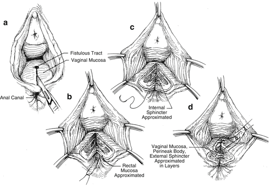
When there is an intact sphincter muscle, this repair is termed a perineo-proctotomy. When the fistula is unroofed, the sphincter is divided. Figure 14.2 describes and illustrates the procedure.

Fig. 14.2
Perineo-proctotomy. (a) For a perineo-proctotomy, there is intact sphincter muscle. The fistula is still unroofed to create a defect like a fourth-degree obstetrical laceration. (b) The tract is excised, and the vaginal and rectal walls are dissected away from the sphincter muscle. The rectal mucosa is approximated. (c) The internal sphincter muscle is sutured together. (d)The external sphincter muscle and then the vaginal mucosa are reapproximated. The perineal body is reconstructed and the skin closed
The deliberate division of an intact sphincter muscle should be approached with caution (the perineo-proctotomy) as the impact on continence has not been well studied. Success rates have been reported from 87 to 100 %.
Inversion of Fistula
Usually performed through the vagina, the vaginal mucosa around the fistula is mobilized and the tract is excised. A purse-string suture is used to invert the fistula into the rectum. The vaginal wall is closed over this inverted tissue.
Ligation of the intersphincteric fistula tract (LIFT) uses a similar technique. An intersphincteric dissection is carried out, and the fistula tract is identified and divided. The openings on the rectal and vagina side are ligated and the wound closed. Success rates of 60–94 % have been reported.
Patients with a complex transsphincteric fistula and an intact sphincter may be the best candidate for the LIFT.
Tissue Interposition
General Considerations
Tissue interposition allows well-vascularized tissue to be placed between the vagina and rectum. Sources include rectus, bulbocavernous, gracilis, gluteus, and sartorius muscle.
Stay updated, free articles. Join our Telegram channel

Full access? Get Clinical Tree


