Author
Year
Port
Appendectomy
Operative time
Pain score (VAS score)
Complications
Hospital stay (h)
SILA
3-port
SILA
3-port
SILA
3-port
SILA
3-port
St Peter
2011
Fascial puncture
Extracorporeal
35
30
9.6
8.5
3.30 %
1.70 %
23
22
Frutos
2013
SILS port
GIA 45 mm
38
32
2.76
3.78
4/91
4/93
19
21
Sozutek
2013
SILS port
2/0 suture
33
30
2.9
3.4
1
1
26
29
Kye
2013
Glove
Endoloop
37
38
3.22
3.9
1.96 %
1.96 %
75
117
Perez
2013
Fascial puncture
GIA 45 mm
46
34
ns
ns
1
0
40
37
29.2.2 SILA for Complicated Appendicitis
The difficulty in keeping instrument triangulation limits the indication of SILA in cases with a retrocecal appendix or perforated appendix. It has been shown that operative time was significantly longer with complicated appendicitis (gangrene, abscess, perforation, and/or peritonitis) [6]. In addition, retrocecal appendicitis often necessitates a second, or even a third port [10]. Therefore, it is conceivable that complicated and retrocecal appendicitis can be considered as a relative contraindication for SILA. However, Kye et al. [7] performed SILA for cases of complicated appendicitis and showed comparable results in operative time and even better results in hospital stay and recovery time to daily life in comparison with standard 3-port appendectomy. Therefore, it is possible that advance in surgical instruments for SILA and maturation in our surgical techniques to perform SILA could broaden the indication of SILA for complicated appendicitis in the near future.
29.2.3 Indications for SILA
In obese patients, it is even more difficult to keep the instrument triangulation in SILA; therefore, they do not benefit from SILA [11]. SILA for patients with a body mass index >95th percentile shows longer operative time, increased postoperative pain, and increased complication rates (wound infection and intraabdominal abscess). In addition, patients with previous lower abdominal surgery are almost inevitable to have abdominal adhesions that could limit the indication for SILA. Taken together, relative contraindications for SILA include retrocecal appendicitis, complicated appendicitis, obese patients, and past history of lower abdominal surgery. However, technical and instrumental progress in the single-port approach could expand the current indication. Given the fact that almost all the clinical outcomes are equivalent between SILA and standard 3-port appendectomy, the major advantage of SILA over standard 3-port appendectomy is focused mostly on cosmesis. In this regard, good populations that notably benefit from SILA include children and premenopausal women.
29.3 Technique
29.3.1 Patient Positioning and Room Set-up
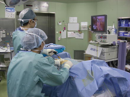
Operation room is set up and the patient is placed in a fashion analogous to conventional 3-port laparoscopic appendectomy. Briefly, the patient is placed in the supine position with the legs together, the left arm is tucked, and a Foley catheter inserted to decompress the bladder. The surgeon and camera assistant both stand on the left of the patient facing the monitor on the right side at the patient’s hip. The operating table is tilted in the Trendelenburg position with the right side up for 15–20°. To perform single-incision surgery, the laparoscope should have a co-axial, not perpendicular, light cable to avoid crowding. A 30-degree 5-mm laparoscope is often used.
29.3.2 Umbilical Port
29.3.2.1 Fascial Puncture Method (Direct Access Method)
After local anesthetic infiltration, a 2.0-cm vertical transumbilical incision is made and a subcutaneous pocket is created to expose the anterior fascia. A Veress needle is inserted to establish pneumoperitoneum and removed after insufflation. Three laparoscopic ports (one 10-mm and two 5-mm ports or three 5-mm ports) are placed by a closed access method or an optical trocar access method. The first trocar is at the base of the umbilical stalk with the second at the inferior edge along the linea alba and the third at the superior edge along the linea alba or the right hand side of the first trocar. The first trocar can be inserted without pneumoperitoneum through a small umbilical fascial defect at the base of the umbilical stalk. Three ports should be of low-profile (preferably threaded and different in length) in order to avoid clashing.
29.3.2.2 Multi-Channel Port Method (Access Device Method)
After local anesthetic infiltration, a 2.0-cm vertical transumbilical incision is made down to the peritoneum under direct vision. A multiple-entry port from various manufactures (Table 29.2) is inserted through the incision by an open access method.
Table 29.2
Multiple-entry ports
SILS™ port, Covidien, New Haven, CT, USA |
Gelport, Applied Medical, Rancho Santa Margarita, CA, USA |
Unix-X, Pnavel Systems, Brooklyn, NY, USA |
Triport, Advanced Surgical Concepts, Wicklow, Ireland |
SSLAS, Ethicon, Cincinnati, OH, USA |
X-Cone, Karl Storz-Endoskope, Tuttlingen, Germany |
OCTO port, Dalim Surgnet, Seoul, Korea |
Spider surgical system, TransEnterix, Durham, NC, USA |
29.3.2.3 Glove Method (Home Made Port Method)
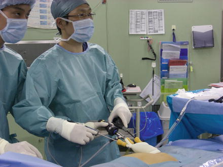
After local anesthetic infiltration, a 2.0-cm vertical transumbilical incision is made for Alexis wound retractor™ XS size (Applied Medical), which is inserted by an open access method and a surgical glove (size 5.5) is attached. Three low-profile laparoscopic ports (all 5-mm trocars) are inserted through the holes of the surgical glove with cut fingertips (Figs. 29.1 and 29.2).


Fig. 29.1
Glove method using Alexis wound retractor™ (Applied Medical)

Fig. 29.2
Positioning of the surgeon and assistant
29.3.3 Appendectomy
29.3.3.1 Extracorporeal Appendectomy
The appendix locates at the posteromedial border of the cecum and can be identified by following the anterior taenia coli to its confluence with the other two taeniae. The cecum is mobilized by incising the lateral attachment (Jackson’s membrane and Lane’s band) and the fusion fascia of Toldt. Complete mobilization of the appendix is confirmed by withdrawing the appendix towards the left upper quadrant. The appendix can be easily exteriorized if the instrument reaches a halfway point between the left costrochodral margin and the umbilical port. The appendix is withdrawn through the umbilicus and exteriorized to perform a conventional “open” appendectomy. If the appendix is perforated, the operating surgeon prefers to perform an intracorporeal appendectomy. Extracorporeal appendectomy is less expensive compared to intracorporeal appendectomy and can be a good alternative to expensive intracorporeal devices.
29.3.3.2 Intracorporeal Appendectomy
Exteriorization of the appendix is not always possible in cases of more inflamed appendicitis or obese patients. For such cases, the appendix must be divided intracorporeally. The identification and mobilization of appendix can be performed in the same manner as in extracorporeal appendectomy. The following appendectomy is done in a purely laparoscopic approach without exteriorizing the appendix. This method has the potential to be more technically demanding than the extracorporeal method, accounting for the higher intraoperative and postoperative complication rates, such as hemorrhage, wound infection, and intrabdominal abscess [12].
Stay updated, free articles. Join our Telegram channel

Full access? Get Clinical Tree


