Adenocarcinoma of the Prostate


Adenocarcinoma of the Prostate


GLADELL P. PANER

CRISTINA MAGI-GALLUZZI

MAHUL B. AMIN

JOHN R. SRIGLEY


INTRODUCTION

Prostatic adenocarcinoma, the commonest noncutaneous malignancy in humans, is a significant cause of morbidity and mortality in men. Adenocarcinomas account for >99.5% of carcinomas of the prostate gland, and as such, population statistics on prostate cancer essentially reflect adenocarcinoma. The diagnosis and treatment of prostatic adenocarcinoma generates significant workload for the surgical pathologist. Prostatic needle biopsies and radical prostatectomy (RP) specimens are very common specimens in most pathology laboratories. It has been estimated that upwards of a million prostate biopsies are done annually in the United States.1 Adenocarcinoma of the prostate has a wide spectrum of clinical presentations ranging from the identification of a small focus of low-grade carcinoma in the asymptomatic patient with a prostate-specific antigen (PSA) elevation to widely metastatic carcinoma in a patient with bone pain. The architectural patterns of adenocarcinoma are diverse, and certain morphologies are predictive of clinical outcome and help explain, at least in part, the heterogeneous behavior of this tumor. The complexity of reporting both biopsy and radical resection specimens has increased in recent years, and there is an expectation on the part of urologists and oncologists that the surgical pathology report will include not just the diagnostic information but also all relevant prognostic data related to grade, extent, stage, margin status, and other information in selected cases.

In view of the sheer volume of prostatic cancer cases handled by pathologists, the morphologic heterogeneity of adenocarcinoma and the clinical impact of this diagnosis and associated prognostic factors, it was decided to devote an entire chapter to this tumor. Acinar adenocarcinoma and its variations (e.g., atrophic, pseudohyperplastic) and special variants of adenocarcinoma including ductal, mucinous, and signet-ring carcinoma are discussed here. Other rare carcinomas including basaloid, squamous (adenosquamous), sarcomatoid, small cell, large cell neuroendocrine, and urothelial carcinoma are presented in Chapter 10.


EPIDEMIOLOGY


Incidence and Prevalence

Prostate cancer is the second most common and sixth leading cause of cancer death in men with an estimated 899,000 new cases and 258,000 deaths in the world in 2008.2 In the United States, in 2013, it is estimated that there will be 238,590 new cases and 29,720 deaths from prostate cancer with age-adjusted incidence rate of 154.8 cases per 100,000 men.3,4 Due to the high incidence and absolute mortality rates, considerable resources have been allocated to prostate cancer, with approximately $9.9 billion spent each year on treatment in the United States alone.5 In 2009, the National Cancer Institute invested $293.9 million on prostate cancer research.6

Unlike other common visceral organ malignancies, prostate cancer diagnoses far outweigh the number of cancer-related deaths. This is due to its long latency period and the fact that most tumors are low-grade organ-confined tumors. In the United States, the ratio of new prostate cancer diagnosed to total number of deaths in a year is 8:1 in contrast to much higher ratios for other common tumors such as lung cancer (1.4:1) and colon cancer (2:1).4 The high incidence of prostate cancer has been attributed to widespread serum PSA screening introduced in the early 1990s. This had led to increased detection of indolent tumors resulting in long lead-time bias (of at least about 10 years) in survival proportions and no significant effect on the time of death.7,8 Some authors consider this phenomenon as “overdiagnosis,” and it is estimated that 23% to 42% of prostate cancer in the United States and Europe is overdiagnosed because of PSA screening.8,9 Currently, controversy surrounds the use of populationbased PSA screening, discussed later in details.10, 11, 12

Most patients with prostate cancer, due to its long latency, survive the disease, and many eventually die of other nonrelated causes, mostly from cardiovascular disease. As a result, prostate cancer is the most prevalent cancer in men (43%), a figure that is striking in comparison with
the percentage prevalence of colorectal cancer (9%) and melanoma of the skin (7%).13 United States statistics for 2009 indicate that there were approximately 2,496,784 men alive who had a history of prostate cancer.3


Table 9-1 ▪ AGE OF DIAGNOSIS OF PROSTATE CANCER


Age in Years


Diagnosis Rates (%)


<20


0


20-34


0


35-44


0.6


45-54


9.5


55-64


31.6


65-74


35.5


75-84


18.6


85+


4.1


Reprinted from SEER Stat Fact Sheets: Prostate. 2012, with permission.


Age

Prostate cancer is considered a disease of older men, and notably among all cancers, the incidence increases dramatically with age (Table 9-1).3 The median age of diagnosis is 67 years. Incidence is remarkably low in men <50 years old, and about 60% of cases occur in men ≥65 years old. Diagnosis rates peak in men 65 to 74 years old, whereas in men ≤54 years old, the rate is about 10%.


Lifetime Risk

It is estimated that one in six men born today will be diagnosed with prostate cancer at some time during his lifetime.3 The lifetime risk also markedly increases with age, from 1:1,000 in men <40 years old to 1:8 in men 60 to 79 years old. It is estimated that 8.5% of men will develop prostate cancer between their 50th and 70th birthdays.


Ethnic Relationship

In the United States, African-Americans have the high-est incidence rates, which are about 60% higher than in Caucasians, and rates are much lower in Asian Americans and Native Americans and Alaskans (Table 9-2).4 Interestingly, differences among ethnic groups are also documented in other parts of the world, such as in Brazil and Europe. It is not fully understood why the incidence rate is very high in black men in the United States and some Caribbean countries, and perhaps it could be due to inherent genetic factors.14, 15, 16


Table 9-2 ▪ PROSTATE CANCER INCIDENCE BY RACE


Ethnicity


Incidence


African American


228.7 per 100,000


White


141.0 per 100,000


Hispanic/Latino


124.9 per 100,000


American Indian or Alaskan Native


98.8 per 100,000


Asian American or Pacific Islander


77.2 per 100,000


Reprinted from Siegel R, Naishadham D, Jemal A. Cancer statistics, 2013. CA Cancer J Clin 2013;63(1):11-30, with permission.


Mortality

In the United States, the age-adjusted death rate for prostate cancer is 23.6 per 100,000 men per year.3 From 2005 to 2009, the median age at death for prostate cancer is 80 years. Mortality rate is highest in African Americans (54.9 per 100,000 men) and lowest in Asian Americans and Pacific Islanders (Table 9-3). Mortality also increases with age; prostate cancer is the third and second most common cause of cancer death in men 60 to 79 years old and ≥80 years old, respectively, but is not even in the top five causes of cancer death in men 40 to 59 years old.


Worldwide Geographical Distribution


Incidence Rate

The incidence of prostate cancer differs worldwide, and these variations can be attributed to several factors including detection rates of clinically latent tumors, ethnicity, and environmental factors.2 Prostate cancer incidence rates have been increasing in many countries with no notable decrease reported in any nation.17 Incidence rates are highest in more developed or higher-resourced countries such as the United States, Canada, Australia and New Zealand, western Europe and Scandinavia, and the Caribbean and are lowest in south central and eastern Asia and northern Africa (Fig. 9-1).17 Prostate cancer in South Korea, Thailand, and Chennai, India, is as low as <10 per 100,000 men.17 These worldwide disparities can be attributed, at least in part, to variations in practice of PSA screening. Differences may also be related to diet, as incidence is usually low in regions with mainly low fat and plant-based diet and higher with westernized diet.


Mortality Rate

Mortality rates also tend to be higher in less-developed parts of the world including the Caribbean, some countries in southern and western Africa, and in South America, and the lowest
mortality rates are observed in most parts of Asia, northern Africa, and North America (Fig. 9-2).17 Differences overall are less marked for mortality compared to incidence, but are high in Trinidad and Tobago (53.6 per 100,000 men), which are twice the rate of second place Cuba (22.6 per 100,000 men) and 25 times that of lowest place Uzbekistan (1.6 per 100,00 men). During the past several years, prostate cancer mortality rates have been decreasing in many countries, but also increasing in some nations.17 High average increases in mortality rates occurred in Korea (7.8% per year), Moldova (6.5% per year),
and Trinidad and Tobago (4.5% per year), whereas high average decreases occurred in the United States (−4.3% per year), Austria (−4% per year), and Israel (−3.7% per year).


Table 9-3 ▪ PROSTATE CANCER MORTALITY BY RACE


Ethnicity


Mortality


African American


53.1 per 100,000


White


21.7 per 100,000


American Indian or Alaskan Native


19.7 per 100,000


Hispanic/Latino


17.8 per 100,000


Asian American or Pacific Islander


10.0 per 100,000


Reprinted from Siegel R, Naishadham D, Jemal A. Cancer statistics, 2013. CA Cancer J Clin 2013;63(1):11-30, with permission.


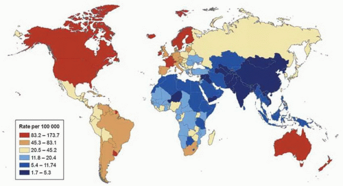
FIGURE 9-1 ▪ Worldwide variation in age-standardized prostate cancer incidence rates 2008. (Reprinted from Center MM, et al. International variation in prostate cancer incidence and mortality rates. Eur Urol 2012;61(6):1079-1092, with permission.)


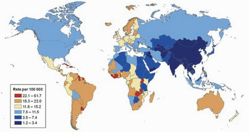
FIGURE 9-2 ▪ Worldwide variation in age-standardized prostate cancer mortality rates 2008. (Reprinted from Center MM, et al. International variation in prostate cancer incidence and mortality rates. Eur Urol 2012;61(6):1079-1092, with permission.)


Time Trends


Incidence Rate

Since the 1990s, in the United States, there was a dramatic 40% decline in prostate cancer deaths and 75% decrease in symptomatic presentation attributed by most experts to earlier tumor detection from widespread PSA screening.18 From 1988 to 1992, there was a large surge in prostate cancer diagnosis with an annual increase of 16.5% followed by an annual increase of 11.6% in 1992 to 1995, followed by relative stability, and then a small annual decline of 1.9% from 2000 to 2009 (Fig. 9-3).3,4 The initial surge essentially paralleled the introduction of PSA screening, which resulted in the identification of a substantial prevalence backlog of asymptomatic cases that later evened out with time.


Mortality Rate

Worldwide, decreasing mortality rates are also seen in many developed and high-resource countries. In the United States, mortality rates from prostate cancer increased from 1975 to 1991 and declined since the mid-1990s that was greatest and most sustained in men >75 years old.19 In both the United States and the United Kingdom, prostate cancer mortality peaked in the early 1990s at almost identical rates, but age-adjusted mortality in the United States subsequently declined by 4.2% per year, four times the rate of decline in the United Kingdom (1.1%).19 Decline in prostate cancer mortality can be attributed to several factors including improved treatments such as RP, radiation, and hormonal therapies (Fig. 9-4).746 Widespread PSA screening
is indirectly linked as a reason for decrease prostate cancer mortality. However, this is currently controversial, as recent large randomized PSA screening studies for prostate cancer in the United States did not show benefit and, in Europe, only 20% reduction in mortality.10,11 Further, decline in prostate cancer mortality was observed even in countries with low PSA screening such as United Kingdom.20


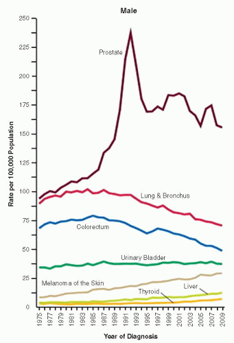
FIGURE 9-3 ▪ Cancer trends for men in the United States from 1975 to 2008. (Reprinted from Siegel R, Naishadham D, Jemal A. Cancer statistics, 2013. CA Cancer J Clin 2013;63(1):11-30, with permission.)


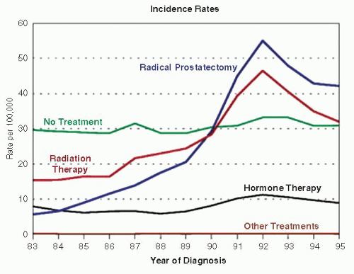
FIGURE 9-4 ▪ Incidence over time of prostate cancer treatment. (Reprinted from Stanford JL, Stephenson RA, Coyle LM, et al. Prostate Cancer Trends 1973-1995, SEER Program, National Cancer Institute, NIH Pub. No. 99-4543, Bethesda, MD, 1999, with permission.)


ETIOPATHOGENESIS


Introduction

The exact cause of prostate cancer remains elusive despite extensive research carried out in this field. Currently, older age, ethnicity (black race), and family history, which are all nonmodifiable factors, are well-established risks for prostate cancer.21 Epidemiologic studies show that immigrants from low-incidence regions acquire intermediate-risk level after migrating to high-risk areas suggesting also a role for environmental factors.

There is no single identified genetic event that can cause prostate cancer, and perhaps, neoplastic growth arises from multiple genetic alterations and/or epigenetic factors. It remains poorly understood why most of the prostate cancer diagnosed exhibits long latency and only a subset unpredictably transforms into clinically aggressive disease. Similar to nodular hyperplasia, growth and development of prostate cancer is dependent on androgen signaling. However, a subset of prostate cancer may progress even with an absent or suppressed level of androgen hormone (castration-resistant state), and these are generally incurable. The mechanism of evolution to androgen independence by prostate cancer is currently an intense area of research.


Risk Factors


Age

As discussed above, the incidence of prostate cancer sharply increases with age with about 60% of cases diagnosed in men >65 years old. Interestingly based on these statistics, if men live up to 100 years old, almost all will have prostate cancer.


Ethnicity

As discussed above, the incidence of prostate cancer is much greater in African-American men, which is 1.6 and 2.8 times higher than in whites and Asian Americans, respectively. Likewise, death from prostate cancer in African-American men is less than two times and about five times higher than in whites and Asian Americans, respectively. There is also an increased likelihood of a high Gleason score and more rapid tumor growth with earlier transformation from latent to aggressive disease in black than in white men.22,23

Several studies suggest that the higher incidence and mortality from prostate cancer in black men are partly due to genetic susceptibility.21,24 Genetic differences such as variants in alleles on chromosomes including 8q24 and 17q21-22, which are associated with increased risk for prostate cancer, are suggested to be higher in men of African descent.25, 26, 27, 28 Microsatellite marker DG8S737 on chromosome 8q24 has a population attributable risk of 16% in African Americans, which is higher than in European men (5% to 11%).28 The frequency of risk allele in 17q21 is about 5% in men of African descent, whereas it is rare in other races (<1%).25 Variant G allele of CYP3A4 associated with aggressive prostate cancer progression is much more frequent in African-American men (81%) than in white men (8%).29,30 Polymorphism of CYP17 may have a role in the susceptibility to prostate cancer in men of African but not of European descent.31 Mutation in ephB2 gene is associated with prostate cancer risk in African-American men with a family history of prostate cancer but is not found in white men.32


Family History

Familial association in prostate cancer is well documented.33,34 There is a twofold increased risk if 1 first-degree relative and a 9 times increased risk if 3 first-degree relatives are diagnosed with prostate cancer. Risk also increases inversely with age of diagnosis in relatives. If the age of onset is 50 years old, the relative risk is increased 7 times.

There are compelling evidence to suggest a genetic basis for familial predisposition in prostate cancer. High-risk alleles are identified with either autosomal dominant or X-linked mode of inheritance. Implicated genetic factors for hereditary prostate cancer include BRCA2 on chromosome 13q12, ELAC2 on chromosome 17p, RNASEL on chromosome 1q25, MSR1 on chromosome 8p22-23, NBS1 on chromosome 5, and CHEK2 on chromosome 22q.35,36


Diet

There is a positive association of prostate cancer with consumption of animal products, particularly red meat cooked at high temperatures.37, 38, 39 This association is suggested to be due to heterocyclic amine content in meat.38 A recent large prospective study suggested little or no association between fruit and vegetable intake and prostate cancer risk.40 In the large Selenium and Vitamin E Cancer Prevention Trial (SELECT) of 35,533 men, neither vitamin E nor selenium supplementation was shown to be significantly associated with prostate cancer risk.41


Obesity and Metabolic Syndrome

Several studies suggest obesity as a risk factor for prostate cancer.42,43 Elevated body mass index (BMI) increases the risk of prostate cancer mortality in prospective cohort studies and biochemical recurrence in prostate cancer patients.43 Adipose tissue may influence circulating levels of several bioactive substances that may affect the risk of developing prostate cancer.42 It is also suggested that obesity may modify genes of periprostatic adipose tissue to promote a favorable environment for prostate cancer progression.44 Overall,
the highly prevalent metabolic syndrome including central obesity, insulin resistance, dyslipidemia, and hypertension is suggested to have an association with prostate cancer.45


Environmental Factors

Increased risk for prostate cancer has been proposed with exposures to cadmium, pesticides, rubber, textile, and chemicals.46, 47, 48, 49 Vitamin D deficiency has been implicated as a risk for prostate cancer and may explain geographic differences in incidence due to light exposure, as ecologic studies have shown that mortality rates from prostate cancer are inversely correlated with levels of ultraviolet radiation.50 Earlier mortality analysis suggested a higher risk for prostate cancer in atomic energy workers, but this was seen to be declining.51 Farming is suggested as a risk factor for prostate cancer, but this increased risk may not be due to pesticide exposure.52


Vasectomy

There is conflicting evidence suggesting vasectomy as a risk factor for prostate cancer, although more recent data support a lack of association.53, 54, 55, 56, 57, 58


Others

There is a controversial association of xenotropic murine leukemia-related virus (XMRV) to prostate cancer after XMRV DNA was isolated in 6% of prostate cancer.59, 60, 61, 62, 63 Other later studies however did not show XMRV infection in prostate, and it is being suggested that the identification may be due to sample contamination.


Cell of Origin

The luminal acinar secretory cells were thought to be the cell of origin of prostate cancer since this tumor is characterized by phenotypic similarity to acinar cells (PSA positive, p63/HMWK negative) and absence of basal cells. Recent in vitro studies in animal models however suggest that basal cells are more likely the precursor cells of prostate adenocarcinoma.64 The cooperative effects of AKT via loss of PTEN, ERG, and androgen receptor (AR) in basal cells were able to reproduce the histologic and molecular features of prostate cancer including absence of basal cells and overexpression of alpha-methyl-CoA racemase (AMACR).64


Molecular and Genomic Basis

Prostate cancer, like most other cancers, exhibits multiple genomic alterations such as point mutations, microsatellite sequence changes, and chromosomal rearrangements (e.g., translocations, insertions, duplications, and deletions).36,65, 66, 67, 68, 69 The most common chromosomal alterations in prostate cancer are losses at 1p, 6q, 8p, 10q, 13q, 16q, and 18q and gains at 1q, 2p, 7, 8q, and Xq.66,70 Rearrangement in chromosome 22 between TMPRSS2 and ERG is seen in about half of prostate cancers and is discussed below in detail.71,72

Multiple putative prostate cancer susceptibility loci have been identified by linkage analysis including the HPC1 locus on 1q23-25 (RNASEL), HPC2 locus on 17p (ELAC2), PCAP locus on 1q42-43, CAPB locus on 1p36, HPC20 locus on 20q13, and HPCX locus on Xq27-28.30,73, 74, 75, 76, 77, 78, 79 Analysis among 426 families with hereditary prostate cancer identified a susceptibility region at 17q22 (BRCA1).80 Genome-wide association study identified a variant on 8q24 containing MYC oncogene that confers risk for prostate cancer.28 Nine SNP loci identified at this chromosome region were independently associated with prostate cancer risk.65 Another genome-wide association study identified 7 new prostate cancer susceptibility loci at 2p11, 3q23, 3q26, 5p12, 6p21, 12q13, and Xq12.59.81 About 50 prostate cancer susceptibility loci have now been identified.65

Multiple somatic genetic alterations in prostate cancer may result in inactivation of tumor suppressor gene or activation of oncogenes (Table 9-4).30,36,65 Among implicated in prostate carcinogenesis includes tumor suppressor genes GSTP1, PTEN, CDKN1B, NKX3.1, KLF6, Rb, and p53 and oncogenes c-myc, bcl-2, c-kit, and STAT5.36

PTEN is mutated in about 20% to 40% of prostate cancers (often 10q23 deletion), more identified in advanced prostate cancer suggesting a role in cancer progression.66,69,82 PTEN is present in normal prostatic epithelial cells and high-grade prostatic intraepithelial neoplasia (HGPIN) and is reduced in prostate cancers with high grade and stage.83,84 There is a significant co-occurrence of TMPRSS2:ERG and PTEN loss in prostate cancer, suggesting the possibility of PTEN loss as a late genetic event or “second hit” after ERG rearrangement.85 SPOP is the most frequent nonsynonymous mutated gene in prostate cancer, detected in 6% to 13% of prostate cancers.69 Interestingly, unlike PTEN loss, SPOP alterations are present in TMPRSS2:ETS-negative prostate cancer and may represent a different class of prostate cancers.65 The mutually exclusive occurrence of TMPRSS2:ERG and SPOP forms the basis for a possible molecular classification of prostate cancer (Fig. 9-5).

Alterations in AR may lead to AR activation in prostate cancer that may allow them to survive in an environment deprived of androgen hormone (castration resistant).86 AR is mutated or amplified in 20% to 30% of castration-resistant prostate cancers.87,88 Polymorphisms are also observed in genes associated with androgen biosynthesis.65

Hedgehog signaling pathway has been shown to play a role in growth and metastasis of prostate cancer.89,90


Gene Fusion

The first chromosome translocation identified in prostate cancer was t(6;16), which results in the TPC:HPR fusion.91

Fusion of TMPRSS2 and ETS gene family is specific for prostate cancer detected in about 50% of cases.71,72,92, 93, 94 TMPRSS2 encodes for serine protease secreted by prostatic cells in response to androgen exposure, and this explains why the gene fusion leads to androgen-responsive expression of
ETS transcription factors.95,96 ETS family of transcription factors includes ERG, ETV1, ETV4, and ETV5 as 3′ end fusion partners, and ERG is the most commonly fused gene at 5′ end with TMPRSS2 comprising about 90% of cases. Several other 5′ end fusion partners of ETS gene have been identified (Table 9-5). With fusion, ERG is brought under the control of an androgen-regulated promoter causing overexpression. Fusion of the two genes occurs by intra- and interchromosomal genetic rearrangements. In about two-thirds of cases, fusion results from deletion of the intervening 3 Mb between TMPRSS2 and ERG (Fig. 9-6).94 Fusion may also occur by more complex rearrangements such as translocation. The most commonly reported fusion transcript is between exon 1 of TMPRSS2 and exon 4 of ERG. About 20 TMPRSS2:ERG transcripts have now been identified. Morphologic features of prostate cancer associated with TMPRSS2:ERG include blue-tinged mucin, cribriform pattern, intraductal spread, macronucleoli, and signet ring cells.97 Only 24% of tumors without any of these features displayed TMPRSS2:ERG, whereas 93% of cases with three or more features harbor the fusion.97 The clinical significance of this gene fusion on prostate cancer behavior is not yet fully understood, and studies correlating it with outcome and pathologic variables have conflicting results.98, 99, 100, 101, 102, 103, 104, 105, 106, 107


Table 9-4 ▪ MUTATED GENES IN PROSTATE CANCER


Gene


Chromosome


CHD5


1p36.31


SDF4


1p36.33


EPHB2


1p36.12


SPTA1


1q23.1


SRD5A2


2p23.1


THSD7B


2q22.1


SCN11A


3p22.2


PLXNB1


3p21.31


PRKCI


3q26.2


PIK3CA


3q26.32


ZNF595


4p16.3


CHD1


5q15-q21.1


APC


5q22.2


HSP90AB1


6p21.1


DLK2


6p21.1


HDAC9


7p21.1


EGFR


7p11.2


BRAF


7q34


HSPA5


9q33.3


KLF6


10p15.1


PTEN


10q23.31


CDKN1B


12p13.1


KRAS


12p12.1


MLL2


12q13.12


RB1


13q14.2


GPC6


13q31.3-32.1


FOXA1


14q21.1


HSPA2


14q23.3


DICER


14q31.13


MYH11


16p13.11


ZFHX3


16q22.2-q22.3


TP53


17p13.1


CDK12


17q12


SPOP


17q21.33


ASXL1


20q11.21


CHEK2


22q12.1


KDM6A


Xp11.3


AR


Xq12


MED12


Xq13.1


HPRT1


Xq26.2-q26.3


Reprinted from Boyd LK, Mao X, Lu YJ. The complexity of prostate cancer: genomic alterations and heterogeneity. Nat Rev Urol 2012; 9(11):652-664, with permission.


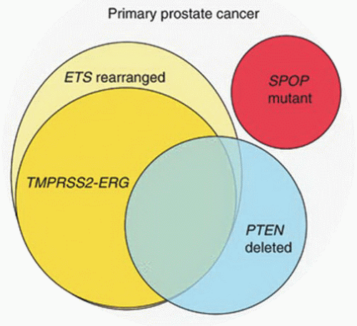
FIGURE 9-5 ▪ Molecular classification of prostate cancer. About 50% of prostate cancers harbor ETS rearrangement, and majority of these are TMPRSS2:ERG. PTEN is deleted in 20% to 40% prostate cancers, with significant overlap to ETS rearrangements. SPOP mutation is mutually exclusive with ETS rearrangement. (Reprinted from Barbieri CE, Demichelis F, Rubin MA. Molecular genetics of prostate cancer: emerging appreciation of genetic complexity. Histopathology 2012;60(1):187-198, with permission.)


CLINICAL FEATURES


Symptoms and Signs

In the United States, the vast majority of prostate cancers are diagnosed in asymptomatic patients through early detection programs. The main reasons for performing prostate
biopsies leading to cancer diagnoses are elevated PSA and/or abnormal digital rectal examination. Prostate cancer is also a common incidental finding in 28.5% cystoprostatectomy specimens, of which only 25.3% are considered clinically significant, defined by most experts as tumors with a volume of 0.5 mL or more and Gleason score of 7 or higher.106,107


Table 9-5 ▪ DETECTED ETS GENE FUSIONS IN PROSTATE CANCER


5′ Partner


3′ Partner


TMPRSS2


ERG


HERPUD1


ERG


SLC45A3


ERG


NDRG1


ERG


FKBP5


ERG


TMPRSS2


ETV4


DDX5


ETV4


CANT1


ETV4


KLK2


ETV4


TMPRSS2


ETV5


SLC45A3


ETV4


SLC45A3


ETV1


SLC45A3


ETV1


TMPRSS2


ETV1


SLC45A3


ETV1


C15orf21


ETV1


HNRPA2B1


ETV1


FLJ35294


ETV1


ACSL3


ETV1


EST14


ETV1


HERVK17


ETV1


HERVK22Q11.23


ETV1


FOXP1


ETV1


KLK2


ETV1


FUBP1


ETV1


SNURF


ETV1


Reprinted from Spans L, et al. The genomic landscape of prostate cancer. Int J Mol Sci 2013;14(6):10822-10851, with permission.


In symptomatic patients, prostate cancer usually manifests with symptoms indicative of advanced disease. Presentations may include irritative (e.g., frequency, urgency) or obstructive (e.g., hesitancy, dribbling) voiding symptoms. Cancers located in transition zone may manifest earlier because of its proximity to the urethra. These lower urinary tract symptoms, however, are not specific for prostate cancer and, when encountered, are more often attributed to hyperplasia. About 10% to 15% of transurethral resection specimens performed for hyperplasia however may contain incidental prostate cancer. Local extension of tumor to structures adjacent to prostate may produce pelvic pain. Skeletal metastasis may result to bone pain and tenderness, spinal cord compression, weakness of lower extremities, and urinary or fecal incontinence. Metastasis to lymph nodes may produce adenopathy, and lower extremity lymphedema in there is inguinal lymph node involvement. Rarely, advanced prostate cancer may present as disseminated intravascular coagulation, nonbacterial thrombotic endocarditis, malignant ascites, or pleural effusion from tumor dissemination. Paraneoplastic syndrome (e.g., syndrome of inappropriate ADH secretion, hypercalcemia, DIC, thrombotic thrombocytopenic purpura, neurologic syndromes) may occur with prostate cancer and is seen more often, but not always, when the histology contains small cell carcinoma component.

The hallmark physical finding for prostate cancer is the presence of palpable prostatic nodule or firmness on rectal examination. It should be noted that most patients with prostate cancer have normal rectal exams. In about 25% of cases, serum PSA is not elevated, and abnormal rectal exams is the reason for prostate biopsy. Other signs of prostate cancer are related to advanced stage such as hyperreflexia and increased bulbocavernosus reflex from cord compression of bone metastasis.

Aside from serum PSA elevation detailed below, abnormal laboratory findings in prostate cancer may reflect presence of advanced disease. These include azotemia (increase BUN and creatinine) from bilateral urinary obstruction, increased alkaline phosphatase from bone metastasis, and anemia and rarely low platelets from DIC or TTP.


Prostate-Specific Antigen

Prostate-specific antigen (PSA) is currently the most widely used tumor biomarker in medicine. PSA is an androgen-regulated serine protease of the human kallikrein (hK) family located on chromosome 19q13.4.108 PSA is synthesized by secretory cells of normal, hyperplastic, or malignant prostatic acinar cells. Thus, while detectable levels are considered specific for prostatic origin, it is not a specific marker for cancer of the prostate. Nonneoplastic causes of PSA elevation include BPH, inflammation or prostatitis, ejaculation, and injury or manipulation (e.g., recent needle biopsy, bicycle ride).

When prostate cancer disrupts the basement membrane, there is leakage of PSA into the peripheral blood. Most of the circulating PSA (about 70% to 90%) is bound to α1-antichymotrypin and a subset with other protease inhibitors such as α2-macroglobin and α1-antityrpsin. Most anti-bodies currently used in PSA assays detect free and most of the bound form, except for that, which is complexed with α2-macroglobin. Detection by PSA elevation is more sensitive than rectal examination in detecting prostate cancer. The introduction of PSA as an oncologic marker and its integration into practice with rectal examination and transrectal ultrasound allow for the detection of early-stage curable prostate cancer. Serum PSA level is also a good measure in monitoring response after treatment and to diagnose disease recurrence.

Certain factors may influence PSA level. 5α-reductase inhibitors (finasteride) may cause artificial lowering of PSA level by about 50%. Patients with a high BMI may have lower PSA values because of hemodilution.109 Some modifications of measurement and interpretation in PSA are being
used to enhance sensitivity and specificity in diagnosis of prostate cancer discussed below.


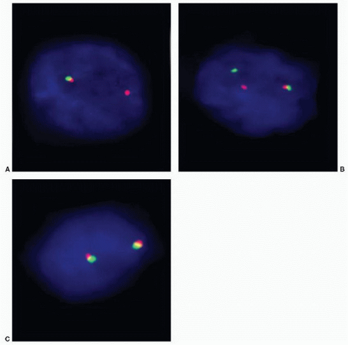
FIGURE 9-6 ▪ FISH for TMPRSS2:ERG gene rearrangement in prostate carcinoma showing (A) deletion (1 Edel) and (B) split (Esplit). The (C) normal pattern is observed in about half of prostate cancers. (Courtesy of Glen Kristiansen, MD and Sven Perner, MD, PhD.)


PSA Screening

The aim of screening programs for a particular cancer is to ultimately reduce cancer-related deaths. PSA screening in asymptomatic men is currently controversial because of the lack of definitive evidence in reducing prostate cancer mortality.10, 11, 12,110, 111, 112 Amidst the ongoing controversy, most urology experts advocate PSA screening mainly because (a) there was a 40% decline in prostate cancer mortality since the widespread application of PSA screening from the early 1990s, (b) most tumors detected by PSA screening are low-grade low-volume tumors that can be cured, and (c) treatment options are available for these tumors.

In 2009, the two largest randomized studies on population-based PSA screening in the United States and Europe showed no or only limited benefit in reducing prostate cancer mortality.10,11 The Prostate, Lung, Colorectal, and Ovarian Cancer Screening Trial (PLCO) in the United States included 76,693 men randomized for PSA screening (38,343 men) and usual care as control (38,350 men).10 After 7- to 10-year follow-up, the death rate from prostate cancer in the PCLO study was very low and did not differ significantly between the
study and control groups. In a larger European Randomized Study of Screening for Prostate Cancer (ERSPC) study that involved 7 countries, 162,243 men were randomized to care with and without PSA testing.11 Results of the ERSPC study with median follow-up of 9 years showed that PSA-based screening reduced prostate cancer death rates by 20% but was associated with high detection and treatment of indolent tumors. In practical terms, 1,410 men must be screened, and additional 48 men must be treated to prevent one death from prostate cancer.11

Another study from Sweden of 9,026 men with longer follow-up of 20 years also did not show a significant difference in deaths between men with and without PSA screening.12 In ERSPC, a subset study in Rotterdam showed that disease-specific survival of men with interval cancer screened every 4 years was similar with and without PSA screening.111 Few other, but lesser quality, studies showed benefits of PSA screening such as in Quebec City involving 46,486 men showed significant reduction in prostate cancer mortality in the PSA-screened group versus the nonscreened group.113

In great part due to the concurrent PCLO and ERSPC studies, the US Preventive Services Task Force (USPSTF) issued a grade D recommendation or essentially recommending against population-based PSA screening for prostate cancer.114 The USPSTF recommendation is to avoid unnecessary over treatment of prostate cancer leading to complications; the latter should be weighed against the benefit of PSA screening. This recommendation received strong opposition from urology advocate groups such as the American Urological Association (AUA), Prostate Cancer Foundation, Large Urology Group Practice Association, Men’s Health Network, and others.115,116 Arguments against the studies included high (50%) contamination rate by PSA measurement in the PCLO control group (essentially making the study one of regular versus inconsistent PSA screening), whereas the ERSCP did not specify contamination, many subjects with abnormal PSA tests did not have prompt biopsy, the follow-up was relatively short considering the long latency of prostate cancer, and that there was no consideration of high-risk groups particularly black men.116


PSA Cutoff

Currently, most guidelines recommend PSA screening in men between 40 and 50 years old, and the younger age will have less confounding effect of hyperplasia.117, 118, 119, 120 Some recommend PSA screening only in men with longer life expectancy (>10 years expectancy or <75 years old), since these patients will likely die of other reasons and not from prostate cancer.117,120 Yearly PSA testing is recommended, although may opt for follow-up testing at longer intervals if PSA level is low (<1 ng/mL).

The traditional cutoff for PSA elevation to do prostate biopsy is 4 ng/mL. This cutoff has a sensitivity of about 20% and specificity of 60% to 70% for prostate cancer. Lower PSA cutoffs are considered in higher-risk individuals such as African-American men or those with family histories of prostate cancer. Higher cutoff increases the predictive value; a cutoff of 10 ng/mL has a positive predictive value of 42% to 71%. Of note, no level of PSA is risk free and that the PSA value represents a risk continuum. The AUA in particular does not recommend any PSA cutoff value to prompt a biopsy and recommends that other factors such as family history, ethnicity, and rectal examination findings be taken into account.


Posttherapy PSA Monitoring

After therapy of localized prostate cancer, PSA is expected to decline to a nadir of <0.2 ng/mL, which is used to determine cure. PSA nadir is usually achieved 6 weeks after RP since all benign glands and cancer are removed, but is much longer with radiotherapy (about 27 weeks) as it takes time for cancer to degenerate. PSA elevation afterward (≥0.2 ng/mL on two occasions) is considered biochemical recurrence.121


Other PSA Measurements


Age-Specific PSA Ranges

Age-specific PSA ranges are considered because they increase the sensitivity in detection in younger men and increase the specificity for prostate cancer in older men. The prostate is usually larger in older male because of hyperplasia; thus, higher PSA value is permissible (e.g., 6.5 ng/mL for men 70 to 79 years old versus 2.5 ng/mL for men 40 to 49 years old) to perform biopsy. However, use of age-specific PSA ranges is controversial particularly in older men because increasing the cutoff value may lead to missing significant numbers of clinically important cancer.


PSA Dynamics (Velocity, Doubling Time)

Changes in PSA level over time may be used to stratify risk for prostate cancer including in predicting response to therapy. PSA velocity refers to the relative change in time of PSA value.122, 123, 124 An increase of >0.75 ng/mL/y is considered a significant risk factor for prostate cancer that would prompt a prostate biopsy. It is important that the same laboratory is used to measure PSA levels over a period of at least 18 months. A very rapid rise in PSA value can be seen in prostatitis.125 PSA doubling time refers to the amount of time required for PSA to double.126,127 Use of pretreatment PSA velocity and doubling time is currently controversial. While both are associated with outcome, there is no clear evidence that they improve outcome prediction beyond pretreatment PSA alone.125,128


PSA Density

PSA density takes into account the variation in size of the prostate that may influence the PSA level. PSA density is serum PSA divided by the prostate gland volume, and a higher value suggests increase risk for prostate cancer. A PSA density of >0.15 ng/mL/cm3 would be the recommended
cutoff to perform a biopsy. Others however have found this test to be inaccurate. The problems are that the prostate may have a larger size also because of increase in stromal volume and TRUS is not accurate in measuring prostate volume. The positive predictive value of PSA density is only slightly higher than the 4 ng/mL cutoff for serum PSA. Further, performing TRUS is still an uncomfortable procedure for the patient even without biopsy. A modification of PSA density adjusts for the transition zone volume.


Free PSA (fPSA)

About 90% of PSA in circulation is bound to α1-antichymotrypsin, and a smaller amount is bound to other serum protease inhibitors. Unbound PSA is known as fPSA. Low level (<10%) of fPSA is associated with a higher risk of prostate cancer and can be useful in monitoring patients after therapy. A large multicenter study in men with normal rectal examinations and PSA of 4 to 10 ng/mL showed that fPSA with cutoff of 25% was able to detect 95% of cancers while avoiding 20% of unnecessary biopsies.129 Another approach is to determine the ratio of fPSA to total PSA, and lower value increases the specificity in diagnosing cancer.


PUTATIVE PRECURSOR LESIONS


High-Grade Prostatic Intraepithelial Neoplasia


Introduction

Since 1926, the model for the transition of benign acinar epithelium to malignant cellular change via intermediate “atypical glands” in the prostate has been proposed by several authors. In 1949, Andrews illustrated precancerous conditions, which he identified in 70% of the glands in the prostate containing carcinoma compared to only 26% of glands without carcinoma.130 The atypical glandular foci were characterized by cellular stratification, papillary formation, nuclear enlargement, and mitotic activity—many features that in contemporary practice would be identified in prostatic intraepithelial neoplasia (PIN). McNeal’s landmark work in 1965 described the morphogenetic origin of prostate carcinoma that became the foundation for our current understanding of PIN.131,132 In 1986, McNeal and Bostwick133 characterized this premalignant lesion as “intraductal dysplasia” describing it to be present in more than half of carcinomatous prostates. One year later, Bostwick and Brawer134 introduced the now preferred term “prostatic intraepithelial neoplasia,” in part, to recognize that this lesion may arise from either the prostatic acini or ducts, which are often indistinguishable from each other. In 1989, the term PIN was endorsed in a consensus conference and since then became the terminology uniformly used in the literature.135

PIN is defined as a noninvasive neoplastic transformation of the epithelium of preexisting prostatic ducts and acini.136, 137, 138, 139, 140, 141 Originally, PIN was divided in three grades (PIN I, II, and III) based on a spectrum of architectural and cytologic abnormalities,133 with the suggestion that PIN III was equivalent to carcinoma in situ. At the 1989 consensus meeting, PIN was condensed into low grade (I) and high grade (II and III) because of poor diagnostic reproducibility.135 The separation of low- and high-grade PIN was based on a number of criteria but especially the presence of nucleolar prominence in the latter. The reporting of low-grade PIN eventually fell into disfavor due to its poor diagnostic reproducibility and the lack of clinical relevance including its questionable association with prostate cancer.142,143 Nowadays, the word PIN is used almost synonymously to refer to HGPIN.


Epidemiology

HGPIN is a common finding in routine prostate pathology specimens, identified as an isolated diagnosis in up to 16% of needle core biopsies (NCBs) (usually 5% to 10%) and 1% to 5% of TUR specimens.144,145 Approximately, 115,000 new cases of isolated HGPIN are diagnosed each year in the United States.137,146 There is a significant variation in the reporting of HGPIN of 0.7% to 20% in needle biopsies and 3% to 33% in TURs that can be attributed to interpretation discrepancies and varied application of morphologic criteria.139 The incidence is much higher at 80% to 100% in prostatectomies harboring adenocarcinoma compared to 43% of age-matched nontumorous controls. The incidence of HGPIN, like that of adenocarcinoma, increases with age although with somewhat earlier onset, beginning in the third decade of life reaching about 67% in white men by the eighth decade of life.147,148 Like prostate adenocarcinoma, the incidence is higher in African Americans compared to other races.149, 150, 151, 152 The lesion is usually more diffuse and presents earlier in African Americans compared to Caucasian Americans. HGPIN is detected with increasing incidence in African Americans with rates of 7%, 26%, 46%, 72%, 75%, and 91% from the third through the eight decade.148 This strong association with race is not limited to the United States, as similar higher incidence is reported among black Brazilian men compared to white Brazilian men.153


PIN as a Precursor for Cancer

There is much evidence supporting PIN as a precursor lesion for prostate cancer,145 including (a) the spatial relationship of HGPIN to foci of carcinoma, including the budding off of early invasive glands; (b) HGPIN is more common and multifocal in prostates containing carcinoma154, 155, 156, 157; (c) a significant subset of HGPIN and carcinoma harbor similar molecular alterations (e.g., TMPRSS2:ERG fusion,72,158 AMACR overexpression159,160); (d) both HGPIN and carcinoma incidence increase with age and are more common in black men; and (e) appearance of HGPIN in the prostate precedes carcinoma by about 10 years.

Historically, the diagnosis of isolated HGPIN in a prostate biopsy would prompt repeat biopsy, with carcinoma
being detected in 40% to 60% of cases.161, 162, 163, 164, 165, 166, 167, 168 However, contemporary data in the era of extended prostate biopsy sampling have shown that the risk of cancer following diagnosis of HGPIN is between 21% and 27.5%.144,169 This risk of finding cancer after unifocal HGPIN is not significantly different from the risk following a benign diagnosis.144,155,156 Extended biopsy sampling has resulted in higher sensitivity for cancer detection, and therefore, less carcinoma is detected on subsequent samples.155

However, patients with multifocal HGPIN (i.e., present in more than 2 cores), bilateral HGPIN, and those associated with an atypical small acinar proliferation (ASAP) diagnosis on biopsy have a higher risk of harboring concomitant prostate carcinoma and should be more aggressively followed.154, 155, 156, 157,170, 171, 172, 173 Multifocal and bilateral HGPIN are considered adverse features significantly increasing the risk of prostate cancer detection; other clinical variables such as serum PSA and abnormal rectal examination are taken into account.174 The estimated probabilities in detecting cancer 1 and 5 years after a benign diagnosis are 3.7% and 22.5% and are much higher with multifocal HGPIN at 9.1% and 47.8% and bilateral multifocal HGPIN at 12.5% and 57.8%, respectively.174


Molecular Biology

There is genetic evidence associating HGPIN and prostate carcinoma. HGPIN and cancer share similar chromosomal anomalies, telomere shortening, alterations in members of the bcl-2 gene family, decreased expression of NKX3.1 and p27, and overexpression of p16, p53, MYC, and AMACR.160,175, 176, 177, 178, 179, 180, 181, 182, 183, 184, 185, 186 TMPRSS2:ERG fusion, which is seen in about half of clinically localized prostate cancers, can also be detected in 16% to 21% of HGPIN that is usually intermingling with cancer foci.72,158,187 When TMPRSS2:ERG is detected in cancer and in HGPIN in the same prostate, the matching lesions usually share the same fusion pattern.158 Sixty percent of TMPRSS2:ERG fusion-negative prostate cancer has also concomitant fusion-negative HGPIN.158

DNA aneuploidy is common in both HGPIN (65%) and cancer (62%).185 There is high correlation (75% of cases) in ploidy and pattern of cytogenetic alterations between HGPIN and paired prostate cancer foci in the same specimen.185 Numeric alterations of chromosomes 7, 8, 10, 12, and Y are common in both HGPIN and carcinoma, although the mean overall number of alterations is higher in carcinoma.175 Chromosome 8p deletion is the most commonly detected allelic loss, present in both HGPIN and carcinoma.146 A genetic pathway for prostate carcinogenesis has been proposed with two distinct initiating events, namely, 8p and 13q losses.188 Loss of 8p leads to development of HGPIN and carcinoma, whereas loss of 13q leads to carcinoma without the presence of HGPIN. These primary chromosomal imbalances are then preferentially followed by 8q gain; 6q, 16q, and 18q losses; and a set of late events that make recurrent and metastatic prostate cancers genetically more complex.188 Nuclear morphometric studies also show no significant difference in nuclear volume between HGPIN and cancer, but both show significant increments from benign prostatic glands.189,190 p53 mutation is suggested to be an early change in prostate carcinogenesis, and p53 overexpression associated with chromosomal instability is greater in HGPIN foci intermingled with cancer compared to HGPIN away from cancer.186 Hypermethylation of glutathione S-transferase, considered to be a major event in prostate carcinogenesis, can be detected in HGPIN.191,192


Clinical Features

Isolated HGPIN does not cause any specific symptoms. The lesion is encountered as an incidental finding in prostate specimens, often in biopsies performed for an abnormal serum PSA level. It is however debatable whether or not HGPIN is the actual cause of the elevated PSA since it is difficult to exclude an undetected coexistent carcinoma, and in some cases, the PSA elevation may relate to BPH or inflammation. By ultrasound, it has been suggested that HGPIN may be associated with a hypoechoic pattern in the peripheral zone similar to carcinoma.193,194

No treatment (e.g., surgery, radiotherapy, hormonal therapy) is warranted for HGPIN, unless a concomitant carcinoma is identified. Several clinical trials are being undertaken targeting HGPIN using different agents, including antiandrogens, 5α-reductase inhibitors, and even dietary nutrients and supplements, as chemopreventive measures for the development of prostate carcinoma.137

As discussed above, the current clinical practice is to perform a repeat biopsy after a diagnosis of HGPIN when it is multifocal, bilateral, or associated with ASAP.155,157 If carcinoma is detected on repeat biopsy, it may or it may not be present in or adjacent to quadrant where HGPIN was detected.165,167,195 In 40% to 74% of cases, the carcinoma will be detected at the area of HGPIN.165,167,196 On average, 30% of carcinomas are found on the contralateral side.144 The incidence of detection of subsequent carcinoma in patients with isolated HGPIN increases when rebiopsy is performed at 1 and 3 years. At mean biopsy intervals of 34 months (first rebiopsy) and 66 months (second rebiopsy), cancer was detected at 22% and 23%, respectively, with a high likelihood for organ-confined and clinically significant disease.197 Thus, while rebiopsy within 1 year may not be that crucial, biopsy at longer intervals can be beneficial and should be considered as a valid follow-up option.


Pathology

HGPIN is not associated with any recognizable gross findings. Histologically, PIN consists of preexisting ducts and acini, usually of medium to large size, lined by crowded secretory cells with abnormal cytologic features. PIN is divided into low and high grade. Low-grade PIN exhibits tufted or micropapillary patterns with nuclear crowding,
stratification, and irregular spacing. Nuclei are mildly enlarged, and, in particular, nucleoli are inconspicuous to only rarely prominent.

HGPIN shows proliferation of medium- to large-sized glands. The cells have increased basophilia or amphophilia that can readily be detected at low-power magnification. The nuclei are larger, are round, show overlapping, are hyperchromatic, have nuclear membrane irregularity, and, most importantly, show prominent nucleoli easily appreciable at 20× magnification (Fig. 9-7). Multiple nucleoli may be present and, similar to carcinoma, are occasionally peripherally situated close to the nuclear membrane. The diagnostic threshold for HGPIN varies, as some individuals require all cells to be atypical and others require at least 10% of the cells to have prominent nucleoli. If the cytologic threshold for diagnosis is doubtful or borderline, our practice bias is not to consider the lesion as HGPIN. A preserved or discontinuous basal cell layer may be readily identified on routine slides or with use of basal cell-specific immunostains.

PIN may exhibit several architectural and cytologic features (Table 9-6). There are four major architectural patterns described for HGPIN, namely, tufted, micropapillary, cribriform, and flat HGPIN (Fig. 9-8).198 Multiple patterns of HGPIN may be seen concurrently within a specimen. Tufted PIN is the most common pattern seen in 87% of PIN and is characterized by stratification of acinar cells imparting luminal undulations or folds (Fig. 9-8A). Micropapillary PIN is also common (85%), which shows nuclear stratification forming intraluminal slender filiform projections and cellular budding (Fig. 9-8B). Cribriform PIN (32%) shows complex intraluminal proliferation resulting in multiple irregular or round punched-out lumina (Fig. 9-8C). Cribriform PIN in particular may show “cellular maturation” wherein peripheral cells show greater nuclear atypia (i.e., nucleomegaly, prominent nucleoli) than do cells at the luminal or central aspect. Flat PIN (25%) lacks significant cellular stratification and is composed of only 1 or 2 cell layers (Fig. 9-8D). Other uncommon types have been described such as foamy (Fig. 9-9), inverted or “hobnail” (Fig. 9-10), small cell (Fig. 9-11), signet ring, mucinous, and PIN with squamous differentiation.199, 200, 201, 202 Rarely, PIN may involve large cystic glands and glands in nodular hyperplasia and exhibit mucinous metaplasia.198 Central necrosis is not a feature of HGPIN, and its presence should raise concern for an invasive or intraductal carcinoma (IDC).


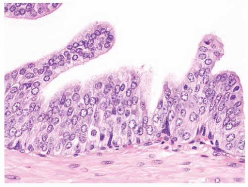
FIGURE 9-7 ▪ HGPIN is characterized by presence of high-grade nuclei.


Table 9-6 ▪ COMMON AND UNUSUAL PATTERNS OF HGPIN


Basic Architecture


Tufted


Micropapillary


Cribriform


Flat


Other Patterns


Foamy


Inverted or “hobnail”


Small cell


Signet ring


Mucinous


PIN with squamous differentiation


Miscellaneous Features


Apocrine snouts


Cytoplasmic lipochrome


Paneth cell-like change


Several other features may be present in HGPIN including luminal cytoplasmic blebs, epithelial arches, cellular trabecular epithelial bars, “Roman” bridges, partial gland involvement, and basal cell layer disruption with glandular budding. Additionally, luminal cytoplasmic blebs, cytoplasmic lipochrome deposits (Fig. 9-12), and scattered Paneth-like cells (Fig. 9-13) may be present in HGPIN. Small round blue-tinged vacuoles (so-called blue blobs) are present in increased frequency in HGPIN compared to nonneoplastic glands. Rarely, HGPIN may also exhibit apical snouts (Fig. 9-14). A variety of luminal features may also be observed in HGPIN such as amorphous proteinaceous secretions, corpora amylacea, exfoliated cells, and rarely crystalloids. The basic approach for diagnosis is to screen for HGPIN at low-power magnification and to confirm the cytologic features at high-power, similar to cancer. Interobserver reproducibility for the diagnosis of HGPIN is fairly high among urologic pathologists and is moderate among general surgical pathologists.144 HGPIN is noninvasive and must be associated with of basal cells (Fig. 9-15). The basal cells can be continuous and readily apparent on hematoxylin and eosin (H&E) sections or discontinuous. A discontinuous basal cell layer may not be appreciable in a particular plane of section, compounding the differential diagnosis with carcinoma. Use of basal cell markers (p63, CK5/6, HMWK [keratin 34βE12]) may highlight the discontinuous basal cell layer around the involved acini or ducts.203 AMACR stains the dysplastic or neoplastic cells, but immunostaining can be variable at 44% to 90%.159,204, 205, 206, 207 HGPIN cells stain positively for PSA, prostate-specific acid phosphatase (PSAP), prostate-specific membrane antigen (PSMA), and pro-PSA.204,208,209 Small cell HGPIN does not show reactivity for synaptophysin and chromogranin, suggesting that it is not a manifestation of neuroendocrine differentiation.210


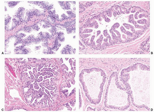
FIGURE 9-8 ▪ Basic patterns of HGPIN include (A) tufted, (B) micropapillary, (C) cribriform, and (D) flat architectures. Note the presence of cell maturation in cribriform HGPIN.


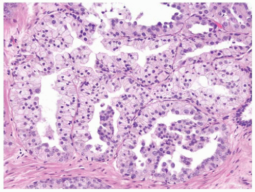
FIGURE 9-9 ▪ HGPIN with foamy cells.


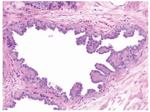
FIGURE 9-10 ▪ HGPIN with inverted pattern.


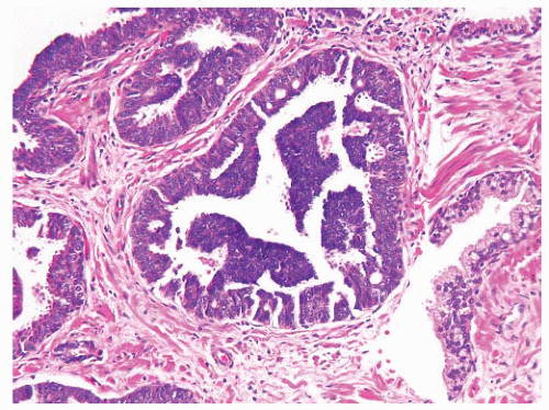
FIGURE 9-11 ▪ Small cell HGPIN. The small cells do not express neuroendocrine markers.

If HGPIN (without invasive foci) is found in prostate needle biopsy specimens, it is prudent to consider performing deeper sections particularly if HGPIN is extensive or associated with ASAP (glandular atypia). If isolated HGPIN is detected in a TUR, further sampling of the specimen may be considered depending on factors such as the patient’s age and PSA value. The presence of HGPIN in biopsies without concomitant invasive foci should be reported. The site and number of biopsy cores involved by HGPIN should also be documented. Further, the number of foci of HGPIN (e.g., isolated, focal, multifocal, extensive) should be reflected in the report. In the presence of carcinoma in or a needle biopsy (resection), reporting of HGPIN becomes optional. However, in the setting of a small focus of carcinoma, the presence of extensive HGPIN may be taken into account in determining treatment.


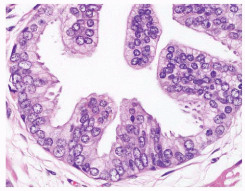
FIGURE 9-12 ▪ Lipochrome pigments may sometimes be seen in HGPIN.


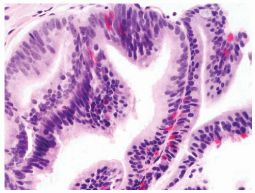
FIGURE 9-13 ▪ HGPIN with scattered Paneth-like cells.


Differential Diagnosis

A variety of carcinomas especially adenocarcinoma variants enter the differential diagnosis of HGPIN, and distinction among these can be difficult, particularly in limited samples. Of note, acinar adenocarcinoma with a cribriform pattern, ductal adenocarcinoma, and IDC (intraductal spread of cancer) may cause diagnostic difficulties. High-grade acinar carcinoma with a cribriform pattern (Gleason grade 4 or 5) shows architectural overlap with cribriform HGPIN. Unlike HGPIN,
cribriform adenocarcinoma exhibits more complexity including the presence of large expansile glands (bigger than preexisting glands), confluence or back-to-back glands, consistent cribriform architecture, and associated solid nests or may have intraluminal necrosis (Gleason pattern 5). Basal cells are absent in invasive cribriform carcinoma, but this may need to be confirmed with basal cell immunostains. Ductal adenocarcinoma may resemble the different architectures of HGPIN. Ductal adenocarcinoma is characteristically composed of tall columnar cells that are frequently pseudostratified and can be mitotically active. Unlike HGPIN, ductal adenocarcinoma may have large expansile glands and papillae with true fibrovascular stalks, and the cells do not exhibit luminal “maturation.” Invasive features such as crowded back-to-back glands, stromal fibrosis, perineural invasion (PNI), and extraprostatic extension (EPE) are present in ductal adenocarcinoma. Basal cells are absent in the majority of ductal adenocarcinomas that can be confirmed with basal cell immunostains. IDC of the prostate can be very difficult to distinguish from HGPIN due to the lack of a consistent morphologic cutoff separating these two entities. IDC like HGPIN involves large glands containing markedly atypical cells and notably has preserved basal cells (see subsequent section). Unlike HGPIN, the glands of IDC can be expansile and show extensive cribriform change, sometimes with comedonecrosis. A lumen-spanning proliferation of cells with marked atypia and mitoses is usually seen associated with high-volume and high-grade carcinoma (Gleason pattern 4 or 5). However, without an invasive focus, IDC can be very difficult to distinguish from HGPIN in limited biopsy material. Sometimes, a descriptive term such as “markedly atypical cribriform proliferation” or “atypical cribriform lesion” can be used and the differential diagnosis explained in a comment. Generally such cases require additional biopsies.


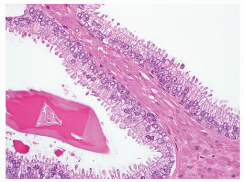
FIGURE 9-14 ▪ HGPIN with cells with apical snouts.


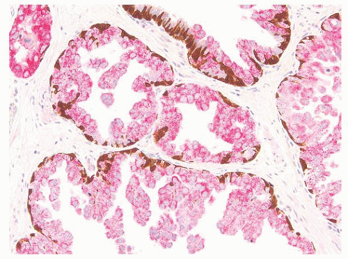
FIGURE 9-15 ▪ Cocktail of AMACR, HMWK, and p63 on HGPIN shows the presence of basal cells (brown) and increased AMACR expression (red).


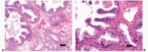
FIGURE 9-16 ▪ Examples of (A) HGPIN with adjacent Gleason pattern 3 carcinoma (arrow) and (B) HGPIN with adjacent atypical glands that may represent outpouching (HGPIN-ATYP).

In needle biopsy, when an atypical small glandular focus is identified next to HGPIN (so-called PIN-ATYP or PIN-ASAP), distinction should be made between a tangentially sectioned outpouching of HGPIN (so-called transitive glands) and a small focus of low-grade acinar adenocarcinoma (Gleason pattern 3) (Fig. 9-16). Immunostains are useful only if basal cells are demonstrated in the small glands, confirming outpouching of HGPIN. Even if no basal cells are present, they still could represent HGPIN, as the basal cell layer can be discontinuous or markedly attenuated. If a definitive diagnosis cannot be reached, our approach is to label the entire focus as “HGPIN with an atypical small acini suspicious but not diagnostic for carcinoma” or similar terminology.

Other malignant differential diagnoses for PIN include stratified or “PIN-like” adenocarcinoma, basal cell/adenoid cystic carcinoma, and urothelial carcinoma spreading through prostatic ducts and acini. “PIN-like” adenocarcinoma has stratified epithelium and may form medium-sized glands and, as the name implies, resemble HGPIN particularly flat or tufted patterns. Unlike HGPIN, “PIN-like”
adenocarcinoma has no basal cells and may be associated or mingled with typical acinar carcinoma. Basal cell/adenoid cystic carcinoma contains basaloid-appearing cells with smaller nuclei and exhibits solid or adenoid cystic patterns and basement membrane material. Unlike HGPIN, basal cell/adenoid cystic carcinoma expresses p63 and keratin 34βE12 and is usually negative for PSA and PSAP. Urothelial carcinoma originating in the urethra or prostate may spread within the prostatic ducts and acini without invading stroma and thus mimic HGPIN. Urothelial carcinoma, however, tends to fill and expand the ductal or acinar lumen exhibiting solid growth and has significant cell pleomorphism and increased mitotic activity. The cells of urothelial carcinoma may have dense eosinophilic cytoplasm and may show squamoid features. Randomly distributed atypical cells with a pagetoid pattern may be present in glands involved by urothelial carcinoma. Urothelial carcinoma, unlike HGPIN, expresses GATA3 positivity and p63, HMCK (34βE12) staining in the nonbasally situated cells. PSA and PSAP stains are negative.

Several benign structures and processes in the prostate may also mimic HGPIN. Prostate central zone glands show architectural complexity including cribriform and Roman bridges, but lack the typical nuclear changes of HGPIN. Seminal vesicle/ejaculatory duct epithelium shows more pleomorphism than HGPIN with spotty cellular atypia, nuclear pseudoinclusions, degenerative nuclear changes, and coarse cytoplasmic lipofuscin pigment. The distinction between HGPIN and reactive atypia requires stringent criteria especially when infarction, inflammation, or a history of radiation is present. The architectural features of HGPIN tend to be absent in reactive glandular mimics. Urothelial metaplasia has multilayered cells or solid nests lacking the typical patterns of HGPIN. Cells are uniform and smaller with nuclear grooves, and a native secretory cell layer may be focally present. Nodular hyperplasia with prominent papillary infolding is often located in the transition zone, has background hyperplastic stroma, and lacks the nuclear changes of HGPIN. Cribriform hyperplasia is also in the transition zone, frequently shows clear cytoplasm without amphophilia, and lacks the nuclear changes of HGPIN. Atypical basal cell hyperplasia shows atypical nuclei in basal cells and not the secretory cells, and lumina are frequently obliterated not demonstrating the usual architecture of HGPIN.


Atypical Adenomatous Hyperplasia


Introduction

Atypical adenomatous hyperplasia (AAH), also known as adenosis, has been described under different terms such as atypical adenosis, small acinar atypical hyperplasia, and atypical hyperplasia. A consensus statement in 1994 recommended the use of the term AAH, although both AAH and adenosis are still used interchangeably.211 AAH is characterized by small to medium size acini usually forming a well-circumscribed nodule in the transition zone associated with hyperplasia, but does not fulfill the cytologic criteria of carcinoma.212, 213, 214, 215, 216, 217 Unlike PIN, the evidence linking AAH to prostate carcinoma is weak at best and is still debated. It has been suggested that some well-differentiated adenocarcinomas (Gleason patterns 1 and probably some 2) originally described before the era of immunohistochemistry were perhaps examples of AAH.218 The greater significance of AAH in current practice is as a differential diagnosis or potential pitfall for cancer in the setting of small atypical glandular lesions in needle biopsy.203,219


Epidemiology

AAH is detected more commonly in older men (mean 64 to 70 years old), similar to carcinoma and BPH.214, 215, 216 AAH is relatively common in resection specimens and can be present in up to 19.6% of TUR and 23% of RP specimens.215,219 The lesion is usually found in the transition zone and less commonly in peripheral zone and can be multifocal. Only about 3% of AAH foci are exclusively seen in nontransition zones.215 AAH is seen in only 2% of biopsies mainly because the transition zone is often less sampled.216


Molecular Biology

One study on chromosomes 7q, 8p, 8q, and 18q demonstrated allelic imbalance in 47% of AAH, which are also genetic changes present in early prostate carcinogenesis that suggested a link.220 One study showed a lower rate of allelic imbalance at 12%, with loss only within chromosomes 8p11-12.221 By fluorescence in situ hybridization (FISH), chromosomal anomalies were seen in 9% of AAH cases compared to 55% in prostatic carcinoma.222


AAH as Risk for Carcinoma

There is minimal evidence to suggest that AAH is a precursor of adenocarcinoma, including low-grade transition zone carcinoma.145 Thus far, evidence is circumstantial, mostly based on morphologic findings, with little supportive molecular or clinical data. It was reported in an earlier study that 6.4% of patients with AAH developed carcinoma compared to only 3.7% for patients with nodular hyperplasia; however, the diagnosis of AAH in some cases was thought by experts to represent carcinoma.223 The estimation of nuclear volume is able to discriminate AAH and nodular hyperplasia from well-differentiated prostate adenocarcinoma suggesting that AAH is probably a variant of BPH.224 AAH, nodular hyperplasia, and carcinoma show similar DNA ploidy status (diploid), and these three lesions are not discriminated by a panel of Ki-67, mib1, bcl-2, and c-erbB-2.225 Nuclear Ki-67 labeling discriminates AAH from nodular hyperplasia but not from carcinoma, and microvessel density is different for AAH and carcinoma but not for AAH and nodular hyperplasia.225 Interestingly, about half of AAH foci exhibit stronger and more extensive AMACR expression when associated with prostatic adenocarcinoma.226


Clinical Features

AAH is an incidental histologic finding that usually comes to attention in routine practice as mimicker of prostate cancer in needle biopsy. There is no specific gross abnormality for AAH other than the presence of associated nodular hyperplasia in the transition zone. Reporting of AAH is optional, and no treatment or follow-up is warranted.


Pathology

At low-power, AAH exhibits a relatively well-circumscribed nodular proliferation of tightly packed small- to mediumsized glands sometimes associated with a parent duct (Fig. 9-17). The lesion is in the transition zone and is admixed with typical hyperplastic nodules. Nuclear and nucleolar enlargement if present are only mild. Some glands at the periphery of the nodule infiltrate the surrounding stroma, tending to merge with adjacent benign glands. Usually the glands at the centers of the nodules are larger or dilated. Secretory cells with pale or clear cytoplasm, round uniform nuclei, and inconspicuous nucleoli line the glands (Fig. 9-18). The basal cells are discontinuous and may not be easily discernible, often requiring immunostains to be detected (Fig. 9-19). The lumina of glands occasionally contain eosinophilic secretions, crystalloids, and, uncommonly, basophilic mucin, features that are often seen in cancer. In addition, corpora amylacea may also be present in 43% of cases.216

By immunostaining, basal cell markers (p63, HMWK) frequently show a discontinuous layer of basal cells, which may also be absent in occasional to many acini within the focus.226,227 AMACR is focally positive in 10% of cases and can be diffusely positive in up to 7.5% of AAH.227 There is generally less luminal accentuation of AMACR staining in AAH compared to adenocarcinoma.


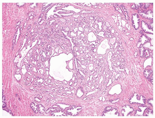
FIGURE 9-17 ▪ Low-power view of AAH.


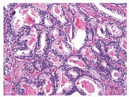
FIGURE 9-18 ▪ Glands of AAH are often crowded and may contain luminal eosinophilic materials and crystalloids, mimicking carcinoma.


Differential Diagnosis

The diagnosis of AAH in prostate resection specimens is usually straightforward since the entire nodule can be appreciated as well as background hyperplasia and the transition zone location. However, the diagnosis may be difficult when only part of the nodule is sampled in needle biopsy. The periphery of an AAH focus may show glands with limited infiltration, and further individual glands may contain intraluminal mucin, crystalloids, or eosinophilic secretions, mimicking microacinar (Gleason pattern 3) adenocarcinoma. In contrast, carcinoma exhibits nucleomegaly, prominent nucleoli, and often amphophilic cytoplasm. Basal cell immunomarkers should be carefully interpreted in AAH since some of the acini may have patchy or absent staining. All atypical glands within a focus of concern on H&E
should have complete abscence of basal cell immunostaining in order to support a diagnosis of adenocarcinoma.


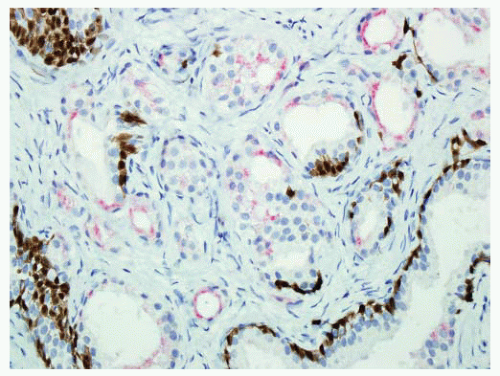
FIGURE 9-19 ▪ Cocktail of AMACR, HMWK, and p63 on AAH shows patchy basal cell immunostaining (brown). AMACR can be positive in a small subset of AAH like in this case.

Other benign processes in the prostate may mimic AAH (see Chapter 8). Usual nodular hyperplasia has better circumscribed nodules with no stromal infiltration of the glands at the periphery. Ordinary hyperplastic glands are larger in caliber, more uniform in size and shape and usually have luminal papillary infoldings. Stromal proliferation is usually present. Sclerosing adenosis is associated with dense fibroblastic spindle cell stroma. The glands are more irregular, angulated, or pointed and may have budding, and the basal cells are usually easy to recognize. The glands exhibit thickened basement membranes and may have surrounding rims of hyalinized stroma. Mesonephric remnants are lined by cuboidal to flattened cells and contain dense eosinophilic colloid-like material in the lumina. Mesonephric glands are less circumscribed and are more haphazardly arranged. Verumontanum mucosal gland hyperplasia is often subjacent to urothelium, and the glands often contain dark orange or brown concretions.


Atrophy and Proliferative Inflammatory Atrophy

Some authors have previously considered focal glandular atrophy as a possible precursor of prostate cancer. Focal atrophy similarly occurs commonly in the peripheral zone where most cancer arises. But so far, there is no strong evidence linking pure glandular atrophy to carcinoma. However, atrophy associated with inflammation has been raised as a risk for prostate carcinoma development.228 Simple atrophy or postatrophic hyperplasia occurring in association with inflammation that appears to be regenerating or proliferating is referred to as “proliferative inflammatory atrophy” (PIA) (Fig. 9-20). A topographic study showed that PIA appears to merge with HGPIN in 42.5% of HGPIN lesions and is seen adjacent to carcinoma in 30%.229 Areas of presumed low-grade PIN are also found in association with HGPIN and PIA suggesting progression from PIA to PIN and/or carcinoma.229 Clusters of atypical epithelial cells with nuclear enlargement, hyperchromasia, and enlarged nucleoli can also be associated with PIA.230 Cells that are phenotypically intermediate between basal and secretory cells (CK5, GSTP1, c-MET, and C/EBPbeta expression) are enriched in PIA lesions.230,231 Somatic inactivation of glutathione S-transferase-pi gene (GSTP1) via CpG island hypermethylation occurs early during prostate carcinogenesis, which is present in about 70% of HGPIN and more than 90% of carcinomas. GSTP1 CpG island hypermethylation can also be detected in some PIA lesions but not in normal or hyperplastic epithelium.232 Half of p53-positive PIA epithelial cells express diffuse GSTP1 immunostaining and may play a role in induction of p53 overexpression.233 NKX3.1 and p27, which are lost or down-regulated in cancer and HGPIN, are also down-regulated in atrophy.182,228 Currently, more evidence is required to definitively establish or disprove a relationship between PIA and cancer.


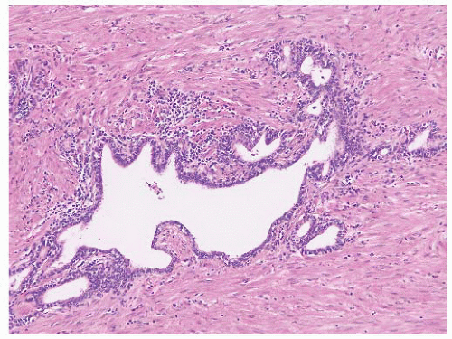
FIGURE 9-20 ▪ PIA lesion shows glandular atrophy with associated chronic inflammation. Ki-67 shows increase nuclear labeling (not shown).


PATHOLOGY OF ACINAR ADENOCARCINOMA


Macroscopic Features

Prostatic adenocarcinomas are often not recognized at the macroscopic level especially when they are small. In TUR samples, gross examination of chips is of little value.234,235 In rare cases, usually with extensive tumor, the carcinomatous chips may be harder and have a yellow or orange color compared to the spongy tan-brown chips of nodular hyperplasia.236 The color and texture of chips, however, are non-specific, and conditions such as granulomatous prostatitis can be associated with firm yellow fragments.

In RP specimens, carcinomas are sometimes visualized but usually only when the tumor is large. In general, lesions, <5mm in diameter are not detectable.237 Carcinomas were commonly visualized in the pre-PSA screening era when many patients presented with large-volume stage cT2 disease. In one study from 1998, 63% of carcinomas were identified in a consecutive series of RPs.238 Importantly, there was a false-positivity rate of 19%. The detected tumors were larger and of higher stage and grade than the more subtle ones. In recent years, with the higher proportion of cT1c disease, carcinoma is grossly recognized in a minority of cases, perhaps 30% of cases in our experience. Palpation of the gland is generally not helpful unless there is a significant peripheral nodule.

When grossly recognized on transverse sections, carcinoma usually involves the posterior or posterolateral peripheral zone just beneath the capsule (Fig. 9-21). Anterior involvement may be seen, but this is often difficult to recognize because of admixed stromal tissue. It is useful to compare the right and left sides of the gland in transverse sections looking for asymmetry and subtle color differences.
The tumor tissue has a smoother and more solid appearance and firmer texture than the spongy or sometimes cystic benign tissue. Carcinoma often has a yellow, yellow-orange, white, or gray color compared to the tan or beige appearance of benign prostate. The edges of the lesion are often indistinct, and microscopy usually reveals more tumor than was appreciated grossly. Extensive tumor can be overlooked since the whole gland may have a uniform appearance (Fig. 9-22). Many benign lesions such as stromal nodules, infarcts, and areas of prostatitis and atrophy may be grossly mistaken for carcinoma attesting to the nonspecificity of the macroscopic findings.236 Sometimes, extension of tumor beyond the confines of the gland may be seen macroscopically, but the observation may not be accurate, and it always requires histologic confirmation (Fig. 9-23). In one study,238 only one-half of carcinomas thought to show gross extension demonstrated EPE or margin positivity microscopically. In another study, 80% of cases with extraprostatic or margin involvement were correctly identified on gross assessment.239 Necrosis and hemorrhage are rarely seen in prostate carcinoma.


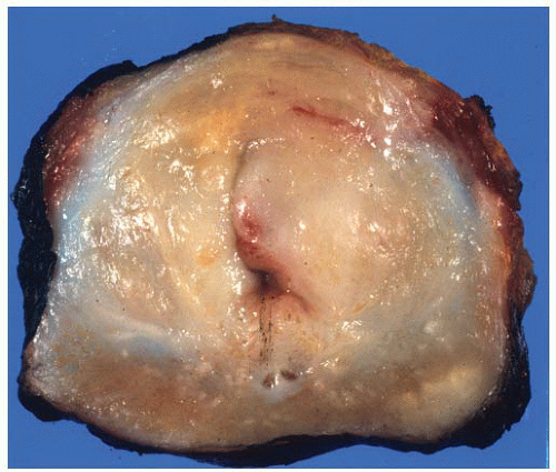
FIGURE 9-21 ▪ Transverse section of RP specimen showing peripheral nodule, right. Periurethral nodularity corresponding to a nodular hyperplasia is also noted.


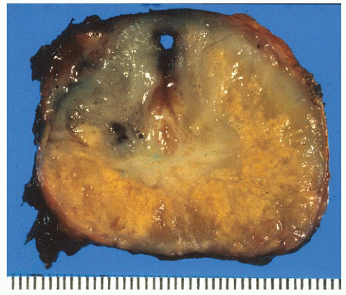
FIGURE 9-22 ▪ Transverse section of RP. Note widespread irregular yellow areas corresponding to extensive adenocarcinoma.


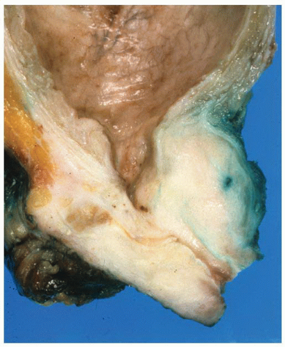
FIGURE 9-23 ▪ Parasagittal section of radical cystoprostatectomy showing adenocarcinoma extending around the seminal vesicle.


Tumor Location

About 70% of prostatic adenocarcinomas are located in the peripheral zone, mostly in posterior and posterolateral locations.240,241 (A small number are located in the anterior horns of the peripheral zone.)242 Ten percent to twenty-five percent of adenocarcinomas have an epicenter in the transition zone, and some of these also have peripheral zone involvement.240,241 Tumors uncommonly arise in the central zone, about 5% in two studies.240,241 In some cases, especially where there is more extensive tumor, the zone of origin cannot be determined.

There are clinicopathologic differences between tumors arising in the peripheral and transition zones.241,243 Peripheral zone carcinomas are more commonly detected by needle biopsy, show higher Gleason scores, and are more likely to be extraprostatic than transition zone carcinomas.244,245 The latter are more likely to be detected in TUR specimens, have lower Gleason scores and be organ confined. Nevertheless, some high-grade transition zone tumors may be encountered, and some may be difficult to detect because of biopsy
sampling protocols.246 Additionally, low-grade, high-volume transition zone tumors may be found, and these cases may be present with a high serum PSA (Fig. 9-24).247 Some have suggested that transition zone tumors have a distinct appearance with acini lined by columnar cells with basally located nuclei, but this latter can be seen in peripheral zone carcinomas as well.248 Interestingly, there have been some papers discussing differences in biomarker and molecular genetic findings between zones, but all of these changes may be related to the underlying Gleason score.249 The outcome data on differences between peripheral and transition zone tumors are mixed, some studies showing better outcomes and others showing no difference at all.249,250 Central zone carcinomas are uncommon and, in at least one study, have been associated with high Gleason score and high rates of extraprostatic involvement.251 The specific location of a tumor does not appear to have any independent prognostic significance.


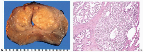
FIGURE 9-24 ▪ Transverse section of RP showing prominent periurethral nodular lesions, which corresponded to extensive transition zone adenocarcinoma. A: Transverse section of RP specimen. Note yellow-tan nodular lesions in transition zone corresponding to extensive adenocarcinoma. B: Low-grade adenocarcinoma (Gleason score 6) involving transition zone.


Tumor Focality

Adenocarcinomas are commonly multifocal in 50% to >90% of cases.252, 253, 254, 255 The additional tumors are often small and are often located at the apex. Commonly, the multifocal tumors involve both peripheral and transition zones (Fig. 9-25). Analysis of histologic and molecular features in multifocal tumors shows significant heterogeneity.256,257 There is a suggestion based on comparative genomic hybridization and gene rearrangement studies (TMPRSS2) that multifocal prostatic adenocarcinomas arise from independent clones.258


General Approach to Microscopic Diagnosis


Major Diagnostic Criteria

The diagnosis of adenocarcinoma of the prostate is based on the careful analysis of H&E-stained slides using a systematic approach. The slides are derived from a variety of specimen types including thin-core needle biopsies, prostatic chips, suprapubic and RPs, and, at times, metastatic sites. Time-tested morphologic criteria are analyzed in an algorithmic fashion starting from scanning power to high magnification. The histologic diagnosis of adenocarcinoma involves the assessment of overall glandular architecture, the cellular composition of individual architectural structures, and the cytologic features of constituent cells. The criteria advanced by Arthur Purdy Stout et al.259 are as applicable today as they were in the 1950s. They include (a) glandular pattern usually manifest by small irregular glands without any particular relation to adjacent glands or stroma, (b) arrangement of glandular epithelium with a lack of basal cells, and (c) cellular details including large deeply staining nucleoli. This morphologic triad constituted the major diagnostic criteria proposed by Algaba.260 In
that study, minor criteria included basophilic luminal secretions, pink amorphous secretions, intraluminal crystalloids, amphophilic cytoplasm, mitotic figures, and adjacent high-grade PIN. Our approach to diagnosing prostatic adenocarcinoma also utilizes major and minor criteria (Table 9-7). In addition to architectural pattern, absence of basal cells, and nuclear abnormalities including nucleolar enlargement, we include tinctorial alteration as a major criterion for diagnosis. Tinctorial changes are usually appreciated on scanning magnification, and like architectural disarray, they often draw one’s attention to sometimes subtle glandular abnormalities.


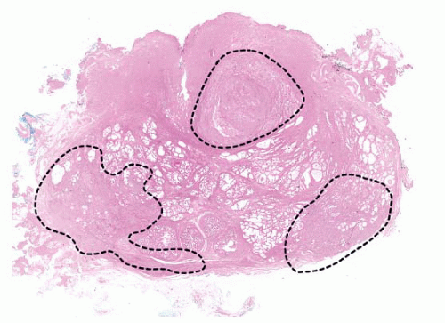
FIGURE 9-25 ▪ Diagram of RP showing multifocal adenocarcinoma with involvement of transition and peripheral zones. (Reprinted from Eggener S, et al. Focal therapy for prostate cancer: possibilities and limitations. Eur Urol 2010;58(1):57-64, with permission.)


Table 9-7 ▪ MICROSCOPIC DIAGNOSIS OF ADENOCARCINOMA


Major Diagnostic Criteria


  1. Disordered glandular architecture


  2. Tinctorial alteration


  3. Absence of basal cells


  4. Nuclear atypia including nucleolar enlargement


Minor Diagnostic Criteria


  1. Eosinophilic luminal secretion


  2. Basophilic luminal mucus


  3. Luminal crystalloids


  4. Mitotic figures


  5. Apoptotic bodies


  6. Smooth rigid luminal borders


  7. Associated high-grade PIN


The usual architectural patterns of prostatic adenocarcinoma are best described by referring to the classical Gleason drawing (Fig. 9-26).261 This diagram not only has formed the historical basis for grading prostate cancer (see page 64) but also is important in discussing the fundamentals of diagnosis and differential diagnosis. In this context, the architectural features are presented focusing on the original nine patterns (1; 2; 3A,B,C; 4A,B; 5A,B) described by Gleason. The subsequent section on Gleason grading deals with the substantial modifications of the original system for the purposes of contemporary grading.

Most prostatic adenocarcinomas are composed of separate small- to medium-sized atypical acini permeating between nonneoplastic glands (Gleason pattern 3A, 3B). Less commonly, separate acini are arranged in tight or loose clusters or more randomly in stroma without admixed nonneoplastic glands. When individual acini are uniform in size and shape and show little intervening stroma and the edge of the proliferation is rounded and “pushing” without admixed non-neoplastic glands, the pattern conforms to Gleason pattern 1. This diagnosis of adenocarcinoma, of course, would require other criteria including an absence of basal cells and other cytologic criteria (see later section). Gleason pattern 1 is nonexistent in needle biopsies and vanishingly rare in other specimen types, and many experts deny its existence arguing that the application of Gleason criteria as strictly written essentially eliminates the possibility of making this diagnosis since there is almost always some degree of nonuniformity in the small gland proliferation. Nevertheless, one occasionally encounters a tightly packed proliferation of uniform small neoplastic acini in the transition zone in TURs or RP specimen (Fig. 9-27).236


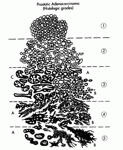
FIGURE 9-26 ▪ Gleason diagram showing 9 distinct patterns arranged in 5 grades. (Reprinted from Gleason DF. Histologic grading of prostate cancer: a perspective. Hum Pathol 1992;23:273-279, with permission.)

Gleason pattern 2 also involves small- to medium-sized acini without admixed nonneoplastic ones. There is more variation in acinar size and shape than grade 1, and the edge
of the lesion is more irregular. Furthermore, there is more intervening stroma between proliferating glands in Gleason pattern 2 compared to pattern 1 (Fig. 9-28). Gleason pattern 2 is an uncommon pattern of carcinoma that may be seen in the transition zone and is therefore identified in prostatic chips, enucleations, and RP specimens. In modern practice, Gleason grade 2 is virtually never diagnosed in prostate needle biopsy specimens.262


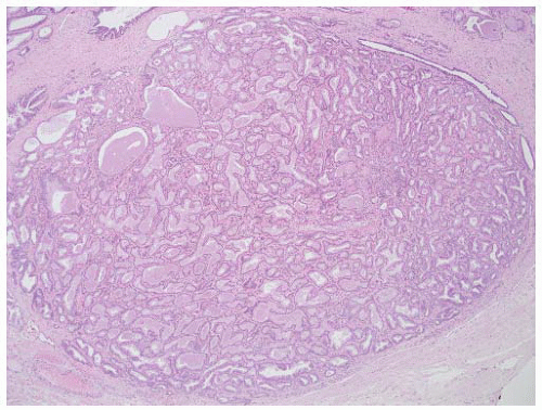
FIGURE 9-27 ▪ Gleason pattern 1. Closely packed small- to medium-sized acini show only slight size and shape variation. The edge of the lesion is visualized as a smooth pushing interface with stroma.


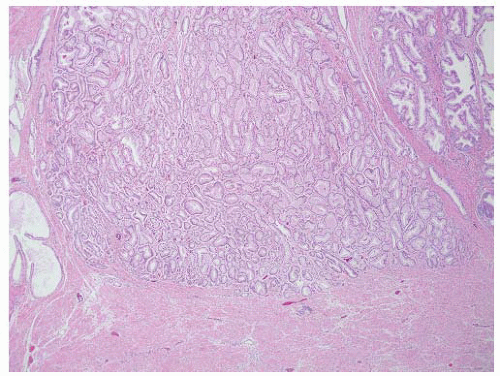
FIGURE 9-28 ▪ Gleason pattern 2 adenocarcinoma in the lesion is well circumscribed but contains acini showing some size and shape variation with more interweaving stroma than in Gleason pattern 1.

The adenocarcinomas that constitute Gleason patterns 3A and 3B show a greater degree of infiltrative growth than pattern 2 tumors, and in fact, they are the commonest patterns of prostate carcinoma. There is a greater degree of size and shape variation compared to Gleason pattern 2 tumors especially in the 3A tumors, and there is greater separation of acini (Fig. 9-29). The 3A tumors include small to mostly medium and sometimes large acini that are irregular and commonly show angulation and branching (Fig. 9-30). The 3B tumors are composed of small to tiny acini, some with lumina and others without, displaying an infiltrative growth pattern (Fig. 9-31). The most frequent pattern of invasion for these 3A/3B tumors is irregular infiltration between nonneoplastic glands (Fig. 9-32). This is the typical pattern recognized as a cardinal diagnostic criterion by Stout and others in the early days. Generally, the pattern is easily appreciated on low power especially if there is a reasonable amount of tumor. Another pattern seen with these tumors is a haphazard infiltration of stroma without any nearby nonneoplastic glands (Fig. 9-33). There is an irregular distribution of glands and splitting of stroma that is readily appreciated on low power (Fig. 9-34). The neoplastic acini of Gleason grade 3 tumors can be associated with periacinar clefts leaving a peculiar halo-like effect (Fig. 9-35).236,263, 264, 265 This observation while uncommon is more likely associated with carcinoma than benign glandular processes.


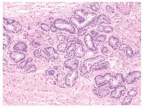
FIGURE 9-29 ▪ Gleason pattern 3A. Note proliferation of single separate acini showing size and shape variation. This lesion is more irregular and infiltrative than Gleason pattern 2.


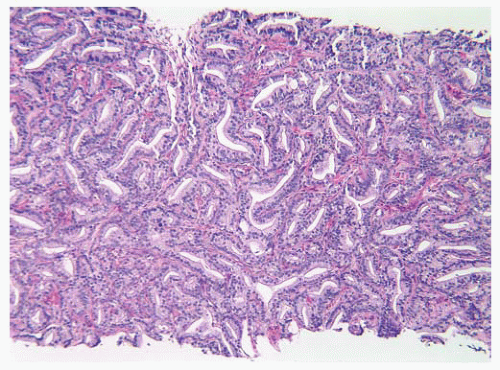
FIGURE 9-30 ▪ Pattern 3A adenocarcinoma showing focal glandular angulation.


FIGURE 9-31 ▪ Gleason pattern 3B. Note small irregular acini infiltrating between nonneoplastic ones. All acini have at least partially discernable lumina.


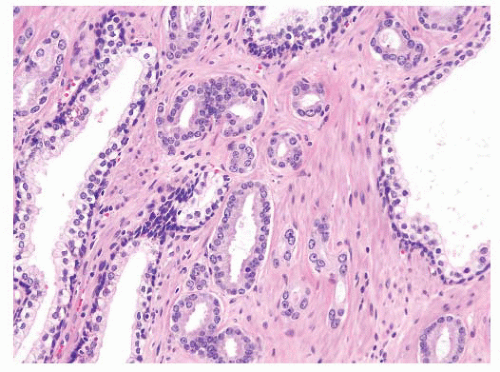
FIGURE 9-32 ▪ Gleason pattern 3 adenocarcinoma showing infiltrations between nonneoplastic glands.

While separate acini constitute the commonest morphology of adenocarcinoma, there are additional architectural patterns including cribriform, papillary, fused acinar, cord-like, solid, and single cell that must be recognized. These account for the intermediate- and high-grade patterns in the Gleason system. Gleason pattern 3C in the original description consists of smoothly contoured glandular aggregates with punched-out relatively uniform lumina creating a sieve-like pattern (Fig. 9-36). Similarly rounded glands with a papillary architecture were also included in this category. Confluence of aggregates, solid cylinders, and necrosis was not allowable in the 3C category. Sometimes, there is difficulty in separating Gleason pattern 3C from other patterns including cribriform high-grade PIN and cribriform hyperplasia (Fig. 9-37). Immunohistochemistry stains looking for the presence of basal cells may be required in some cases.203,266,267 While it is important to appreciate the diagnostic significance of the cribriform pattern of adenocarcinoma, recent studies suggest that pathologists, for the most part, include any significant amount of cribriform tumor in a higher-grade (Gleason 4) category for the purpose of prognostication268 (see grading section).


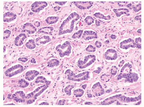
FIGURE 9-33 ▪ Adenocarcinoma characterized by irregular permeation of stroma without any nearby nonneoplastic glands.


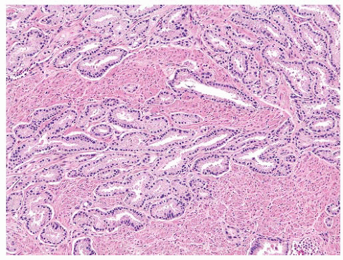
FIGURE 9-34 ▪ Gleason pattern 3 adenocarcinoma showing splitting and permeation between stromal muscle elements.

The original Gleason grade 4 comprised two patterns, 4A and 4B. Pattern 4A consists of fused glandular aggregates or chains and cords of neoplastic cells with little or no intervening stroma (Figs. 9-38, 9-39 and 9-40). The fused glands can have a rounded edge or one that is more ragged. Stromal invasion is usually readily seen in Gleason pattern 4. Large and irregular cribriform masses are also part of the original Gleason pattern 4 (Fig. 9-41). The cells comprising most Gleason 4A tumors have relatively scant cytoplasm, which is usually amphophilic or sometimes eosinophilic. Rarely, Gleason 4 tumors consist of masses and sheets of fused or poorly formed glands with abundant clear cytoplasm. These so-called hypernephroid carcinomas (Gleason pattern 4B) bear a passing resemblance to renal clear cell carcinoma (Fig. 9-42). Interestingly, the nuclei in such cases may be small and dark and have inconspicuous nucleoli.


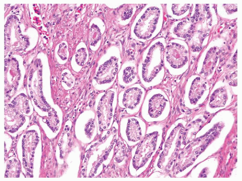
FIGURE 9-35 ▪ Gleason pattern 3 adenocarcinoma showing prominent periglandular spaces.


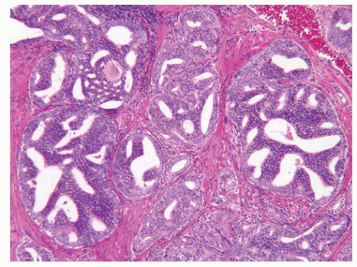
FIGURE 9-36 ▪ Classic Gleason pattern 3C rounded cribriform glands with punched-out lumina. In contemporary useage, this pattern is considered grade 4.


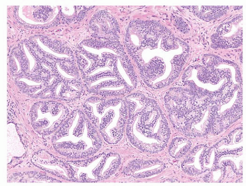
FIGURE 9-37 ▪ Rounded cribriform structures showing stromal infiltration. No basal cells identified. These cribriform structures are considered Gleason pattern 4.


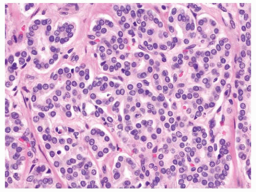
FIGURE 9-38 ▪ Gleason pattern 4A. Note prominent acinar fusion.


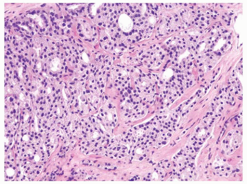
FIGURE 9-39 ▪ Gleason pattern 4A. Note striking fusion of acini and formation of occasional elongated acinar chains (lower right).


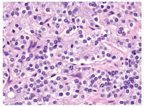
FIGURE 9-40 ▪ Gleason pattern 4A. Note prominent fusion with virtually no lumen formation.


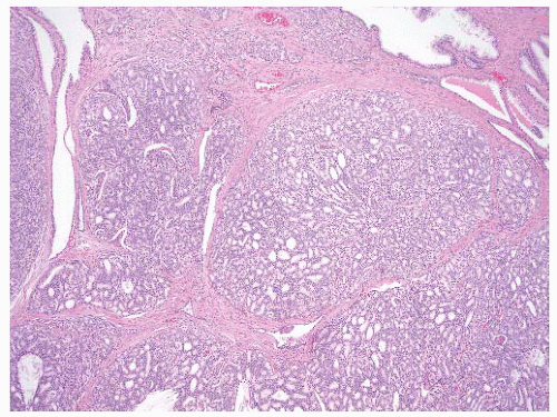
FIGURE 9-41 ▪ Gleason pattern 4A. Note large irregular and consulate cribriform structures with extensive infiltration of stroma.


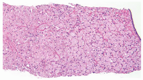
FIGURE 9-42 ▪ Gleason pattern 4B in needle biopsy. Note a fused acinar morphology with little or no gland formation. Individual cells have abundant clear to foamy cytoplasm and small dark nucleoli.

The highest grade in the Gleason system includes a variety of patterns including tumors composed of rounded solid cylinders and cribriform structures with central comedo-like necrosis (Fig. 9-43). The comedocarcinoma may look similar to ductal carcinoma of the breast (Fig. 9-44). Often, there is a spectrum of cribriform tumor ranging from cribriform pattern 4 blending imperceptively into more solid cribriform areas (Gleason 5A). The most poorly differentiated tumors are composed of solid sheets, aggregates, cords, and single cells sometimes with vacuolated cytoplasm (signet ring cells) but without well-defined lumens (Figs. 9-45, 9-46 and 9-47). Solid masses, trabeculae, and sheets of tumor with rosettelike structures may also be seen. Grade 5 tumors often show extensive stromal invasion.

The architectural patterns of adenocarcinoma as described above show varying degrees of stromal disruption; however, stromal reaction is unusual especially in the lower grades of carcinoma. A desmoplastic reaction may rarely be seen in Gleason grade 3 carcinomas but is more commonly noted in higher-grade patterns (Fig. 9-48). Stromal reaction may be difficult to appreciate on routine slides.269 Lymphocytic reactions are rarely seen in prostate carcinoma. A neutrophilic response is highly unusual except if there is superimposed acute bacterial prostatitis. A granulomatous stromal reaction is rarely seen. Sometimes, one can see a layered fibrous reaction at the advancing edge of the carcinoma especially in RP specimens when the tumor is beyond the prostatic capsule (Fig. 9-49).


FIGURE 9-43 ▪ Gleason pattern 5A. Note rounded cribriform structures with prominent central comedonecrosis.


FIGURE 9-44 ▪ Cribriform carcinoma. Note extensive infiltration of stroma by cylindrical and sheet-like cribriform structures.

In addition to the above patterns that conform to the original Gleason diagram, there are other uncommon patterns of prostate adenocarcinoma that may be underrecognized because of their resemblance to common nonneoplastic conditions including atrophy and hyperplasia. These variations, which include atrophic, pseudohyperplastic, cystic, foamy gland, and stratified (PIN-like) adenocarcinoma, are detailed in the next section of the chapter.


FIGURE 9-45 ▪ Gleason pattern 5B. Note extensive tissue involvement by Indian file-like cords and spindle cells.


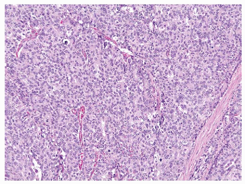
FIGURE 9-46 ▪ Gleason 5B. Note solid sheet of tumor with only focal lumens.

While the architectural pattern is the most common and most important low-power manifestation of prostatic carcinoma, tinctorial alteration is another frequent phenomenon that can be appreciated at scanning magnification. Sometimes, the tinctorial change is more obvious than any architectural disturbance, and we consider the glandular color change to be a major diagnostic criterion of adenocarcinoma. The tinctorial quality of glands is somewhat dependent on fixation and staining. In applying this criterion, it is important to have a histology laboratory that produces consistent H&E staining. Tinctorial alteration is always judged in relation to nearby nonneoplastic glands, and while the change is usually manifested by increasing degrees of amphophilia (Fig. 9-50), this is not always the case. Sometimes, the neoplastic glands are clearer than the nearby nonneoplastic ones (Fig. 9-51). Rarely, the neoplastic glands have an eosinophilic hue. In addition to the general difference in “color temperature,” the neoplastic cells may show more specific cytoplasmic changes including Paneth cell-like alteration, which usually indicates neuroendocrine differentiation (Fig. 9-52). Paneth cell-like change when present is usually focal, but in rare cases, it may be extensive. Neoplastic glandular cells may have a microvacuolated appearance (so-called foamy gland or xanthomatous change) or a macrovacuolated (signet ring-like cell) morphology (see later section). In very rare instances, the cells may display an oncocytic look with abundant granular eosinophilic cytoplasm (Fig. 9-53).


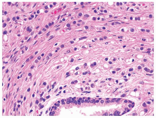
FIGURE 9-47 ▪ Gleason pattern 5. Note poorly differentiated cells arranged in cords, and individually. Many cells show prominent signet ring cell change.


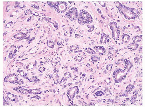
FIGURE 9-48 ▪ Adenocarcinoma showing a desmoplastic stromal reaction.

A third major criterion for the diagnosis of adenocarcinoma is an absence of basal cells, often but not always resulting in single cell-layered glands. Most normal and hyperplastic glands show a double layer with luminal secretory cells enveloped by basal cells.270 The basal cell layer may be complete or discontinuous, a feature that is often
difficult to discern with an H&E-stained slide. Most benign mimickers of adenocarcinoma have complete or incomplete layers of basal cell.203,266,271 The usual patterns of complete atrophy show complete basal cell layers, while partial atrophy, postatrophic hyperplasia, and AAH typically have a discontinuous layer.214,216,272, 273, 274, 275, 276, 277 Some benign glands especially in areas of partial atrophy totally lack a basal cell layer, and therefore, an absence of basal cells is not a specific criterion of malignancy.274, 275, 276,278 Nevertheless, in adenocarcinoma, basal cells are absent, and this feature is an important aid in establishing a malignant diagnosis (Fig. 9-54).


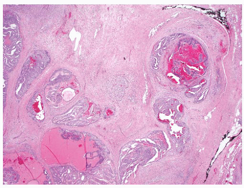
FIGURE 9-49 ▪ Fibrous reaction at advancing edge of carcinoma in extra-prostatic extension.


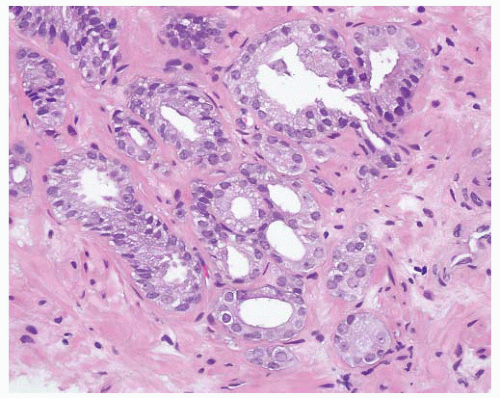
FIGURE 9-50 ▪ Gleason pattern 3 adenocarcinoma showing cytoplasmic amphophilia.

Careful microscopy at medium to high power is often required to appreciate the basal cell layer. Basal cells are darker than secretory cells, have relatively high nuclear to cytoplasmic ratios and may have a variety of shapes including cuboidal, triangular, and elongated forms (Fig. 9-55). The last may be difficult to separate from periglandular stromal cells, which may be closely apposed to glands. Additionally, distorted or poorly preserved neoplastic glands may simulate basal cells. Most small acinar carcinomas display a single layer of abnormal cells; however, some carcinomas show cellular overlapping creating a double layer with an apparent basal cell component (Fig. 9-56). Careful inspection in these cases shows nuclear atypia in all cells including the basally located ones, thus helping to confirm a malignant diagnosis. While it is clear that the absence of basal cells is important in acinar carcinoma, it is also critical in the analysis of atypical cribriform and papillary proliferations. Invasive cribriform carcinoma may be difficult to separate from high-grade intraepithelial neoplasia and intraductal spread of adenocarcinoma (see later section). The complete lack of basal cells is important in arriving at a correct diagnosis.


FIGURE 9-51 ▪ Gleason pattern 3 adenocarcinoma showing abundant pale cytoplasm.


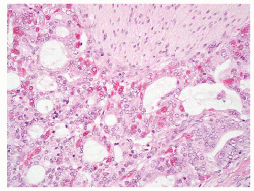
FIGURE 9-52 ▪ Adenocarcinoma with prominent Paneth-like cells.

The observation of basal cell absence can be judged on routine slides in most cases; however, one may need to
employ basal cell markers (see later section) in some cases especially when the issue of periglandular stromal cells is a problem or when there is more than one layer with cellular overlapping or in situations where there are only very few glands on which to base a judgment (Fig. 9-57). A basal cell marker should not be considered a “malignant stain” since one is looking for a negative observation, a finding that may also be rarely seen in benign glands.203,266


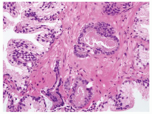
FIGURE 9-53 ▪ Adenocarcinoma composed of cells with abundant eosinophilic cytoplasm.


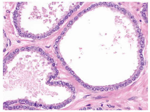
FIGURE 9-54 ▪ Adenocarcinoma composed of single-layered glands with no basal cells identified.

Nuclear atypia is the fourth major criterion for diagnosing malignancy. It is usually manifested by nuclear and nucleolar enlargement. Historically, the latter has been emphasized as a key feature of adenocarcinoma; however, the finding while helpful may not always be present.279, 280, 281, 282, 283 Prominent nucleoli are generally defined as ones measuring 1 to 3 µm in diameter, and they have an amphophilic or eosinophilic coloration (Fig. 9-58). The absence of prominent nucleoli may result from a number of factors including poor fixation or processing, type of fixative, thick sectioning, and overstaining. Some fixatives such as Bouin or B5 can lead to nucleolar prominence even in normal and hyperplastic glands. The use of such fixatives is discouraged in prostate biopsy pathology. While enlarged nucleoli are typical of carcinoma, they may be seen in other conditions such as high-grade PIN, radiation effects, inflamed glands, and basal cell hyperplasia.284,285 Multiple nucleoli are seen in malignancy more often than in benign lesions; however, they are not considered a specific malignant feature (Fig. 9-59).279,286 In addition to the technical factors that may obscure nucleoli in malignant glands, there are some carcinomas that are considered nucleolus poor (Fig. 9-60).280 In one study of limited carcinoma, one-quarter of cases lacked prominent nucleoli.282 In another analysis of low-grade adenocarcinoma, 8% of cases lacked nucleoli, and a further 20% only showed rare prominent nucleoli.280 Furthermore, certain
patterns of adenocarcinoma, especially hypernephroid carcinoma (Gleason pattern 4B), and the nuclei are often dark and have a pyknotic appearance with inconspicuous nucleoli (Fig. 9-61). The key point here is that the nuclear atypia does not always equate to nucleolar prominence.282 In the absence of the latter, there are other important attributes of nuclear atypia to look for including nuclear enlargement, nuclear shape and membrane irregularities, hyperchromasia, and parachromatin clearing (Fig. 9-62). These features need to be assessed using high-power microscopy.


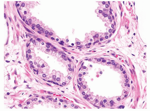
FIGURE 9-55 ▪ Nonneoplastic prostatic glands showing prominent basal cells with elongate and triangular forms.


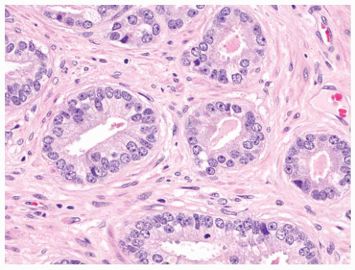
FIGURE 9-56 ▪ Adenocarcinoma showing cellular overlapping with no basal cells.


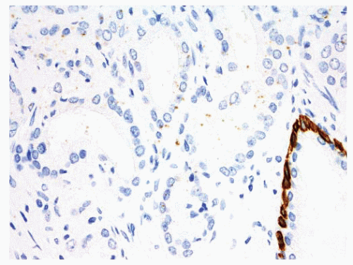
FIGURE 9-57 ▪ Gleason pattern 3 adenocarcinoma showing absence of basal cells with basal cell marker. Note the useful internal positive control of positive staining in basal cells of a nonneoplastic gland (lower right).


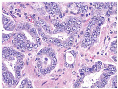
FIGURE 9-58 ▪ Adenocarcinoma showing prominent cytoplasmic amphophilia and enlarged nuclei with large inclusion-like nucleoli.


Minor Diagnostic Criteria

The minor diagnostic features generally occur more frequently in adenocarcinoma than in benign lesions and are useful as sentinels for a malignant diagnosis especially when other criteria may not, at least initially, be obvious. A minor feature may draw one’s eyes to an abnormality and prompt a closer look at a given group of glands. The minor criteria are listed in Table 9-8.


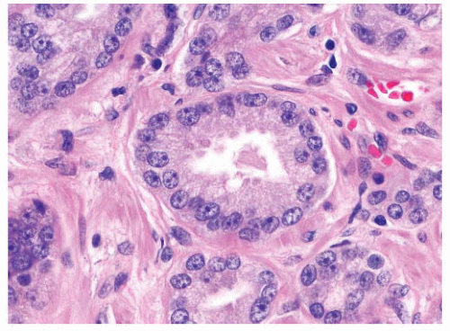
FIGURE 9-59 ▪ Adenocarcinoma showing prominent nucleoli including spindle cells with multiple nucleoli.


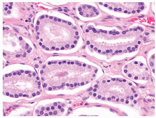
FIGURE 9-60 ▪ Gleason pattern 3 adenocarcinoma showing mildly enlarged nuclei without prominent nucleoli.

Amorphous eosinophilic secretions are more commonly seen in carcinomatous glands than in benign ones.281 The flocculent secretions are pink to red and often have a granular appearance (Fig. 9-63). In occasional cases, nuclear debris may be seen but not to the extent seen in comedocarcinoma (Fig. 9-64). The secretions do not have the well-defined structure of corpora amylacea, and the exact nature of the secretory material or its relationship with neoplasia is unclear. Eosinophilic secretions may be found in up to 84% of adenocarcinomas in prostatectomy specimens and 53% to 73% of minimal carcinomas in needle biopsies.282,287 Eosinophilic secretions are commonly seen in high-grade PIN and occasionally in benign glands including atrophic ones.

Basophilic luminal mucin (blue mucin) is more frequent in malignant glands than in benign ones.288 The mucin is often focal and present as wispy small strands, or it may fill lumina
and distend glands (Fig. 9-65). On occasion, there is extensive involvement of acini (Fig. 9-66). The mucin stains with Alcian blue at pH 2.5 indicating its acidic nature. The blue mucin can be present on its own, or it may be accompanied by granular eosinophilic secretions or crystalloids. Blue mucin is present in up to 72% of completely embedded RP specimens and in 18% to 32% of limited adenocarcinomas in needle biopsies.282,287 Blue mucin is not specific for carcinoma and may be seen in a variety of other processes including mucinous metaplasia, basal cell hyperplasia, sclerosing adenosis, and high-grade PIN.288 Importantly, basophilic mucin may also be seen occasionally in atrophy and AAH (adenosis), both of which are common mimickers of small acinar carcinoma289 (Fig. 9-67).


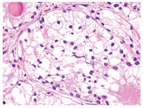
FIGURE 9-61 ▪ Gleason pattern 4B adenocarcinoma composed of cells with small dark nuclei with no nucleoli.


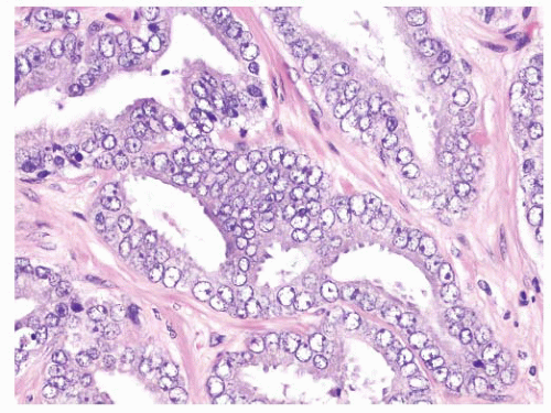
FIGURE 9-62 ▪ High-power photomicrograph showing marked nuclear atypia. Note nuclear membrane irregularities, prominent parachromatic clearing, and focally prominent nucleoli.

Intraluminal eosinophilic crystalloids occur more often in malignant than in benign glands.282,287,290,291 They are nonbirefringent brightly eosinophilic structures, which have a variety of shapes including needle-like, rectangular, and triangular ones (Fig. 9-68). Rarely, a crystalloid is seen in the prostatic stroma. They differ from corpora amylacea, which are laminated structures usually but not always with rounded contours. Corpora amylacea may have odd triangular or rectangular shapes, but they tend to conform to the shapes of glandular lumina and they are less brightly eosinophilic than crystalloids. Corpora amylacea are uncommon in acinar carcinomas but are regularly seen in benign glands (Fig. 9-69). Crystalloids are commonly associated with flocculent eosinophilic secretions and may be associated with basophilic mucin as well. When all three luminal products are present, there is a very high likelihood that the glands are neoplastic (Fig. 9-70). Crystalloids are more commonly present in low-grade acinar carcinomas and are rarely seen in Gleason grade 4 and 5 tumors. The composition and mechanism of formation of crystalloids are poorly understood although they are known to contain a high content of sulfur.292,293 Like other minor diagnostic criteria, crystalloids often draw the pathologist’s attention to an area of abnormality.


Table 9-8 ▪ SPECIFIC DIAGNOSTIC CRITERIA OF CARCINOMA


  1. Atypical glands in extraprostatic location


  2. Lymphovascular space invasion


  3. Circumferential perineural invasion


  4. Collagenous micronodules (Mucinous fibroplasia)


  5. Intraglandular glomerulations


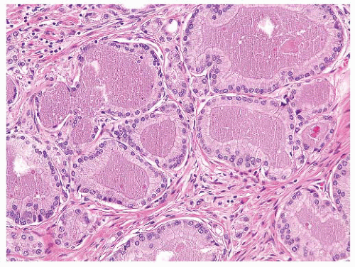
FIGURE 9-63 ▪ Gleason pattern 3 adenocarcinoma with prominent granular eosinophilic secretions. A crystalloid is seen on the right.

Mitotic figures are rarely detectable in benign glands but may occur more often in reactive epithelium and high-grade PIN. In needle biopsies, they are present in about 10% of Gleason 6 adenocarcinomas and more commonly in high-grade tumors.282,287 Mitotic figures are uncommon in the low- to intermediate-grade (Fig. 9-71) (Gleason scores 2 to 7) carcinomas and more likely to be seen in Gleason grade 8 to 10 tumors.294 Atypical mitotic figures are almost never identified except in high grade tumors (Fig. 9-72). Overall, the finding of a mitotic figure is neither sensitive nor specific for a diagnosis of adenocarcinoma, but it should prompt the pathologist to take a closer look at the involved gland and nearby ones.


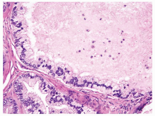
FIGURE 9-64 ▪ High-powered photomicrograph of carcinomatous glands showing eosinophilic luminal secretions and scattered nuclear debris.


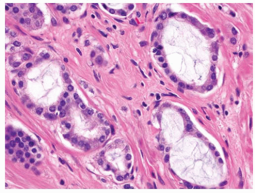
FIGURE 9-65 ▪ Adenocarcinoma Gleason pattern 3 showing luminal basophilic mucin.


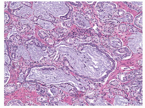
FIGURE 9-66 ▪ Adenocarcinoma showing extensive luminal mucin.


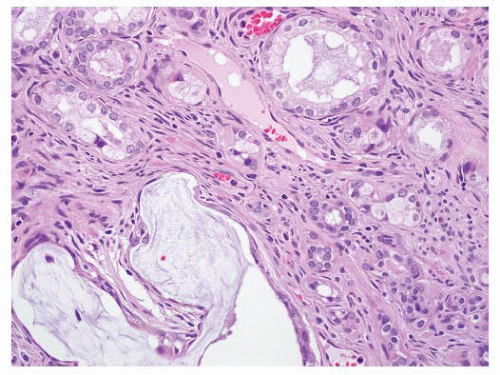
FIGURE 9-67 ▪ Luminal blue mucin in benign glands.


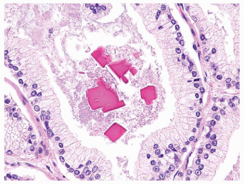
FIGURE 9-68 ▪ High-power photomicrograph showing prominent crystalloids of varying shapes.


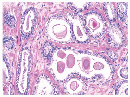
FIGURE 9-69 ▪ Adenocarcinoma, Gleason pattern 3 with numerous corpora amylacea.


FIGURE 9-70 ▪ Admixed luminal products—eosinophilic material, basophilic mucin, crystalloids.


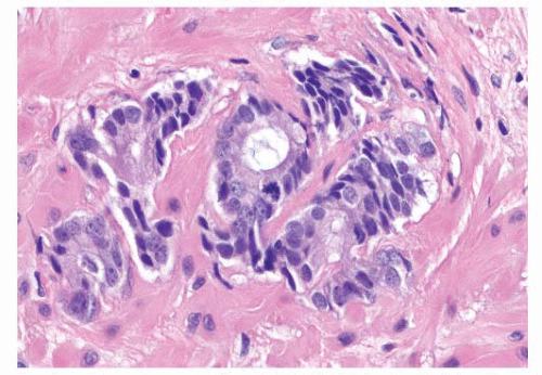
FIGURE 9-71 ▪ High-powered photomicrograph of adenocarcinoma showing a mitotic figure.

Some authors have found the presence of apoptotic bodies useful (Fig. 9-73).286 Apoptotic bodies are found in decreasing frequency in carcinoma, high-grade PIN, and benign glands, respectively.295, 296, 297

With respect to cytoplasmic membrane features, it has been suggested that neoplastic acini have a more “rigid” appearance with smooth sharp cytoplasmic borders (Fig. 9-74). While this feature is common, it is not specific for carcinoma and indeed some benign glands, especially in atrophy will also demonstrate smooth luminal borders. Furthermore, some carcinomas have irregular and undulating luminal edges.

The presence of high-grade PIN should heighten the pathologist’s suspicion that carcinoma may be present in the immediately adjacent tissue or in a separate fragment. In one study, 47% of patients with high-grade PIN in one core had carcinoma in a separate core compared to 31% of cases without high-grade PIN.


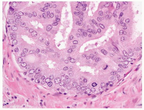
FIGURE 9-72 ▪ Atypical mitotic figures in high-grade carcinoma.


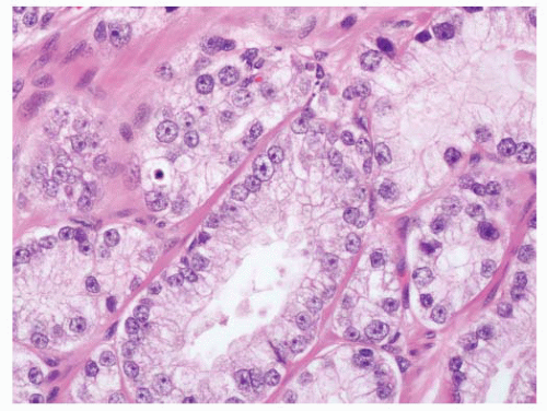
FIGURE 9-73 ▪ Adenocarcinoma showing a few apoptotic bodies.


Putatively-Specific Diagnostic Criteria

The presence of abnormal prostatic acini outside the prostate gland is a definitive indicator of malignancy with a few caveats. Ectopic prostate tissue may be found in a variety of sites including bladder, seminal vesicle, retrovesical space, and pericolonic fat, to name a few. The prostate epithelium in these cases can appear normal, atrophic, or hyperplastic. Therefore, it is important to recognize that the glands in question have some degree of abnormality, as defined above, in order to use this criterion. When neoplastic acini are outside the prostate gland, they usually involve periprostatic connective tissue, which generally contains fat. On rare occasions, one may see atypical acini in fibroadipose tissue at the tip of a core biopsy that is otherwise free of carcinoma (Fig. 9-75). While no tumor is present in the prostate
proper, this finding is generally diagnostic of prostatic carcinoma and suggests that the intraprostatic tumor has not been sampled. One must be cautious in such situations since metastatic carcinoma (e.g., gastrointestinal signet-ring carcinoma) may also present in this fashion, so prostatic marker studies may be useful. It is important to be aware that intraprostatic fat can rarely be seen. Involvement of seminal vesicle or bladder tissue by atypical prostatic glands is a good indicator of malignancy.


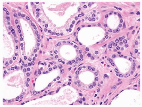
FIGURE 9-74 ▪ Adenocarcinoma, Gleason pattern 3. Note smooth “rigid” luminal cytoplasmic borders.


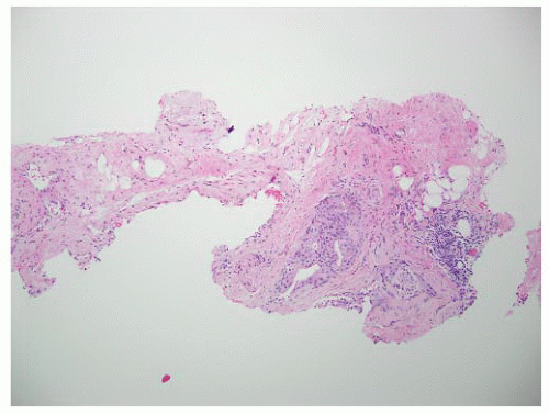
FIGURE 9-75 ▪ Low-power photomicrograph showing adenocarcinoma in periprostatic fibroadipose connective tissue. No actual involvement of prostate parenchyma was present.

Lymphovascular space invasion is considered a specific criterion for malignancy; however, it is rarely seen and, when present, is usually associated with high-grade carcinoma that is diagnostically obvious (Fig. 9-76). We are not personally aware of a case in which lymphovascular involvement by tumor was the only criterion of malignancy, but it remains a theoretical possibility.

With the exception of seeing abnormal acini clearly beyond the confines of the prostate or as an isolated finding in lymphovascular spaces, there are three reasonably specific criteria diagnostic of carcinoma. They are circumferential PNI, collagenous micronodules, and intraglandular glomerulations.


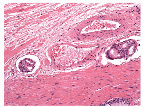
FIGURE 9-76 ▪ Intralymphatic adenocarcinoma.

PNI has historically been recognized as a common feature of adenocarcinoma and one that is important for diagnosis. In RP specimens, PNI is present in up to 94% of cases. A recent pooled analysis of biopsy studies shows that 22% of adenocarcinomas exhibit PNI.298 The likelihood of seeing perineural invasion is related to the grade and amount of tumor. In minimal carcinomas, the frequency of PNI has been reported to be as low as 2%. In rare cases, a small focus of perineural carcinoma is the only feature identified (Fig. 9-77). Originally, it was thought that PNI represented perineural lymphatic space invasion, but this hypothesis has been disproven. The nerves represent a pathway of least resistance for spread of carcinoma, and in fact, there may be an active process resulting in a tropism of carcinoma cells for nerves.299 Perineural “invasion” can have a number of patterns ranging from tumor cells abutting or indenting nerves to true circumferential involvement (Figs. 9-78 and 9-79). It is only the latter that should be considered a specific diagnostic criterion of malignancy.300,301 Normal, hyperplastic, and occasionally atrophic glands may abut or indent intraprostatic nerves. Likewise, while intraneural involvement suggests malignancy, however, there are rare examples of intraneural benign glands, so one should exercise caution in using this situation.300

PNI can have many forms. Circumferential involvement can be seen in transverse or longitudinal sections (Fig. 9-80). Partial circumferential involvement is sometimes manifested as crescentic extension of neoplastic glands around nerves. Peninsular or knob-like patterns of PNI may also be noted (Fig. 9-81). Neoplastic glands can directly invade nerves and can be completely surrounded by nerve tissue. Involvement of ganglia may also accompany neural invasion (Fig. 9-82). The perineural involvement may involve separate acini or fused glands and cribriform structures. In some cases, the involvement is subtle and may resemble hyperplasia or PIN (Fig. 9-83). The latter may have a micropapillary appearance with a nerve being in a papillary core. On occasion, the glands around an involved nerve may show cystic dilatation. Crushed cells can be seen around nerves, and this can lead to problems in differential diagnosis. Lymphocytes can be distributed in a perineural location, and when crushed, they can mimic the appearance of distorted tumor cells. Immunohistochemistry may be required to make a correct diagnosis. Sometimes stromal cells or aggregates of extracellular material such as one sees in collagenous micronodules may simulate PNI and, in selected cases immunostains for S-100, may be useful to confirm the presence of neural tissue (Fig. 9-84).


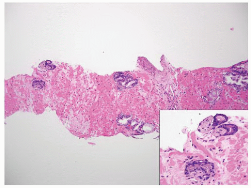
FIGURE 9-77 ▪ Low-power photomicrograph of prostatic core. Note perineural carcinoma near tip on left (inset).


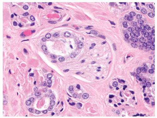
FIGURE 9-78 ▪ Adenocarcinoma, Gleason pattern 3 indenting a small nerve.


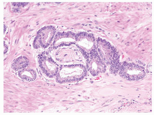
FIGURE 9-79 ▪ Circumferential perineural involvement by atypical glands is diagnostic of adenocarcinoma.


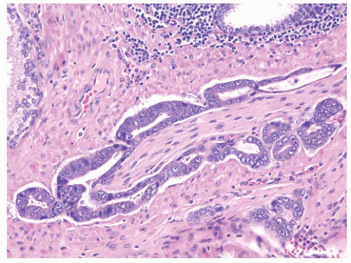
FIGURE 9-80 ▪ Circumferential perineural invasion in longitudinal section.


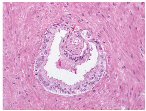
FIGURE 9-81 ▪ Partial encasement of a small nerve by adenocarcinoma creating a peninsular pattern.


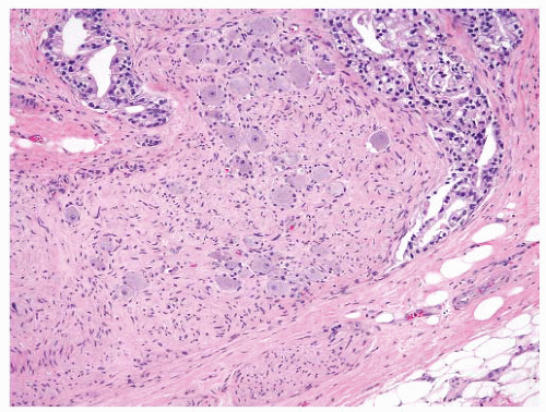
FIGURE 9-82 ▪ Adenocarcinoma infiltrating around a large ganglion.


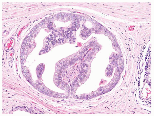
FIGURE 9-83 ▪ Perineural adenocarcinoma with PIN-like pattern.


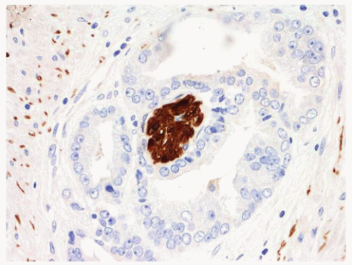
FIGURE 9-84 ▪ Perineural invasion confirmed by S100 stain.

Collagenous micronodules also referred to as musinous micronodules consist of rounded collections of hyalinized stroma admixed with or surrounded by abnormal acini.302,303 Sometimes, the micronodules are actually within the lumina of neoplastic glands. They are composed of collagen and often show a few admixed fibroblast nuclei (Fig. 9-85). Collagenous micronodules are commonly associated with mucin-producing areas and likely represent a peculiar fibrous organization of mucus. While usually rounded, the collagenous formations may be present as strands or larger lobulated masses (Fig. 9-86). Collagenous micronodules have not been identified in benign glandular lesions and are considered pathognomonic of adenocarcinoma. Unfortunately, this feature only presents in 1% to 5 % of needle biopsies with carcinoma and is rarely present in cases of limited carcinoma.271 Collagenous micronodules are more commonly found in totally embedded prostatectomy specimens.


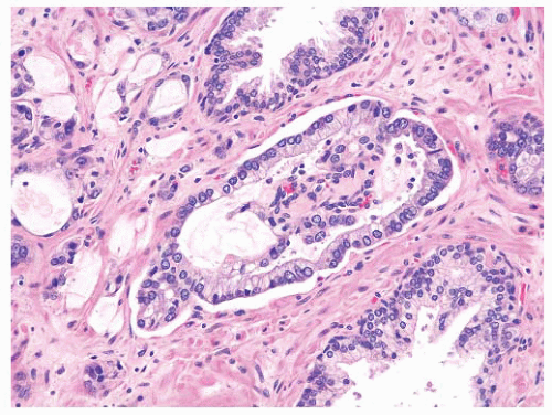
FIGURE 9-85 ▪ Adenocarcinoma with fused small collagenous micronodules.


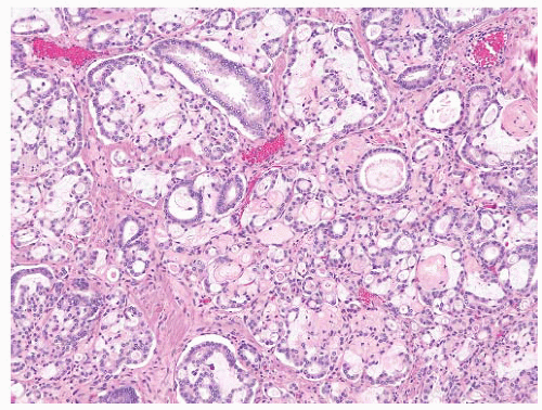
FIGURE 9-86 ▪ Adenocarcinoma with extensive collagenous micronodules (mucinous fibroplasia).

Intraglandular glomerulations are considered a relatively specific feature of adenocarcinoma. The ball-like tufts of fused cribriform glands project into lumina surrounded by crescentic spaces bearing superficial resemblance to Bowen spaces (Fig. 9-87). One or sometimes multiple points of attachment to the surrounding gland may be seen. Glomerulations are present in 3% to 15% of carcinomas in needle biopsies although our personal experience would be the lower end of that range.304 They are extremely rare in limited carcinoma.287


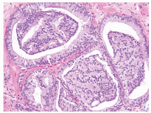
FIGURE 9-87 ▪ Adenocarcinoma with prominent intraluminal glomerulations. This is a specific diagnostic feature of carcinoma.


Ancillary Studies

Immunohistochemistry is a valuable adjunct to routine microscopy in prostatic pathology. In particular, basal cell markers and racemase (AMACR) are useful to confirm the presence of adenocarcinoma, especially in small foci of atypical glands.203,266 It has been recognized for over 25 years that high molecular weight cytokeratin (HMWCK) decorated by antibody 34βE12 [CK903], is preferentially expressed in prostatic basal cells rather than luminal secretory cells.305 In more recent years, other basal cell-specific markers such as cytokeratin 5/6 and p63 have also been successfully used in clinical practice (Fig. 9-88).306, 307, 308 Cocktails including antibodies to p63 and 34BE12 or CK5/6 are sometimes used. The staining is more intense because both nuclear and cytoplasmic domains are being decorated. However, at a practical level, the cocktails have no major advantage over single stains.309 The sensitivity and specificity of p63 and 34BE12 are similar in needle biopsies but, for TURs p63, may have better sensitivity.310 There are numerous factors that may interfere with successful immunohistochemical staining, and it is important to carefully assess internal and external controls when looking at basal cell markers. Factors such as time to fixation, time in fixative, and type of fixation can have an effect on immunostaining. The antibody should be optimized for the conditions within the individual laboratory, and there should be a good system of quality control. Antibodies such as 34BE12 work well in formalin-fixed paraffin-embedded tissues. Antigen retrieval using protease digestion or heat-induced or microwave techniques is generally employed. The stains can be done on stored intervening unstained sections or fresh sections cut from the block. Stains can also be carried out on destained H&E slides although the technique can be tricky and the staining reactions are usually not as strong as one sees with fresh sections.311 Ideally there should be internal nonneoplastic (normal or hyperplastic) glands for comparison. The benign glands should show strong basal cell staining, and it is very helpful when the nonneoplastic glands are close by the abnormal glands in question.


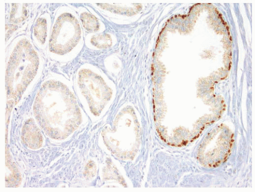
FIGURE 9-88 ▪ p63 staining in adenocarcinoma case. Note absence of p63-positive basal cells in adenocarcinoma (upside) and internal control positivity in nonneoplastic glands on right.

Basal cells markers should be incorporated into an algorithmic approach to the diagnosis of adenocarcinoma. They always need to be assessed in conjunction with the H&E sections and should not be ordered indiscriminately. Minimal adenocarcinoma can be diagnosed in needle biopsies with-out the use of immunostains278; however, in many practices, basal cell markers are used routinely in putative limited carcinomas as a confirmatory measure.

The absence of basal cells is a major criterion used to diagnose adenocarcinoma. Usually, this observation can be made using routine slides, but sometimes, the absence of basal cells must be confirmed. This is especially the case with tiny foci of putative carcinoma in needle biopsies specimens or when the abnormal glands are distorted. Periacinar stromal cells may simulate basal cells as can carcinoma cells when they are distorted, overlapping, or tangentially cut (Fig. 9-89). Basal cell markers will be negative in those situations. A negative basal cell stain on its own is not diagnostic of carcinoma. This observation has to be linked with the other morphologic features as noted earlier. One should exert caution since there may be other technical factors that result in a negative stain. To quote Peter Humphrey, “absence of proof is not proof of absence” (personal communication). While most benign glandular patterns have a continuous or discontinuous pattern of basal cell staining, some small acinar lesions such as partial atrophy show a complete absence of basal cells despite having completely banal cytoarchitectural features. Additionally, lesions such as AAH, partial atrophy, and postatrophic hyperplasia may show mixtures of small acini with basal cells and others without them. Scattered negative acini have been found in 11% to 23% of
atrophic foci.312,313 In an early study, looking at the utility of 34βE12 staining in small foci of atypical glands, the stain established a diagnosis of carcinoma in 14% of cases and was confirmatory for carcinoma in 58% of cases, equivocal in 18%, and of no value in 8%.305


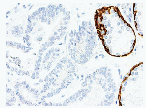
FIGURE 9-89 ▪ 34βE12 stain showing strong control positivity in basal cells in nonneoplastic glands (right) and negative staining in the glands on left, which display nuclear overlapping, a feature that can simulate basal cells on H&E.

Basal cell staining is also useful in the differential diagnosis of carcinoma and can be used to support a diagnosis of atrophy, postatrophic hyperplasia, AAH (adenosis), basal cell hyperplasia, and PIN.203,266,314 Radiation effects can be problematic, and basal cell stains can help to identify residual or recurrent carcinoma. Small atypical pseudoinfiltrative glands can be seen with radiation effects. Nuclear atypia and nucleolar prominence may be present. The presence of basal cell staining in these glands helps to confirm them as altered nonneoplastic acini. Furthermore, basal cell stains can be used in cribriform intraglandular lesions to separate cribriform PIN from invasive cribriform carcinoma (see earlier discussion).

There are rare reports of focal 34BE12 positive cells in prostatic adenocarcinoma.315 Additionally, p63-positive adenocarcinomas have also been identified (Fig. 9-90).316 In the latter cases, the neoplastic cells displaying varying degree of nuclear positivity. Despite these rare exceptions, basal cell markers are important adjunctive stains that are regularly useful to establish or confirm a malignant diagnosis.


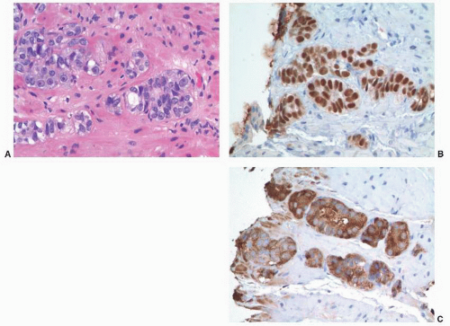
FIGURE 9-90 ▪ p63-positive prostatic adenocarcinoma. A. Note nests and poorly formed glands. B. Diffusely positive p63 nuclear staining. C. Strong racemase staining in atypical cells.

Over the last decade, AMACR (P504S), an enzyme involved in the B-oxidation of fatty acids, has emerged as an important immunomarker in prostate pathology. In contrast to the basal cell markers where one is looking for a negative result, racemase is a “positive cancer stain.” Using high-throughput gene expression studies, the P504S gene was found to be overexpressed in prostatic adenocarcinomas but not in benign prostate glands.317 The protein, identified by immunohistochemistry, is overexpressed in 75% to 95% of adenocarcinomas.318, 319, 320, 321 Racemase can be used as an individual stain or in cocktails. Cocktails of two antibodies (racemase and p63) using single- or dual-color detection systems or three antibodies (racemase, p63, 34βE12) with a dual-color detection kit may be employed.266,322, 323, 324, 325 These cocktails allow the detection of cytoplasmic racemase positivity and an absence of basal cells in the same glands (Fig. 9-91). Racemase staining is cytoplasmic, often with luminal membranous and submembranous enhancement (Fig. 9-92). In carcinomas, the staining is usually extensive with circumferential luminal border enhancement, and the intensity, while varying somewhat from case to case,

Only gold members can continue reading. Log In or Register to continue

Stay updated, free articles. Join our Telegram channel

Jun 10, 2016 | Posted by in UROLOGY | Comments Off on Adenocarcinoma of the Prostate

Full access? Get Clinical Tree

Get Clinical Tree app for offline access