Tubular Sodium Transport


Tubular Sodium Transport


Arohan R. Subramanya

Núria M. Pastor-Solar

Brian W. Reeves

Kenneth R. Hallows


Na+, the primary extracellular cation, is of critical importance to the maintenance of extracellular fluid volume. The kidneys play the dominant role in regulating Na+ excretion. Each day, the glomeruli filter roughly 25,000 mEq of Na+. From this quantity, almost 10 times the total exchangeable Na+ in the body, the kidneys typically absorb over 99%. A remarkable feature of the Na+ absorptive process is the precision with which it is regulated. An individual consuming a typical diet containing 6 g of Na+ will excrete 260 mEq of Na+ per day. The same individual placed on a 2-g Na+-restricted diet will promptly reduce Na+ excretion to 87 mEq per day. Although the fraction of filtered Na+ absorbed by the kidney changes from 99.0% on a standard diet to 99.6% on a Na+-restricted diet, this small change represents the addition or removal of over 1 L per day to extracellular fluid volume. Thus, the kidneys absorb large amounts of filtered Na+ with remarkably precise control.

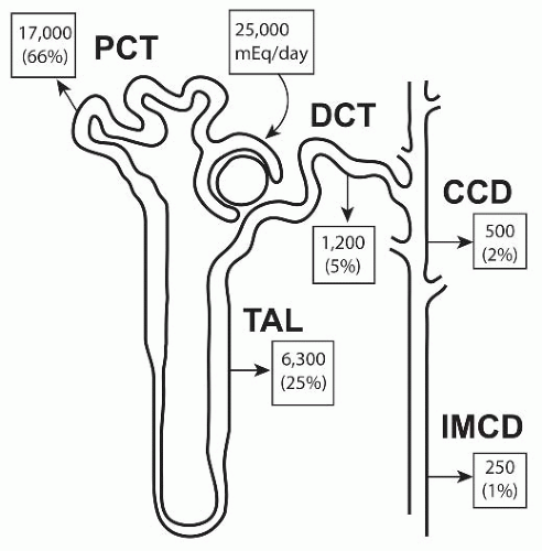
The exquisitely sensitive regulation of Na+ absorption by the kidneys relies on sequential actions of the various nephron segments, each with highly specialized transport capabilities. Figure 5.1 provides an overview of Na+ transport along the nephron. In general, the absolute rates of Na+ reabsorption are greatest in the proximal tubule and fall as the tubular fluid proceeds from proximal to distal segments. Conversely, the ability to transport Na+ against steep tubular fluid to blood gradients and its physiologic control increase along the nephron. For example, the proximal tubule reabsorbs the bulk (60% to 70%) of the filtered Na+ load, but as will be detailed later, does so against at most small electrochemical gradients. Moreover, the ability to alter Na+ transport in the proximal tubule, in relative terms, is rather limited, usually varying by less than 25%. The collecting duct, in contrast, reabsorbs only a minor fraction (˜2% to 4%) of the filtered Na+ load. However, the collecting duct can transport Na+ against a large electrochemical gradient to produce urine, which is almost Na+ free (<10 mEq/L). In addition, the rate of Na+ transport in the collecting duct can vary over a wide range (tenfold) in response to physiologic stimuli. The different nephron segments thus permit both high rates of Na+ transport (proximal segments) and highly regulated Na+ transport (distal segments).

Substantial progress has been made recently in identifying the proteins that mediate Na+ transport in each nephron segment and in defining their interactions and regulation within each segment. Many Na+ transport proteins have been linked to specific genetic disorders (Table 5.1). Given the primacy of renal Na+ transport to the control of extracellular fluid volume, it is not surprising that the majority of these genetic disorders are characterized by either hypotension or hypertension. An updated and curated database of these genetic disorders is maintained at the Online Mendelian Inheritance in Man website (www.ncbi.nlm.nih.gov/entrez/query.fcgi?db=OMIM).

This chapter considers the transepithelial transport of Na+ by the various nephron segments. The discussion of each nephron segment begins with a description of the general features of Na+ transport in that segment along with pertinent structure-function relations. The mechanism of Na+ transport is then considered on a cellular or subcellular level, with emphasis on recent electrophysiologic, biochemical, and molecular findings. Finally, each section includes a consideration of the factors that regulate Na+ transport in the individual segments.


PRINCIPLES OF MEMBRANE TRANSPORT

This section describes physical principles that underlie the movement of ions across individual membranes and epithelia. However, it is not intended to be an extended treatment of the thermodynamic aspects of membrane transport processes.


Diffusion Processes

Solute transport across membranes may occur by diffusion or convection, or by a mediated process. Diffusion is the random Brownian motion of a molecule with respect to adjacent molecules and occurs as the consequence of thermal energy.1 Because the diffusional movement of an
individual molecule is random, a concentration gradient is required for any net transfer of molecules to occur across a membrane. Thus, the concentration gradient represents the driving force for net transport.


FIGURE 5.1 The contribution of various nephron segments to Na+ transport. PCT, proximal convoluted tubule; DCT, distal convoluted tubule; CCD, cortical collecting duct; TAL, thick ascending limb; IMCD, inner medullary collecting duct.

For charged solutes, the driving force for transport is the sum of the chemical and electrical potential gradients. The Nernst equation describes the equilibrium condition for a membrane permeable only to a single ionic species:


where R is the gas constant, T is the absolute temperature, Z is the valence of the solute, F is the Faraday constant, and C and V are concentration and electrical potential terms, respectively. At equilibrium, then, the voltage (Vm) across an ideally selective membrane is defined by the concentrations of the permeant ion on both sides of the membrane, C2 and C1, respectively. For systems containing more than one permeant ion, the equilibrium voltage can be described by the Goldman-Hodgkin-Katz (GHK) equation2,3:


where Px is the permeability of the respective solutes, in this case, Na+, K+, and Cl. Thus, in a system containing multiple charged solutes, the transmembrane voltage is a function of the relative concentrations and permeabilities of each solute on the two sides of the membrane.


Convective Processes

Convection is the vectorial movement of an ensemble of molecules and is driven by an externally imposed force (e.g., hydrostatic pressure). Examples of convective transport include glomerular filtration and solvent drag, a process in which solute movement is coupled to water movement.

Bulk water flow may be driven by hydrostatic pressure and/or osmotic pressure. The familiar Starling equation:


describes net volume flow (Jv) in response to hydrostatic (δP) and osmotic (δπ) pressure differences. The equivalence of osmotic and hydrostatic pressure is explicit in the Starling equation. The degree to which a solute exerts an osmotic pressure depends on the degree to which it permeates membranes. The ratio of the observed osmotic pressure to that predicted if a solute were excluded absolutely from a membrane is termed the reflection coefficient, π:


For impermeant solutes, π = 1; for highly permeable solutes, π approaches 0.

For solutes with π <1, transmembrane solute flux will be accelerated in the direction of volume flow. This acceleration is known as solvent drag.4 Thus, the net passive flux of a permeable solute across a membrane may be driven by both diffusion and entrainment with solvent flow (i.e., solvent drag).


Facilitated Diffusion

Biologic membranes are composed primarily of lipid bilayers. Because the permeability of many hydrophilic solutes through lipid membranes is low, membranes contain proteins that facilitate the transport of certain solutes. Transport proteins, often termed carriers or transporters, have a high degree of specificity for the transported solute. Flux through the limited number of transporters saturates as the solute concentration is increased. An example of carrier-mediated facilitated diffusion is the entry of glucose into renal tubular cells mediated by the hexose transporter, GLUT-1.5 The movement of ions through ion channels represents another form of facilitated diffusion. In this case, integral membrane proteins containing several membranespanning domains form pores in cell membranes through which ions permeate. Ion channels generally have a high degree of specificity for the ions being transported and very high transport rates. Facilitated diffusion mechanisms, like enzymes, serve only to accelerate the rate of transport, but do not affect the equilibrium distribution of solutes. In other words, facilitated diffusion, like simple diffusion and convection, is a passive process that tends to dissipate transmembrane gradients.


TABLE 5.1 Inherited Disorders of Renal Sodium Transport


Disorder


Affected Gene Product


OMIM #


Nephron Distribution


Functional Consequences


Clinical Features


Hypotensive Disorders


Bartter, type 1 (antenatal)


NKCC2 (SLC12A1)


600839


TAL


Decreased Na+-K+-2Cl cotransport


Hypotension, hypokalemia, metabolic alkalosis


Bartter, type 2 (antenatal)


ROMK (KCNJ1)


600359


TAL, CCD


Decreased apical K+ recycling


Same as previous


Bartter, type 3 (infantile)


ClC-Kb


602023


TAL


Decreased basolateral Cl flux


Hypokalemia, alkalosis, hyperreninemia


Bartter, type 4


barttin


606412


tAL, TAL, inner ear


Decreased basolateral Cl flux


As previous, with sensorineural deafness


Bartter, type 5


CaSR


601199


PT, TAL, parathyroid, thyroid, brain


Activating mutation of Ca2+-sensing receptor with decreased K+ recycling and NaCl reabsorption


Hypokalemia, alkalosis, hyperreninemia, hypocalcemia


Gitelman


NCCT (SCL12A3)


600968


DCT


Decreased NaCl cotransport


Hypokalemia, metabolic alkalosis, hypocalciuria, hypomagnesemia


EAST/SeSAME


Kir4.1 (KCNJ10)


612780


DCT


Decreased basolateral K+ recycling; decreased basolateral Na+, K+-ATPase activity


Hypokalemia, metabolic alkalosis, hypocalciuria, hypomagnesemia, epilepsy, ataxia, sensorineural deafness


Pseudohypoaldosteronism type 1 (autosomal recessive)


ENaC subunits (SCNN1A, B, G)


264350


Collecting duct


Decreased amiloride-sensitive Na+ transport


Hypotension, hyperkalemia, metabolic acidosis


Pseudohypoaldosteronism type 1 (autosomal dominant)


Mineralocorticoid receptor (NR3C2)


177735


Collecting duct


Decreased response to mineralocorticoids


Hypotension, hyperkalemia (less severe than recessive)


Liddle


β or γ ENaC (SCNN1B, SCNNIG)


177200


Collecting duct


Increased cell surface expression and activity of ENaC channels


Hypertension, hypokalemia, metabolic alkalosis, responsive to amiloride


Pseudohypoaldosteronism type 2 (Gordon)


WNK1 or WNK4


145260


DCT, collecting duct


Increased NaCl cotransport, increased paracellular Cl permeability


Hypertension, hyperkalemia, metabolic acidosis, responsive to thiazide diuretics


Apparent mineralocorticoid excess


11β-HSD2


218030


DCT, collecting duct


Decreased oxidation of glucocorticoids causing activation of mineralocorticoid receptor


Hypertension, hypokalemia, metabolic alkalosis, responsive to dexamethasone


Other Disorders


Renal glucosuria


SGLT2 (SLC5A2)


233100


Proximal tubule


Decreased Na+-coupled glucose absorption


Decreased threshold for glucosuria


Proximal RTA


NBC1 (SLC4A4)


603345


Proximal tubule


Decreased Na+-coupled HCO3 transport across basolateral membrane


Metabolic acidosis, ocular and dental abnormalities, growth and mental retardation


OMIM #; accession number for Online Mendelian Inheritance in Man; TAL, thick ascending limb; CCD, cortical collecting duct; DCT, distal convoluted tubule; ROMK, renal outer medullary K channel; ClC-Kb, chloride channel Kb; tAL, thin ascending limb; PT, proximal tubule; DCT, distal convoluted tubule; EAST, epilepsy, ataxia, sensorineural, deafness, and tubulopathy; SeSAME, seizures, sensorineural deafness, ataxia, mental retardation, and electrolyte imbalance; RTA, renal tubular acidosis. Updated references available at http://www.ncbi.nlm.nih.gov/entrez/query.fcgi?db=OMIM.


Active Transport Processes

Active transport is a special case of facilitated transport in which chemical bond energy is supplied to the transport process so that the final distribution of the solute is remote from equilibrium. The coupling of solute transport to the energy source can take two forms. In primary active transport, solute transport is coupled directly to an energy-yielding reaction.

The most widely recognized example of primary active transport is the transport of Na+ and K+ by the Na+,K+-ATPase. This enzyme, often referred to as the sodium pump, couples the extrusion of cellular Na+ to cellular K+ uptake.6 In renal tubules, this enzyme is localized to the basolateral membrane. In general, segments with high rates of active Na+ transport have high Na+,K+-ATPase activity.7 The hydrolysis of each ATP molecule ordinarily pumps three sodium ions out of the cell coupled to two potassium ions moving inward.8 Therefore, the pump is electrogenic. The Na+,K+-ATPase is responsible for maintaining the cell Na+ activity at a low level, which provides the energy for the Na+-coupled transport of many other solutes. Thus, the inhibition of Na+,K+-ATPase (e.g., by peritubular ouabain addition) causes a significant rise in the cell Na+ activity.9 The affinity constant (Km) of the pump for intracellular sodium, about 15 to 30 mM,10 is similar to the intracellular sodium activity measured in proximal tubule cells.9 Therefore, the pump is unsaturated with respect to sodium, and pump activity is very sensitive to changes in the intracellular sodium concentration activity.

In secondary active transport, solute movement against its electrochemical gradient is energized by the movement of another solute down its own gradient.11 Na+, because of its steep inward electrochemical gradient maintained by the sodium pump, often participates in the transport of other solutes, either in the same (cotransport or symport) or opposite (exchange or antiport) direction. Thus, by coupling solute transport with sodium movement into cells, cellular metabolic energy generated by the Na+,K+-ATPase is stored in the form of a Na+ concentration gradient, analogous to a battery, and then dissipated in the transport of a variety of different solutes. Some examples of symport processes include Na+-glucose and Na+-amino acid cotransport; Na+-proton and Na+-calcium exchange are two examples of antiport processes.


PROXIMAL TUBULE


General Features

The proximal tubule is the major site for Na+ absorption within the kidney and serves two major purposes. First, the proximal tubule protects the extracellular fluid volume by reclaiming the bulk, approximately 60% to 80%, of the glomerular filtrate.12,13 The proximal tubule with its well-developed brush border membrane is optimally designed to perform the reabsorption of such a large fraction of the filtrate.14 Second, the absorption of sodium in the proximal tubule provides, by way of coupled processes, the driving force for the absorption of other solutes, such as bicarbonate, glucose, phosphate, and amino acids.

Under most circumstances, fluid at any given point along the proximal tubule has virtually the same Na+ concentration and osmolality as plasma.13 The isosmotic nature of proximal tubule fluid absorption derives from the high water permeability of this segment,15 which effectively clamps the osmolality of the tubular fluid at that of plasma. Although Na+ transport in the proximal tubule occurs in the absence of large electrical or chemical gradients, the bulk of Na+ absorption in the proximal tubule involves active transport. For example, Na+ can be reabsorbed against both concentration13,16 and electrical17 gradients. In addition, fluid absorption and sodium transport cease when Na+,K+-ATPase activity is inhibited or when cell metabolism is slowed.18,19

A significant amount of proximal Na+ transport also occurs passively.20 For example, in the late convoluted and straight tubules (S2 and S3 segments), Na+ diffuses passively out of the tubule driven by the lumen-positive electrical potential difference in those segments. This potential difference derives from a Cl concentration gradient across the tubule wall. Even in this case, however, it is the active transport of Na+ in upstream portions of the proximal nephron that ultimately accounts for these gradients and potentials.


Nephron Heterogeneity

Analyses of proximal tubular Na+ transport are complicated by two factors: a nonhomogeneous nephron population and axial changes in fluid composition. There is considerable heterogeneity of both morphologic and functional characteristics along the proximal tubule. The S1 segment cells have extensive basal interdigitations, numerous mitochondria, and a well-developed luminal brush border.21 S3 segment cells, in contrast, are flatter and have fewer mitochondria, lower brush borders, and much less extensive basolateral membranes than S1 cells.21 The Na+,K+-ATPase activity of the S3 segment is only 25% of that for the S1 segments.22 As might be expected on the basis of these observations, the net rates of the Na+ and fluid transport in the S3 straight segment are, in general, lower than in the S1 convoluted tubule.23

Juxtamedullary proximal convoluted tubules have higher rates of volume and bicarbonate absorption than their superficial counterparts,23,24 although this disparity has not been noted between juxtamedullary and superficial straight segments.25

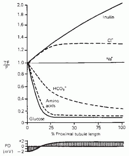
Glomerular ultrafiltrate in the early proximal tubule undergoes axial composition changes. Figure 5.2 illustrates that the chloride concentration rises as a consequence of the preferential absorption of NaHCO3 over NaCl in this segment.26 Glucose, amino acids, and other organic compounds are also absorbed avidly in association with Na+ so that their luminal concentrations in this
segment approach zero.26,27 The omission of glucose and amino acids from luminal fluids reduces both the potential difference and the volume absorptive rate.24,28 In the S3 segment, on the other hand, the omission of glucose and alanine has no effect either on the potential difference or on the fluid absorptive rate,25,29 although the deletion of all organic solutes does reduce volume absorption by 50%.30 The rates of transport of glucose, amino acids, phosphate, and Na+ in the early proximal convoluted tubule exceed those in the proximal straight tubule.31,32,33 These rates correlate well with the relative basolateral membrane areas of the respective segments.34


FIGURE 5.2 The profile of transepithelial voltage and solute concentrations along the mammalian proximal tubule. TF/P, tubular fluid/plasma concentration ratio; PD, potential difference. (From: Rector FC Jr. Sodium, bicarbonate, and chloride absorption by the proximal tubule. Am J Physiol. 1983;244:F461, with permission.)


Electrophysiology of the Proximal Tubule


Transepithelial Potential Difference

The electrogenic nature of the transport of Na+ coupled to glucose and amino acids creates a lumen-negative transepithelial electrical potential difference in the early proximal convoluted tubule. The deletion of glucose and alanine from the luminal fluid reduces the potential difference from about -5.0 mV, lumen-negative, nearly to zero.24,28 This transepithelial potential difference becomes lumen-positive (+2 to 4 mV) when the tubular fluid to plasma chloride concentration ratio is approximately 1.3.35 This lumen-positive voltage is probably a diffusion potential arising from the Cl concentration gradient.


Electrical Resistance

The electrical resistance of the mammalian proximal tubule is remarkably low, making this tubule a classic example of a leaky epithelium, with resistances of 5 to 10 Ω-cm2.36,37 In the proximal tubule, the total cellular resistance (i.e., the sum of the apical and basolateral resistance) is 20- to 70-fold greater than the transepithelial resistance. This indicates that the paracellular resistance is low and that the predominant route for passive ion flows in the proximal tubule involves the paracellular pathway.


Ionic Selectivity

The initial convolution of the superficial proximal tubule is Na+ selective; thereafter, the superficial convoluted and straight tubules are Cl selective.29,38 In contrast, juxtamedullary proximal tubules are Na+ selective throughout their course.38 Because the Cl concentration rises as fluid flows along the convoluted tubule (Fig. 5.2), oppositely directed gradients for Cl and HCO3 in late convoluted and straight segments give rise to a lumen-positive transepithelial potential difference.26 The latter indicates a higher permeability for Cl than HCO3, both in superficial and juxtamedullary proximal tubules.23,39


MECHANISMS OF SODIUM REABSORPTION


Apical Membrane Sodium Entry

In the proximal tubule, Na+ entry into the cell may be coupled to the movement of other solutes, such as glucose, chloride, or protons; or Na+ may enter independently. In either case, the driving force for Na+ entry is the steep electrochemical gradient favoring Na+ influx.

The intracellular Na+ activity ranges from 15 to 35 mM.10,40 The entry of Na+ into cells appears to be rate limiting for transepithelial Na+ transport. Amphotericin B, a polyene antifungal, increases the permeability of the luminal membrane to Na+ and causes a large rise in net sodium absorption.41


Na+/H+ Exchange

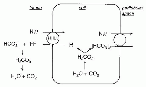
Directly coupled Na+/H+ exchange in the proximal tubular brush border is responsible for most proton secretion and for a large fraction of Na+ reabsorption in the proximal tubule.42 The mechanism whereby Na+/H+ exchange effects Na+ reabsorption is presented in Figure 5.3. Briefly, entry of Na+ is coupled to extrusion of a proton into the lumen. The proton titrates a filtered HCO3 molecule to form carbonic acid. Carbonic acid subsequently is dehydrated to CO2 in a reaction catalyzed by carbonic anhydrase IV in the brush border membrane.43 Within the cell, the reverse process occurs: carbonic acid formed by the hydration of CO2 dissociates into H+ and HCO3. The H+ is extruded into the lumen by Na+/H+ exchange or by the vacuolar H+-ATPase to repeat another cycle while the HCO3 is transported into the blood via a 1Na+:3HCO3 cotransport process (vide infra).


FIGURE 5.3 The scheme of NaHCO3 transport mediated by Na+/H+ exchange. See text for explanation.

The mammalian Na+/H+ exchanger (NHE) is electroneutral with a stoichiometry of one proton for one sodium.44 The exchanger is reversibly inhibited by high concentrations of amiloride.45 Intracellular protons, via an internal activator site,46 increase Na+/H+ exchange in response to intracellular acidosis.47

The apical and basolateral membranes of kidney cells contain different forms of NHEs with different affinities for amiloride.48 The apical Na+/H+ exchanger is involved in urinary acidification and has a low amiloride affinity, whereas the basolateral exchanger has a high affinity for amiloride. The sensitivity of NHE1 to amiloride49 and its basolateral localization50 suggest that it represents the “housekeeping” NHE. In contrast, NHE3 is in the brush border membrane of proximal tubule cells.51 NHE3 knockout mice have significantly reduced rates of Na+ and HCO3 transport in proximal tubules.52 In addition, pharmacologic inhibitors of NHE3 reduce proximal tubule Na+ reabsorption by about one third.53 However, a significant rate of amiloride-sensitive HCO3 transport still persists in the proximal tubules of NHE3 knockout mice,54 indicating that the NHE3 is responsible for much, but not all, proximal tubular Na+-coupled luminal acidification (vide infra). NHE8 is also expressed in the apical membranes of cortical tubules and may contribute to these processes.55


Sodium-Glucose Cotransport

Electrophysiologic studies in kidney proximal tubules show that apical membranes depolarize with addition of glucose to luminal fluids.56 The depolarization occurs because the Na+-glucose transporter is electrogenic. The Na+-glucose transporter is specific for the D-stereoisomers of glucose, galactose, and α-methyl-D-glucoside.57 The Na+-glucose cotransporter has little affinity for cations other than Na+.58 Phlorizin inhibits Na+-glucose cotransport by competing with glucose for its binding site.59

The rate of glucose transport by the early proximal tubule is greater than in late proximal segments.60 In the proximal straight tubule, the Km for D-glucose is 5 to 20 times lower than in the proximal convoluted tubule. Moreover, the transporter in the early cortical proximal tubule has a 1:1 Na+ to glucose stoichiometry,61 whereas the transporter in the straight medullary segment has a 2:1 stoichiometry.62 By coupling the energy from two Na+ ions moving down their electrochemical gradient to the transport of each glucose molecule, the medullary transporter is able to establish a much greater cellular to extracellular glucose concentration ratio than a 1:1 Na+:glucose transporter.27 The 2 Na+:1 glucose transporter is, therefore, well suited to the straight segment, where tubular fluid glucose concentrations have already been reduced by glucose absorption in the more proximal segments.

A Na+-glucose cotransporter (SGLT-1), which belongs to the SLC5 gene family,63 mediates high affinity Na+-glucose cotransport with a sodium-to-glucose coupling ratio of 2:1, whereas SGLT-264 shares 59% homology to SGLT-1 and mediates low-affinity Na+-glucose cotransport with a sodium-to-glucose coupling ratio of 1:1.65 In situ hybridization revealed high levels of a SGLT-2 message in the S1 segment of the proximal tubule.66 Recently, better antibodies have confirmed that, in rat kidney, SGLT-1 immunolocalizes to the brush border membrane of all three segments of the proximal tubule.5 By immunohistochemistry, SGLT-2 was detected at the brush border of the early proximal tubule in mice, which was absent in SGLT-2 knockout animals.67 Thus, it appears that SGLT-2 may represent the low-affinity, high-capacity sodium-glucose cotransporter in the early proximal tubule, whereas SGLT-1 may represent the high-affinity, low-capacity transporter of the proximal straight tubule.65

Mutations in SGLT-2 form the basis for renal glycosuria (Table 5.1), an inherited condition characterized by a lowered threshold for tubular reabsorption of glucose.68 In contrast, the dominant clinical manifestations of inactivating mutations of SGLT-169 relate to the failure to absorb sugars in the intestinal tract (glucose-galactose malabsorption). These findings suggest that SGLT-2 plays a much more significant role, quantitatively, than SGLT-1 in proximal tubule glucose reabsorption. SGLT-2 inhibitors are currently under investigation as potential therapeutic agents for the treatment of diabetes.70


Sodium-Amino Acid Cotransport

The proximal tubule reabsorbs amino acids from the tubular fluid via an active transport step at the luminal membrane.31 Samarzija and Frömter,71 using double-perfusion micropuncture techniques, observed a depolarization of the luminal membrane during amino acid transport, and they were able to identify five classes of amino acid transporters in the luminal membrane. Over the last decade, many of the transport proteins that mediate the different amino acid transport systems have been identified in kidney and intestine. This topic has been recently reviewed.72

Both Na+-dependent and Na+-independent amino acid uptake pathways have been characterized in the kidney.72 Neutral amino acid transport appears to involve at least
three separate transport systems,73 one that transports all neutral amino acids, one specific for imino acids, and one for the β-amino acids. Glycine may also have a specific transporter.74 In the kidney, neutral amino acid transport is driven by a Na+ gradient, as supported by experiments in slices, perfused tubules, and brush border membranes. The neutral amino acid transporter B0AT1 (SLC6A19) cotransports one Na+ per amino acid.75 The Km of the substrate decreases with an increasing cosubstrate concentration and vice versa. The initial step for transport involves the binding of the amino acid to B0AT1, and this binding affinity increases under hyperpolarizing conditions.76

The acidic and basic amino acid groups each have their own transport systems.77,78 At least one amino acid transporter, a Na+-independent transporter for neutral and dibasic amino acids, has been cloned from the kidney.79,80


NaCl Transport

Two basic mechanisms account for NaCl reabsorption in the proximal tubule. In simple electrogenic Na+ entry, sodium is transported actively through the cell, thereby creating a lumen-negative potential difference. Cl reabsorption then proceeds through the paracellular pathway driven by the lumen-negative potential difference. In electrically neutral NaCl transport, both Na+ and Cl move through the cell at equal rates, such that no transepithelial potential and, hence, no driving force for paracellular Cl movement is generated.


Neutral NaCl Transport

Several lines of evidence indicate that a sizable fraction of proximal NaCl transport is transcellular and electroneutral.81 First, by virtue of the coupling of Cl entry to apical Na+ entry, the intracellular Cl activity of proximal tubule cells is greater than predicted from an equilibrium distribution.82 Second, Cl absorption persists even when the driving force for passive, paracellular movement is abolished.83 Conversely, Cl reabsorption is inhibited by cyanide in the absence of any change in the passive driving forces for Cl movement.83 Finally, the luminal application of SITS,84 an anion-exchange inhibitor, or removal of chloride from the tubule perfusate,85 reduces net Na+ reabsorption.

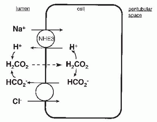
In principle, electroneutral NaCl transport across the apical membrane of proximal tubule cells could occur as directly coupled NaCl cotransport or as parallel Na+/H+ and Cl/base exchangers. There is no good evidence for the former process in the mammalian proximal tubule.81 However, considerable evidence supports the view that electroneutral NaCl transport in the proximal tubule involves parallel exchangers. The coupling of Na+ absorption to Cl absorption in this case occurs because of the relation between cell pH and concentration of base within the cell. With reference to Figure 5.4, the extrusion of H+ in exchange for Na+ results in the liberation of the base for participation in Cl/base exchange. The uphill entry of Cl, then, is indirectly coupled to the downhill entry of Na+, because both are coupled to the transport of an acid-base pair. The model illustrated in Figure 5.4 uses formate/Cl exchange as the anionic component of electroneutral NaCl transport.


FIGURE 5.4 The scheme of neutral NaCl transport mediated by the parallel action of Na+-H+ exchange and formate/Cl exchange. Formate (HCO2) combines with H+ in the tubular lumen to form formic acid (H2CO2), which reenters the cell by nonionic diffusion. A similar scheme applies for oxalate-Cl exchange.

As indicated previously, there is abundant evidence for a Na+/H+ exchanger in proximal tubule brush border membranes. With respect to NaCl transport, the inhibition of Na+/H+ exchange by high concentrations of amiloride86 or more specific inhibitors of NHE353 results in a dramatic fall in transcellular NaCl transport. Likewise, knockout of NHE3 also reduces NaCl and fluid reabsorption in the proximal tubule.52 Several Cl/base exchangers have been implicated in NaCl transport. Recent interest has focused on the role of Cl/formate (HCO2) and Cl/oxalate (C2O42-) exchange in NaCl transport.

A Cl/formate exchanger is present in brush border membrane vesicles.87 A role for Cl/formate exchange in neutral NaCl transport is suggested by the finding that the addition of formate to the luminal perfusate increases the rate of NaCl reabsorption in rabbit proximal tubules.88 As depicted in Figure 5.4, formate is presumed to leave the cell in exchange for Cl. The secreted formate then combines with a proton, which was transported by the Na+/H+ exchanger to form formic acid. The formic acid then reenters the cell by nonionic diffusion and dissociates to supply substrate for the continuation of both exchange processes. CFEX (SLC26A6), a homolog of pendrin, is a protein capable of mediating Cl/formate exchange and is present in apical membranes of the proximal tubule.89

Cl/oxalate (C2O42-) exchange has also been demonstrated in brush border membrane vesicles.90 It has been suggested that Cl/oxalate exchange may mediate neutral NaCl transport in a manner analogous to that described for Cl/formate exchange. It has also been suggested that NaCl absorption proceeds via the operation of three parallel transporters: the Na+-sulfate cotransport, the sulfate/oxalate exchange, and the Cl/oxalate exchange.91 Indeed, the CFEX protein, in addition to Cl/formate exchange, is also able to mediate Cl/oxalate, oxalate/formate, oxalate/oxalate, and oxalate/sulfate exchange.92


Other Na+-dependent transport processes have been described in the apical membrane of the proximal tubule. However, they do not contribute significantly to Na+ reabsorption because of the low concentrations of substrate present.


Simple Electrogenic Na+ Entry

The classic Ussing model for salt reabsorption involves passive entry of Na+ across apical membranes and extrusion across the basolateral membrane by Na+,K+-ATPase. A problem in assessing the contributions of electrogenic processes to Na+ transport in the proximal tubule is the presence of other mechanisms of Na+ entry. However, when the contributions of Na+/H+ exchange and Na+ cotransport to net Na+ absorption are minimized by deleting glucose, amino acids, and bicarbonate from the perfusate, a fraction of fluid absorption in isolated perfused straight segments persists and the transepithelial potential is – 1.0 mV.30 These results indicate that in the proximal straight tubule simple electrogenic Na+ transport constitutes a mechanism for Na+ absorption. A conductive Na+ pathway has been demonstrated in brush border membrane vesicles.93 Unlike the Na+ channel found in the distal nephron segments, the Na+ channel in the proximal tubule is not blocked by amiloride.93

In the proximal convoluted tubule, however, the deletion of glucose, bicarbonate, and amino acids completely abolishes fluid absorption.23 Consequently, simple electrogenic proximal Na+ transport may be limited to straight segments.


Passive NaCl Absorption

The rise in tubular fluid Cl concentration, and the attendant lumen-positive voltage (Fig. 5.2), provides a mechanism for passive NaCl absorption in late regions of the proximal nephron. In the Cl-selective superficial pars recta, approximately one-third of net NaCl absorption can be accounted for by this mechanism.20


Basolateral Membrane

The proximal tubule, particularly the S1 segment, possesses high Na+,K+-ATPase activity in the basolateral membrane. The Na+,K+-ATPase pumps Na+, which entered cells apically, across basolateral membranes. In other words, the pump keeps the cell Na+ activity low and maintains the electrochemical gradient for Na+ entry across the apical membrane. Consequently, the inhibition of Na+,K+-ATPase activity with ouabain decreases transepithelial Na+ reabsorption and increases the intracellular Na+ activity in the proximal tubule.94

Na+ also exits across the basolateral membrane in concert with HCO3. Studies in intact tubules and in membrane vesicles have demonstrated an electrogenic, stilbene-sensitive Na+-HCO3 cotransporter in the basolateral membrane of rat and rabbit proximal tubules.95,96 The cotransporter transfers two net negative charges across the basolateral membrane. The stoichiometry of this process is 1 Na+: 1 HCO3: 1 CO32- (or SO32-).97 Thus, this transport moiety for Na+ extrusion, [Na+(HCO3)3]2- is electronegative, with the lumen-negative cell interior providing a major driving for Na+ extrusion. A number of Na+-HCO3 cotransporters have been cloned, along with different splice variants.98 One of these, NBC1 (encoded by SLC4A4 and subsequently renamed NBCe1-A) is localized to the basolateral membrane of the S1 and S2 segments of the proximal tubule.99 As illustrated in Figure 5.3, the net result of these steps is the reabsorption of Na+ and HCO3, accounting for the bulk of HCO3 reabsorption and about 20% of Na+ reabsorption in the proximal tubule. It is believed that this cotransport process accounts for the basolateral transport of most of the bicarbonate reclaimed from the luminal fluid.97 Mutations in NBCe1-A cause proximal (type II) renal tubular acidosis and other defects in the eye, teeth, and mental development (Table 5.1). The regulation and role of NBC in acid-base transport in the proximal tubule is an area of active research that has been reviewed recently.100

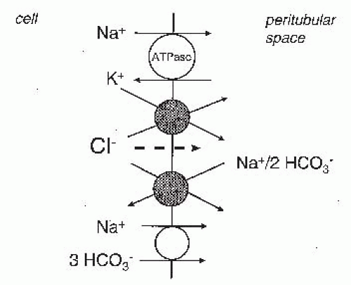
The pathways for Cl exit across the basolateral membrane are less well defined. Several pathways for Cl exit across the basolateral membrane have been proposed: conductive Cl channels, KCl cotransport, and Na+(HCO3)2/Cl exchange (Fig. 5.5). Studies of rat proximal convoluted tubules101 and rabbit convoluted and straight proximal tubule segments102,103 using intracellular microelectrodes have indicated that the proximal tubule cell has a very low Cl conductance. Thus, in normal proximal convoluted tubule and proximal straight tubule segments, conductive Cl efflux across basolateral membranes appears to play a minor role in NaCl absorption. Under hypotonic conditions, however, cell swelling dramatically increases the basolateral membrane Cl conductance.104

Because the chemical gradient for K+ to leave cells exceeds that for Cl entry, KCl cotransport can mediate basolateral Cl exit from proximal tubule cells. Ion-selective microelectrode studies have demonstrated KCl cotransport in basolateral membranes of rabbit proximal tubule cells.103,105

Stilbene-sensitive, Na+-dependent Cl/HCO3 exchange has been demonstrated in rat106 and rabbit107,108 proximal tubules. In this case, the entry of 1 Na+ and 2 HCO3 across the basolateral membrane is coupled to the efflux of Cl. The Na+
and HCO3 that enter the cell are thought to be recycled through the [Na+(HCO3)3]2- cotransporter (see previous). Indeed, Na+(HCO3)2/Cl exchange may account for much more Cl movement than KCl transport.108 Na+-independent Cl/HCO3 exchange is also present in the basement membrane of proximal tubules.106,107,108 However, under physiologic conditions, this process mediates net Cl influx and does not contribute to net NaCl absorption.


FIGURE 5.5 The transport pathways for Na+ and Cl absorption across the basolateral membrane of proximal tubular cells. Cl can leave the cell via KCl cotransport, Na+-2HCO3/Cl exchange, and Cl channels (minor). Na+ exits via the Na+,K+-ATPase and Na+-3(HCO3) cotransport.


CONTROL OF PROXIMAL TUBULAR SODIUM REABSORPTION


Glomerulotubular Balance (GTB)

The proximal tubule responds to an increase in glomerular filtration with an increase in the absolute rate of fluid absorption (APR) to minimize variations in the fractional proximal fluid absorption. This phenomenon is termed glomerulotubular balance (GTB). The efficiency of GTB—that is, the extent to which APR/GFR remains constant—is subject to physiologic and pathologic control. The prime factor modulating GTB in vivo is the effective circulating volume. Thus, at a constant or near constant GFR, volume expansion and volume contraction decrease and increase, respectively, the absolute rate of proximal Na+ absorption. In other words, volume expansion and volume contraction reset GTB upward and downward, respectively.109,110 This section considers some of the factors that modulate proximal Na+ absorption.

Peritubular capillary oncotic pressure is one of the factors regulating the rate of salt and water absorption from the proximal tubule.111 The oncotic pressure of the peritubular proteins favors the movement of fluid across the basement membrane, whereas capillary hydrostatic pressure retards this movement. Thus, at a given renal blood flow, an increase in the glomerular filtration rate and, hence, the filtration fraction, will cause an increase in the oncotic pressure in the postglomerular peritubular capillaries. At constant single nephron glomerular filtration rates (SNGFR), the perfusion of efferent capillaries with hypo-oncotic fluids decreases the absolute rate of proximal fluid absorption, whereas perfusion of the capillaries with hyperoncotic fluids increases proximal absorption.112 The effects of the peritubular protein concentration can also be demonstrated in isolated perfused tubules.113

It is not precisely clear how the peritubular protein concentration modulates proximal fluid absorption. The effect is not simply because of the oncotic pressure exerted by the proteins, because changes in absorption do not occur when active transport is inhibited or from comparable changes in the transtubular hydrostatic pressure.114 The prevailing views are that the peritubular protein may directly affect transcellular Na+ transport115 or the back leak of Na+ through the paracellular pathway.113,116

Luminal factors also contribute to glomerulotubular balance. The flow dependence of proximal absorption has been investigated in the convoluted15 and straight segments30 of isolated, perfused rabbit nephrons. The key observations are (1) the volume absorptive rate is clearly dependent on the perfusion rate, (2) the flow dependence persists in the absence of active transport when anion gradients are present, and (3) the flow dependence is abolished in the absence of active transport and anion gradients.

An explanation for these results lies in a consideration of axial versus radial changes in fluid composition along the tubule.117 The rate of passive Na+ absorption in the late proximal tubule is dependent on the magnitude of the chloride gradient between the lumen and the bath. At low axial perfusion rates, the radial Cl gradient tends to dissipate as a function of distance along the tubule, but this dissipation is minimized at higher axial perfusion rates. Hence, the integrated driving force for passive NaCl absorption increases with the rate of tubule perfusion. In addition, the availability of solutes, such as glucose, amino acids, and bicarbonate, is also partly responsible for the flow dependence of proximal fluid absorption.118

The flow dependence of reabsorption in the proximal tubule has also been tested in the mouse proximal tubule. In this experimental model, which included the use of the NHE3 knockout mouse, the data supported the hypothesis that the “brush border” microvilli act as mechanosensors that transmit fluid dynamic torque to the actin cytoskeleton and thus modulate Na+ absorption.119


Catecholamines

Renal denervation reduces proximal tubule Na+ and fluid absorption.120 Both α– and β-adrenergic receptors exist in the proximal tubule.121,122 The rate of salt and water reabsorption in the proximal tubule is stimulated by α– and β-adrenergic agonists.123 α-Adrenergic agonists increase apical Na+ entry via the stimulation of Na+/H+ exchange122 and also increase basolateral Na+ efflux via Na+,K+-ATPase activity in rat proximal tubules by a pathway that involves the activation of calcineurin.124 The effects of β-adrenergic agonists on Na+,K+-ATPase activity are less clear, with one study showing that they increase Na+,K+-ATPase activity via protein kinase C (PKC).125 However, others have found that β-adrenergic agonists inhibit Na+,K+-ATPase activity via PKA.126

Dopamine, which is produced by proximal tubular cells,127 inhibits Na+ reabsorption. Dopamine inhibits Na+,K+-ATPase activity via its receptors DA-1 and DA-2.128


Parathyroid Hormone

Parathyroid hormone (PTH) causes a 30% to 50% reduction in proximal tubular Na+ and phosphate absorption.129 PTH stimulates adenylyl cyclase, cAMP production, and PKA, which in turn inhibits Na+/H+ exchange in several proximal tubule systems.130 Weinman et al.131 demonstrated that phosphorylation by PKA inhibits Na+/H+ exchange activity via a PDZ domain-dependent interaction with the NHE regulatory factor (NHERF). In the absence of NHERF, cAMP
does not inhibit the exchange activity of NHE3.132 The current model for this inhibition is that NHE3 associates with PKA indirectly via NHERF and the cytoskeletal protein ezrin. PKA, when active, phosphorylates NHE3 at serines 552 and 605, which mediates the inhibition of the exchanger.133 NHE3 is directly phosphorylated by other protein kinases, including calmodulin-dependent protein kinase II, which inhibits Na+/H+ exchange activity and PKC, which stimulates the exchanger.134


Angiotensin II

The systemic administration of low doses of angiotensin II (Ang II) inhibits the excretion of Na+,135 whereas inhibitors of Ang II increase Na+ excretion.136 Systemic Ang II causes changes in renal blood flow, aldosterone secretion, filtration fraction, and catecholamine release from renal sympathetic nerve endings.137 Low concentrations of Ang II (<10-9 M) cause an increase in proximal tubule fluid and bicarbonate reabsorption, effects that are mediated by the AT1 subtype of Ang II receptors present in both the brush border and basolateral membranes of the proximal tubule.138 Higher concentrations (>10-8 M) depress fluid and bicarbonate absorption, presumably via counterbalancing effects mediated by the lower affinity AT2 receptor.138 Studies have demonstrated that the stimulatory effect of Ang II on fluid and bicarbonate reabsorption occurs via enhanced apical Na+/H+ exchange via NHE3 and basolateral Na+-(HCO3)3 cotransport via NBC-1 in the proximal tubule.139 The physiologic effects of Ang II may involve the coupling of these receptors to both phospholipase A2 and inhibitory G proteins.140,141


Thyroid Hormone

On a clinical level, hypothyroidism is associated with a decreased cardiac output, renal blood flow, and glomerular filtration rate (GFR). Clearance studies in hypothyroid rats have documented decreases in GFR, renal Na+ reabsorption, and renal Na+,K+-ATPase activity.142 These changes are reversible after thyroid hormone replacement.143 The thyroid hormone may exert direct effects to stimulate proximal tubular salt and fluid reabsorption via increased basolateral K+ permeability144 and/or direct stimulation of Na+/H+ exchange through an increase in NHE3 transcription.145


Corticosteroids

Although mineralocorticoids do not have an effect on proximal tubular sodium reabsorption,146 there is evidence for glucocorticoid receptors in the proximal tubule.147 Dexamethasone inhibits apical membrane Na+-phosphate cotransport in cultured proximal tubular cells via PKC activation.148 Dexamethasone also enhances the activity of apical NHE3 and the mRNA expression and functional activity of basolateral NBC-1.149 The resulting increase in proximal tubule HCO3 reabsorption could contribute to the maintenance of the metabolic alkalosis that is associated with increased glucocorticoid production in vivo.


Nitric Oxide

Various forms of nitric oxide synthase (NOS) are expressed in the proximal tubule.150 Low basal production of nitric oxide (NO) by the proximal tubule is boosted dramatically by lipopolysaccharide (LPS) and cytokines. Even under basal conditions, the proximal tubule may be affected by NO produced by adjacent cells, such as endothelium or other nephron segments. The overall effect of NO on proximal tubule Na+ transport is controversial and may be biphasic, with acute inhibition and chronic stimulation of Na+ reabsorption as assessed by pharmacologic agents and genetic knockouts of NOS, respectively.151 In vitro, NO decreased Na+/H+ exchange and Na+,K+-ATPase activity in cultured proximal tubule cells.152


MECHANISM OF ISOTONIC FLUID ABSORPTION

Proximal tubule water absorption is coupled tightly to solute absorption, because the measured osmolality of the tubular fluid is generally identical to plasma. Three general mechanisms of solute-solvent coupling have been suggested to account for this isotonic absorption: lateral interspace hypertonicity, effective osmotic gradients because of different reflection coefficients for solutes in the tubular and peritubular fluids, and luminal fluid hypotonicity.

The standing gradient theory argues that active transport of salt into the lateral intercellular space raises the osmolality of the space, thus providing an osmotic gradient for fluid transport from the lumen to the interspace.153 The tight junctions in this model are presumed to be impermeable to water, so that the osmotic flow of water from the cell into the hypertonic interspace raises the hydrostatic pressure in this compartment and forces fluid across the basement membrane.

An alternative explanation117 proposes that an effective osmotic driving force for fluid absorption can exist between solutions of identical osmolalities if the reflection coefficients of the membrane for the solutes in the solutions differ. Specifically, the elevated tubular fluid-to-plasma Cl concentration found in the late proximal convolution and in the pars recta provides an effective osmotic driving force for fluid absorption because πHCO3 exceeds πCl. That is, the bicarbonate in the peritubular fluid is a more “effective” osmotic agent than is the chloride in the tubular fluid, and, thus, net water flows out of the tubule. Although this mechanism may be applicable to the proximal straight tubule, the reflection coefficients for NaCl and NaHCO3 measured across the rabbit proximal convoluted tubule are virtually identical,154 so oppositely directed Cl and HCO3 gradients may make only a negligible contribution to fluid absorption in convoluted segments.

Finally, because of the high osmotic water permeability of the proximal tubule, only small degrees of absolute luminal hypotonicity are needed to provide a sufficient driving
force to account for the observed rates of fluid reabsorption.20 Experimental evidence supports the view that absolute luminal hypotonicity is a significant driving force for fluid reabsorption in the proximal tubule. Thus, when proximal tubules are perfused and bathed by symmetric NaCl solutions, the luminal fluid becomes slightly hypotonic.155 The development of luminal hypotonicity can be amplified by maneuvers that decrease the water permeability of the proximal tubule. The aquaporin 1 (AQP1) water channel is abundantly expressed in the proximal tubule. In AQP1 knockout mice, the osmolality of tubular fluid at the end of the proximal tubule is significantly lower than in normal mice.156 As the luminal fluid becomes more hypotonic, the resorbate becomes more hypertonic, and the degree of resorbate hypertonicity correlates with the rate of volume reabsorption by the tubules.157


THE LOOP OF HENLE

The dissociation of salt and water absorption by the loop of Henle is ultimately responsible for the capacity of the kidney, either to concentrate or to dilute the urine. The active absorption of NaCl in the water-impermeable thick ascending limb of Henle (TALH) serves both to dilute the urine and to supply the energy for the “single effect” of countercurrent multiplication. A functionally similar segment, known as the diluting segment, is found in amphibians and teleosts.158

The mammalian loop of Henle contains the descending thin limb (DTL), the ascending thin limb (ATL), and the thick ascending limb (TAL). The loop of Henle absorbs about 25% to 40% of the filtered Na+ load.159 Furthermore, the fluid leaving the loop is dilute, indicating that more NaCl is absorbed in the loop than water.


SALT TRANSPORT BY THE THIN DESCENDING AND THIN ASCENDING SEGMENTS

As the tubular fluid enters the descending thin limb and flows toward the tip of the renal papilla, it becomes more concentrated.160 Passive models for urinary concentration indicate that this increase in osmotic pressure is attributable to water extraction rather than solute entry.161 Current observations indicate that the aquaporin water channel AQP1 mediates water movement across the luminal surface of the DTL.162 According to some reports, however, AQP1 expression is significantly lower in short loop nephrons than long loop nephrons,163 suggesting that (1) not all DTLs in the kidney extract water to the same degree, and/or (2) water movement in short loop DTLs is facilitated by alternative water channels or via the paracellular route. In contrast to the DTL, in vitro microperfusion studies demonstrate that the ATL is relatively impermeable to water.164

The study of NaCl transport by the DTL and ATL has been complicated by the fact that the transport characteristics of these two nephron segments exhibit significant interspecies heterogeneity.165 Although the Na+ and Cl permeabilities appear to vary widely among rabbits, hamsters, and rats, one consistent finding was that the DTL is relatively less permeable to NaCl than the ATL. Coupled with the DTL’s high permeability to water, the relative lack of solute permeability ensures that the osmotic pressure of fluid entering the renal papilla is greater than that of the fluid leaving it.166 Thus, the formation of dilute urine by the loop of Henle begins in the ATL.

The decrease in osmolality in the ATL is due primarily to a fall in the NaCl content of the luminal fluid. The electroneutral transport of NaCl appears to occur through two key mechanisms. The transepithelial movement of sodium from the ATL lumen occurs via the paracellular pathway,167 whereas Cl diffusion occurs through a transcellular route. Yoshitomi et al.168 detected conductive pathways for C in both the apical and basolateral membranes of ATL cells. The transcellular movement of Cl in the ATL is regulated, because the basolateral Cl conductance is inhibited at low pH168 and by low intracellular Ca2+ concentrations.169 Uchida et al.170 cloned a Cl channel in 1993 from the rat renal medulla, ClC-K1 (termed ClC-Ka in humans), which represents the major mediator of transcellular Cl movement in the thin ascending limb. This channel, which belongs to the ClC family of Cl channels, is expressed exclusively within the kidney and has been localized by immunohistochemistry to both the apical and basolateral membranes of the thin ascending limb of Henle.171 Its activity is dependent on the coexpression of barttin, an accessory protein that forms a complex with ClC-K1, increases its abundance at plasma membranes, and modifies channel gating.172,173,174,175 The expression of ClC-K1 is increased by dehydration.170 Genetic knockout of the ClC-K1 gene in mice produced a urinary-concentrating defect, confirming the role of passive NaCl transport in the thin ascending limb in the urinary concentrating mechanism.176


NaCl ABSORPTION IN THE THICK ASCENDING LIMB


General Features

The studies of Rocha and Kokko177 and Burg and Green178 were the first to investigate salt absorption in the thick ascending limb, and their work defined several key features of this unique epithelium. First, salt absorption in the medullary and cortical TAL generates a lumen-positive transepithelial voltage, which is sensitive to furosemide. Second, the transport of Cl occurs against both electrical and chemical gradients and involves an active transport process that is dependent on intact basolateral Na+,K+-ATPase activity.179 A final important feature of the TAL is that this segment consists of a tight epithelium, which despite its high ionic conductance, possesses a very low permeability to water. The apical membrane of the TAL constitutes the major barrier to transcellular and paracellular water flow.180 The high
ionic conductance and low water permeability effectively further dilutes fluid entering the TAL from the ascending thin limb.


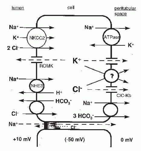
FIGURE 5.6 A model depicting the major elements of the mechanism of NaCl absorption by the thick ascending limb. Dashed lines indicate passive ion movements down electrochemical gradients. ROMK, renal outer medullary K+ channel; ClC-Kb, chloride channel Kb.

Figure 5.6 integrates the results of several electrophysiologic and biochemical studies to provide a model of salt reabsorption in the thick ascending limb. According to this model, net Cl absorption by the TAL is a secondary active transport process. Luminal Cl entry into the cell is mediated by an electroneutral Na+-K+-2Cl cotransport process driven predominantly by the favorable electrochemical gradient for Na+ entry.181 Because the Na+ gradient is maintained by the continuous operation of the basolateral membrane Na+,K+-ATPase pump, the apical entry of Cl via the cotransporter ultimately depends on the operation of the basolateral Na+,K+-ATPase.

In contrast to the electroneutral entry of Cl across the apical membrane, the majority of Cl efflux across the basolateral membrane proceeds through conductive pathways.182,183 A favorable electrochemical gradient for Cl efflux through dissipative pathways has been demonstrated by Greger et al.184 in the rabbit cTAL. Intracellular Cl is maintained at concentrations above electrochemical equilibrium by the continued entry of Cl via the apical Na+-K+-2Cl cotransporter.185

According to the model in Figure 5.6, K+ that enters TAL cells via the Na+-K+-2Cl cotransporter recycles, to a large extent, across the apical membrane via a K+-conductive pathway. This apical K+ recycling serves several purposes. First, it ensures a continued supply of luminal K+ to sustain Na+-K+-2Cl cotransport. Without recycling, the luminal K+ concentration would fall rapidly as a consequence of K+ entry via Na+-K+-2Cl cotransport and would limit net NaCl absorption. Second, the apical membrane K+ current provides a pathway for net K+ secretion by the TAL. In mouse TAL, for example, the rate of K+ secretion amounts to about 10% of the rate of net Cl absorption.182 K+ secretion in this segment is an active process, ultimately driven by the Na+,K+-ATPase, proceeding in the face of a lumen-positive transepithelial potential. Third, under open circuit conditions, the transcellular and paracellular pathways form a current loop in which the currents traversing the two pathways are of equal size, but which traverse in the opposite direction. The potassium current from cell to lumen polarizes the lumen and causes an equivalent current to flow from the lumen to the bath through the paracellular pathway.186 Because the paracellular pathway is cation selective (PNa/PCl = 2 to 6), the majority of the current through the paracellular pathway is carried by Na+ moving from the lumen to the interstitium. This paracellular absorption of Na+ increases the efficiency of Na+ transport by the TAL.187 With reference to Figure 5.6, for each Na+ transported through the cell and requiring the use of ATP, one Na+ is transported through the paracellular pathway without any additional energy expenditure. Finally, the apical K+ current satisfies the continuity requirement imposed by a high degree of conductive Cl efflux across basolateral membranes.182

A small component of Na+ transport by the TAL is accounted for by NaHCO3 absorption.188 In the rat TAL, the rate of NaHCO3 absorption is roughly 5% to 10% of that for NaCl absorption. NaHCO3 absorption appears to be mediated by an apical membrane amiloride-sensitive Na+/H+ exchanger and a basolateral membrane electrogenic Na+-3(HCO3) cotransporter.188

The following sections will describe the individual components of the mechanism for TAL salt transport (Fig. 5.6) in greater detail.


Apical Na+-K+-2Cl Cotransport

Studies of Cl transport across apical membranes of intact TAL segments189 and in isolated membrane vesicle preparations190 established that the predominant mode for Cl entry into the TAL cell is via a Na+-K+-2Cl cotransporter. A characteristic feature of this transporter is its sensitivity to inhibition by furosemide, bumetanide, and other 5-sulfamoylbenzoic acid derivatives.191 The measurement of isotope flux into TAL cells or membrane vesicles prepared from the inner stripe of the outer medulla yielded a stoichiometry of 1 Na+:1 K+:2 Cl cotransport.190 K+-independent NaCl cotransport has also been described under certain conditions.192

The proteins that mediate the Na+-K+-2Cl cotransport have been cloned. An absorptive form of the Na+-K+-2Cl cotransporter, referred to as NKCC2 or BSC1, was initially cloned by Gamba et al.193 based on sequence homology to the thiazide-sensitive Na+-Cl cotransporter (see the following). A second Na+-K+-2Cl cotransporter, NKCC1, was cloned by Payne et al.194 NKCC2 (BSC1) is the primary mediator of apical salt entry in the thick ascending limb. In situ hybridization and single-nephron reverse transcriptase polymerase
chain reaction (RT-PCR) studies demonstrated the expression of NKCC2 in the MTAL and CTAL,195 and immunohistochemical studies indicate that NKCC2 is localized to the apical membrane of these nephron segments.196 The importance of NKCC2 in mediating salt reabsorption in the TAL is illustrated by the fact that loss-of-function mutations of NKCC2 cause Bartter syndrome (Table 5.1),197 a Mendelian salt-wasting disorder characterized by hypokalemia, metabolic alkalosis, hyperaldosteronism, and normal-to-low blood pressure, results from a defect in salt absorption by the thick ascending limb.

The NKCC2 cDNA encodes a glycoprotein containing ˜1100 amino acids and having a predicted molecular weight of 115 to 120 kDa.193 The full-length protein contains 12 transmembrane domains containing a sizable extracellular loop with N-glycosylation sites positioned between transmembrane segments 7 and 8, and large intracellular amino and carboxy termini flank the transmembrane regions. NKCC2 belongs to the SLC12A family of cation chloride cotransporters, which is part of the amino acid polyamine organocation cotransporter (APC) superfamily.198 Based on the homology to other crystallized APC family members, the cotransporter structure probably consists of two clustered groups of five transmembrane helices that are positioned in a symmetric, inverted orientation.199 The details regarding how this fold facilitates the three-ion cotransport remain obscure but surely will provide an initial framework for more detailed structure-function studies in the coming years.

The two cytoplasmic domains of NKCC2 mediate specific regulatory functions. The amino terminus is believed to be unstructured and contains several cytosol-exposed serines and threonines, which are phosphoacceptor sites. These residues are phosphorylated by at least three protein kinases that stimulate NKCC2 activity and/or plasma membrane expression (discussed in detail below).200 The carboxy terminus is large and comprises ˜40% of the total NKCC2 sequence. It may also contain phosphorylation sites, although to date this has not been explored in detail. It is clear, however, that the NKCC2 C-terminus serves as a hub for interactions with proteins that regulate its trafficking, including the glycolytic enzyme aldolase201 and secretory membrane carrier protein 2 (SCAMP2).202 It also serves as the interface for the formation of NKCC2 homodimers,203 which was confirmed recently when the crystal structure of the C-terminus of the related prokaryotic cation chloride cotransporter MaCCC was solved.199

At least six isoforms of NKCC2 have been identified.200 These isoforms are the result of alternative splicing of two regions of the NKCC2 gene: the first region is a 96 base pair region that encodes part of the second transmembrane domain, whereas the second region encompasses the extreme C-terminus. Three variants of the 96 base pair region are encoded by different versions of exon 4 of the NKCC2 gene. These exons are differentially spliced into NKCC2 pre-mRNAs to generate three distinct isoforms (A, B, and F), which alter the amino acid composition of the second transmembrane domain. Each of the A, B, and F isoforms can have either a long C-terminus, or a truncated C-terminus; although to date, the short isoforms have only been described in the murine TAL.204 In addition, several “tandem” transcripts have been described in the human kidney; these contain combinations of exons 4A, 4B, and 4F spliced alongside one another into the NKCC2 pre-mRNA.205 Transcripts containing exons 4A/4F, 4B/4A, and 4B/4A/4F have been reported. Because these tandem transcripts contain redundant sequences encoding for the second transmembrane domain, they probably cause the misfolding of NKCC2, resulting in the formation of nonfunctional isoforms. Because these isoforms may still form oligomers with NKCC2, they likely exert a dominant-negative effect on NKCC2 function and inhibit its activity.206

The A, B, and F isoforms show differential expression within the thick ascending limb. In the rat nephron, the A isoform was found in both the cortical and medullary TAL, the B isoform is restricted to the cortical TAL, whereas the F isoform is present in the medullary, but not the cortical, the TAL, and to a lesser extent, in the outer medullary collecting duct. Although some interspecies discrepancies have been noted, similar findings have generally been observed in the embryonic mouse and human kidney.205,207,208

When the 4A, 4B, and 4F exons are spliced into transcripts containing a long C-terminus, all three products are capable of mediating Na+-K+-2Cl cotransport. However, the A, B, and F isoforms have different transport properties that may have physiologic relevance. Isoforms A and B have higher affinities for Na+, K+, and Cl than the F isoform. The A isoform possesses the highest transport capacity of all three isoforms. Based on the known distribution of the A, B, and F isoforms in the TAL, it is currently thought that the A isoform accounts for the high transport capacity of the medullary TAL, whereas the presence of the more active A and B isoforms in the cortical TAL allows for the continued reabsorption of salt to take place, even though the tubular fluid in this segment is more dilute than plasma. Supporting this is experimental evidence demonstrating that the NKCC2 A and B isoforms can both be strongly activated by Na+, K+, and Cl at concentrations that are much more dilute than the composition of tubular fluid in the cortical TAL.208


Apical K+ Conductance

An important feature of the luminal membrane of the TAL is a barium-inhibitable potassium conductance.185 This apical membrane K+ conductance allows K+ to be recycled from the cell back into the luminal fluid to support further NaCl absorption via the NaCl cotransporter. Using measured values of intracellular K+ activity (rabbit cTAL),209

Only gold members can continue reading. Log In or Register to continue

Stay updated, free articles. Join our Telegram channel

May 29, 2016 | Posted by in NEPHROLOGY | Comments Off on Tubular Sodium Transport

Full access? Get Clinical Tree

Get Clinical Tree app for offline access