■ Specifically review any potential allergy to iodinated contrast agents. Medical preparation to mitigate an allergic reaction may be indicated.
SURGICAL MANAGEMENT
Preoperative Planning
■ The potential need for serial ERCP procedures should be determined. A gastrostomy tube (G-tube) can be placed at the index procedure to facilitate subsequent endoscopic interventions. Potentially affected patients should be prepared for this possibility.
■ These operations require significant coordination between multidisciplinary teams. The authors highly recommend that these procedures be scheduled as the first case of the day.
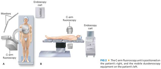
■ The procedure needs to be scheduled in a room that can accommodate two teams, a C-arm fluoroscopy unit, and a mobile endoscopy cart.
Positioning
■ The operative table should be configured so that C-arm fluoroscopy can be readily performed of the upper abdomen (FIG 2).

■ After induction of general anesthesia, a urinary bladder catheter is placed. An orogastric tube is rarely necessary.
■ The patient is positioned supine, ideally with both arms tucked to facilitate access for both C-arm fluoroscopy and the endoscopy team. A footboard is placed. A fixed liver retractor can be positioned on either side of the patient if needed.
■ Venous thromboembolism prophylaxis (both mechanical and pharmacologic) should be used. Antibiotics are administered within the guidelines of Surgical Care Improvement Program (SCIP) criteria for a clean-contaminated procedure.
TECHNIQUES
TROCAR PLACEMENT
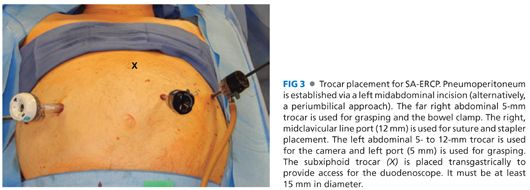
■ Trocar placement is depicted in FIG 3. Four trocars will be required for placement of stay sutures in the stomach, and one of these can be subsequently used for clamping of the small bowel. A subxiphoid, transgastric trocar is also necessary.

■ Initial trocar placement is performed in the left midclavicular line at the level of the umbilicus following insufflation of the abdomen with a Veress needle. The authors prefer the use of an optical separator and a 30-degree scope.
■ Subsequent trocars are placed under direct vision.
GASTRIC PEXY
■ The patient is placed in reverse Trendelenburg position and the location of the transgastric trocar placement determined and marked. Ideally, the trocar will enter the anterior gastric wall at the junction of the body and the antrum of the stomach at the midpoint between the mesentery of the greater and lesser curves.
■ Determine the relationship of an antegastric Roux limb and the associated mesentery to the ideal target for the transgastric trocar. Dissection of the Roux limb or mesentery should be taken with great caution; injury to or vascular compromise of the Roux limb can be catastrophic. It is better to accept a more lateral target, including one that may require taking down some of the greater curve mesentery, than to engage in a significant dissection of the Roux limb from the stomach.
■ Four stab wounds are placed around the planned site of the transgastric trocar.
■
Stay updated, free articles. Join our Telegram channel

Full access? Get Clinical Tree


