Setting Up a Colorectal Physiology Laboratory


Setting Up a Colorectal Physiology Laboratory


Dana R. Sands

Steven D. Wexner


Pathology is the accomplished tragedy; physiology is the basis on which our treatment rests.

— SAMUEL BUTLER

The evaluation of patients with complex functional problems frequently requires multimodality testing for proper diagnosis. Beginning in the 1980s, physicians used the tools of the anorectal physiology laboratory to better understand the pathophysiology behind their patients’ complaints. Over the ensuing decades, many clinicians have established their own physiology laboratories. An appreciation of the available testing modalities and their application to clinical practice is essential for the physician whose aim is the treatment of functional colorectal disorders. No longer is it suitable to rely solely on clinical examination. Sophisticated tools of the colorectal physiology laboratory will offer a greater depth of understanding of numerous disease states, including constipation, evacuatory dysfunction, fecal incontinence, and prolapse. Many of these conditions are underdiagnosed. As physicians become more aware of the prevalence of these conditions and also the incidence of concomitant anterior compartment pathology,136 the use of the colorectal physiology laboratory will continue to increase. A team approach with urology and urogynecology counterparts facilitates accurate diagnosis with combined multimodality treatment regimens.

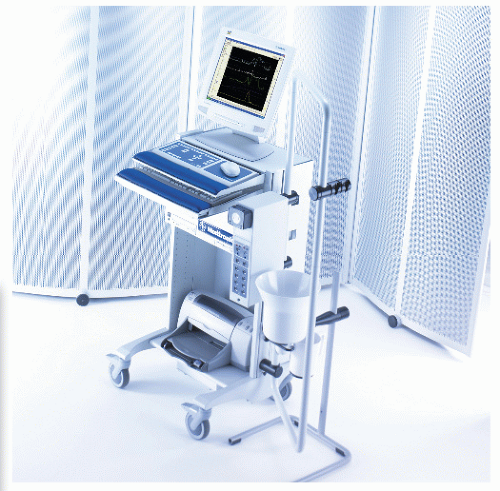
The components of a complete colorectal physiology laboratory include anal manometry, ultrasound of the anus and rectum, neurophysiologic assessment with pudendal nerve terminal motor latency, electromyographic evaluation, defecography, as well as transit studies of the small intestine and colon. No single test can adequately define the status of the anorectum, either in health or when diseased. Accurate diagnosis depends on the integration of a detailed history and clinical examination, along with the appropriate physiologic investigations. Thus, every laboratory must have the resources, space, and personnel to perform a wide array of physiologic studies. Setting up a new laboratory is an expensive venture, and administrative authorities may understandably require a cost-benefit analysis or business plan prior to considering whether this new cost center should be supported. Required data include anticipated case volumes and reimbursement expectations. Still, much of the capital outlay can be minimized if intradepartmental arrangements can be accommodated to share rooms, equipment, and ancillary personnel. For example, the computer hardware equipment, which can be used for both esophageal and anorectal manometry, can be purchased jointly by gastroenterology and colorectal divisions. Specific software packages and catheters can then be acquired. Similarly, an ultrasound scanner can be shared among gynecologists, urologists, and colorectal surgeons, with each using a specific probe. Defecography can be performed in a standard fluoroscopy suite, and electromyography (EMG) equipment can be shared with the neurology department.

Ideally, the various tests should be available in proximity, so that they can be performed in a relatively short time and with minimal inconvenience to the patient. The number of required personnel is dependent on the volume of tests


undertaken each day. Many units have specifically trained nursing staff to perform these studies under appropriate supervision. The same nurses who undertake a given procedure for one department can also perform the analogous procedure for the department sharing the equipment.


▶ MANOMETRY

The first reports of anorectal manometry in the literature date back to 1972.42 Since that time, there has been no standardization of the technique. The basic information provided, regardless of technique, should include an objective assessment of the anal muscular tone, rectal compliance, and anorectal sensation, as well as the determination of the integrity of the rectoanal inhibitory reflex. The lack of standardization creates some difficulty in comparing results from different institutions. In fact, institutions must determine a set of normal values for their patient population. There is often a difference noted between male and female subjects.64

Manometry has been useful in the diagnosis of patients suffering from fecal incontinence,23 constipation,135 Hirschsprung’s disease,127 and anal fissure.57,152 It has been widely used in the investigation of individuals with fecal incontinence to identify the presence of sensory or muscular defects, as well as to define functional weakness of the internal and/or external anal sphincter. Those with fecal incontinence or proctitis may present with significant loss of the ability to sense rectal distension.166 A maximal tolerable volume less than 100 mL may indicate visceral hypersensitivity, poor compliance, or rectal irritability.


Anorectal Manometry Systems

There are three basic types of systems used for performing manometric examinations: air- or water-filled balloon systems, water perfusion systems, and solid-state microtransducer systems.


Air- or Water-Filled Balloon Systems

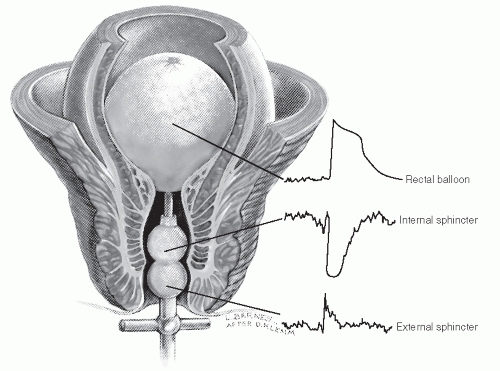
In these systems, fluid- or air-filled balloons are placed within the anus and rectum and are connected to transducers via small catheters. One of the early designs was that of Schuster and colleagues.149 This device consists of a hollow metal tube 4 in. in length surrounded by a latex balloon tied in such a way so as to create two separate compartments. Each balloon communicates with a pressure transducer via two plastic catheters. A further balloon can be placed into the rectum via the metal tube. The apparatus is placed in the rectum so that the internal balloon is surrounded by the internal sphincter, and the external balloon is encircled by the superficial fibers of the external anal sphincter (Figure 7-1).148 The device is set by inflating the rectal balloon to elicit the rectoanal inhibitory reflex (RAIR), which automatically seats the catheter in position. Thereafter, mean resting and squeeze pressures, the presence of the RAIR, rectal sensitivity, and compliance can all be assessed.

Theoretically, this device should be able to assess pressures from the internal and external anal sphincter separately, but in practice the overlap of these two muscles is too great to allow differentiation.149 Furthermore, because pressures within the anus are in part influenced by the distortion of the anal canal, a probe with a large balloon yields a greater pressure than does one with a smaller balloon in the same subject.65,164 In addition, rapid distension of the balloon will generate a higher pressure.65,164 One final area for confusion
with this method is that air is compressible, and the pressures recorded may be somewhat lower than the true values.


FIGURE 7-1. Anorectal manometric tracings indicate the pressure changes at each level after distension of the balloon in the ampulla. (Adapted from Rosen LS, Khubchandani IT, Sheets JA, et al. Management of anal incontinence. J Am Fam Pract. 1986;33:129, with permission.)


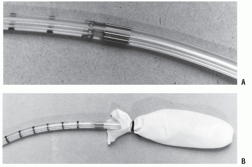
FIGURE 7-2. Water-perfused anorectal manometry catheters. A: Eight-channel radial catheter suitable for vector volume analysis. (Courtesy of Synectics Medical, Irving, TX.) B: Custom-built four-channel radial catheter with two intraballoon channels. (Courtesy of Arndorfer, Inc., Greendale, Wl.)

These disadvantages prompted the development of another balloon system that uses smaller, water-filled balloons (Marquât Company, Boissy-Saint-Léger Cedex, France).65 However, although accurate assessments can be performed with these catheters, they permit measurement of only a limited area of the anal canal.73

Both of these systems are relatively simple, and once placed they do not require further movement. Investigations, therefore, can be performed by one operator. However, the information ascertainable from these systems is relatively limited because the pressure measured is the sum of all forces acting upon the balloon. Thus, only determinations of the global resting and squeeze pressures of the anal canal can be obtained. Indeed, because as implied, larger balloons produce more distortion of the anal canal, basal pressure measurements may be unreliable, with only the changes in pressure being reproducible.65 The presence of the RAIR and information on rectal sensitivity and compliance can be obtained using either system.


Water-Perfused Systems

Water-perfused systems were developed by Arndorfer and colleagues and are the most widely used for performing manometry in the United States.6 These systems function by creating an artificial cavity between the anus and the catheter. As perfusion continues, the full capacity is reached. Thereafter, fluid leaks into the rectal ampulla or out of the anus. The pressure required to overcome initial resistance after the space is filled is termed the yield pressure.68,92 As the pressure in the anal canal increases, the mucosa will be brought into contact with the catheter ports, thereby impeding the flow of water. The yield pressure then becomes the pressure required to overcome this obstruction. This information is then transmitted via nondistensible capillary tubing to transducers that convert this pressure to electrical signals.


Technique of Manometric Evaluation

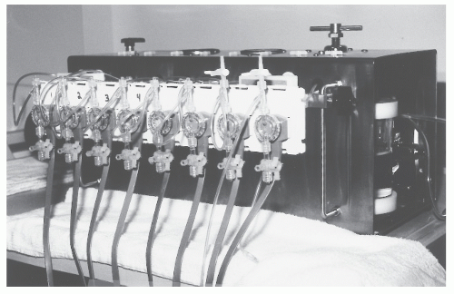
Our preference is to perform a station pull-through technique using a custom-built 4.8-mm diameter polypropylene flexible catheter with four radial ports located 8.5 cm from the tip (Arndorfer, Inc., Greendale, WI).83,84 A further two ports are positioned 2 cm from the end of the catheter and are enclosed within a thin latex rubber balloon fashioned from the finger of an examination glove (Floor/exam latex glove no. 8857; Baxter Health Care Corporation, Valencia, CA). This balloon is secured at 3 cm from the tip of the catheter using a 2-0 silk ligature. An eight-channel hydraulic capillary infusion system (Arndorfer, Inc., Greendale, WI) is used with Medex transducers (M6677; Medex, Inc., Hilliard, OH). Mechanical pressures are transmitted to a PC Polygraf HR (Synectics Medical, Inc., Irving, TX). The resulting electrical impulses are displayed on a computer using Polygram V6.4 software (Synectics Medical, Inc., Irving, TX).

With the patient in the left lateral decubitus position, the catheter is inserted to 6 cm; 20 to 30 seconds are allowed for the sphincter to recover from this insult and for the pressures to equilibrate. It is important to permit adequate time for the small cavity between the rectal wall and the catheter to fill with perfusate so that the yield pressure is reached.

Following this period of equilibration, various wave patterns may emerge that demonstrate the presence of intrinsic cyclic activity attributable primarily to the internal
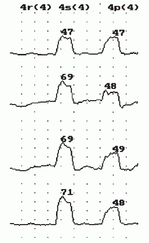
sphincter.175 Three basic patterns are observed. Slow waves are the most frequently encountered. These vary in frequency from 10 to 20 cycles/minute with an amplitude from just above physiologic baseline noise to 15 mm Hg (Figure 7-9).146 They can most often be observed in the region between the proximal border of the sphincter and the area of the maximal average resting pressure.36,130 The clinical significance of these waves is unknown.


TABLE 7-3 Manometry Systems


COMPATIBILITY


MANUFACTURER


SYSTEM


WATER PERFUSED


SOLID STATE


Medical Management Systems, Enschede, Holland


X


Medtronic Functional Diagnostics, Shoreview, MN 55126 USA


X


X


Narco Bio-Systems, Austin, TX 78754, USA


MMS-200


X


X


Sandhill Scientific, Highlands Ranch, CO 80126, USA


BioLAB


X


Synectics Medical, Irving, TX 75038, USA


PC Polygraph


X


X


Synectics Medical AB, Stockholm, S-116 28, Sweden


X


X


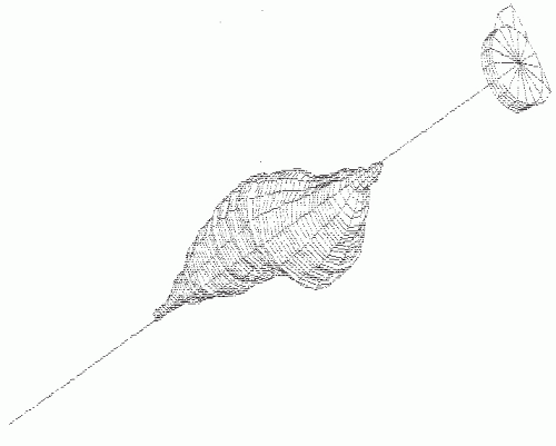
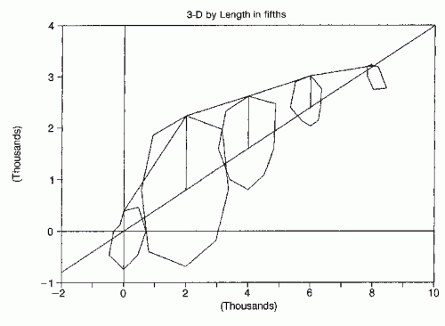
FIGURE 7-8. Three-dimensional reconstruction of same patient illustrated in Figure 7-7 using computer-aided design program. (Courtesy of John A. Coller, MD.)


FIGURE 7-9. Slow waves noted in the region of the high-pressure zone. (Polygram software; courtesy of Synectics Medical, Irving, TX.)

Ultraslow waves are the second most common waveforms recorded. They have a frequency of 0.5 to 1.5 cycles/minute and are of large amplitude (up to 100 mm Hg).146 They are found more frequently in patients with high resting anal pressures,69 such as in those individuals with an anal fissure,66 hemorrhoids,67 or primary anal sphincter hypertonia. They are seen most commonly in the region of maximal average resting pressure.

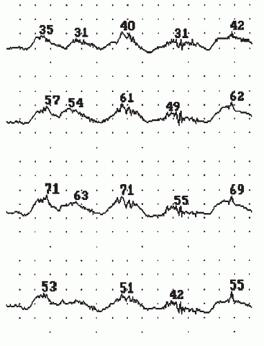
The intermediate wave is the least frequently observed type of oscillation (frequency, 4 to 8 cycles/minute). They are most often noted in patients with neurogenic fecal incontinence or following ileal pouch-anal anastomosis (Figure 7-10).162 When present, they make interpretation of resting and squeeze pressures more difficult. Resting pressure should be assumed to be the mean of the peak and trough pressures at rest.


FIGURE 7-10. Intermediate waves in a patient following colonic pouch-anal anastomosis. (Polygram software; courtesy of Synectics Medical, Irving, TX.)


FIGURE 7-11. Rest, squeeze, and push phases of station pull-through manometry recorded at 4 cm from the anal verge. (Polygram software; courtesy of Synectics Medical, Irving, TX.)


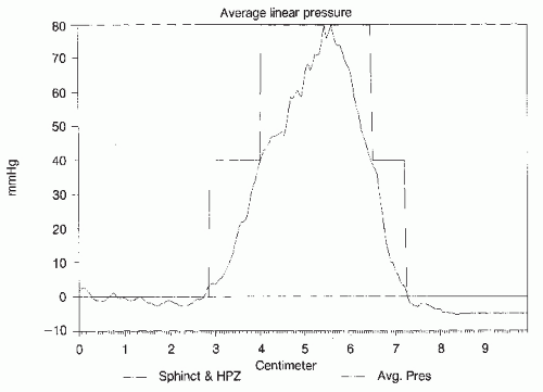
Following equilibration of the pressures, the patient is asked to perform a single maximum squeeze effort followed by a period of rest, and then a maximal push effort (Figure 7-11). These measurements are repeated at a further five stations, separated by 1-cm intervals, as the catheter is progressively moved caudally. Thus, the rest and squeeze pressures over the entire length of the anal canal can be measured, and the mean resting and squeeze pressures over the high-pressure zone (HPZ) can be calculated. The HPZ is defined as that length of the anal canal through which pressures are greater than 50% of the average maximum pressure. This definition is similar to that used when a continuous pull-through technique is employed. Alternatively, the HPZ can be defined as that zone bounded caudally by a rise in pressure of 20 mm Hg and cephalad by a fall in pressure of 20 mm Hg in at least 50% of the channels. This latter definition may present a more accurate picture of the sphincters in incontinent patients because pressures are so low that there is no true HPZ. However, using the former definition, the HPZ will always be at least 1 cm, but this may be an inaccurate value in a patient with a patulous anus.

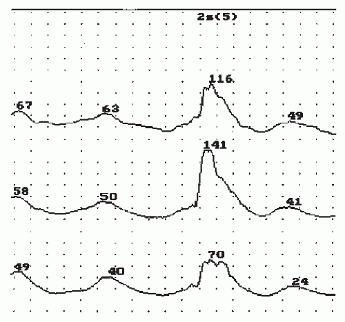
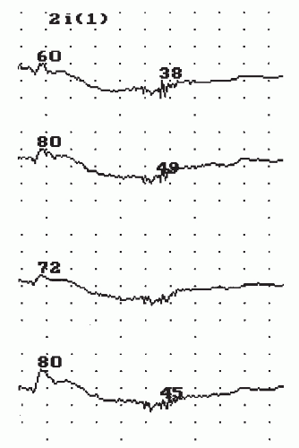
The catheter is then reinserted to a distance of 2 cm from the anal verge, and the latex balloon is insufflated with 40 mL of air over 2 to 3 seconds and kept inflated for 20 seconds in order to elicit the RAIR. In response to distension of the lower rectum and upper anal canal, external sphincter contraction is followed by internal sphincter relaxation (Figure 7-12). If the reflex is not present, it is important to repeat the test with increased insufflation. Some patients, especially those with neurogenic fecal incontinence, decreased anal sensation, or megarectum, may respond only at a higher volume. The air is removed and the balloon reinflated with 50 or 60 mL until a reflex is observed. If the reflex is still undetectable, the catheter is inserted to 3 cm and repeat insufflations are performed. In our laboratory, we record only the presence or absence of the reflex. Some software programs contain the algorithms for calculating various reflex parameters, including duration, percentage of excitation, and excitation latency. Although some authors suggest that delayed, diminutive or absent excitation, or excitation only in response to large volumes may be used as a crude indicator of pudendal neuropathy, the clinical implications of these values are problematic.145 Some prefer a spiral catheter to detect the presence of the RAIR over the entire length of the anal canal. However, the use of a radial catheter at the two stations where the reflex is most likely to be detected avoids this requirement for each investigation. In rare instances in which a reflex cannot be elicited and there is no evidence of megarectum, the spiral catheter may be useful. The most common reasons for absence of the RAIR are megarectum and a prior ileoanal or coloanal anastomosis.

The catheter is then inserted to a distance of 6 cm from the anal verge, positioning the balloon in the rectal ampulla. The balloon is then slowly filled with core temperature water at a
rate of approximately 1 mL/second. The first sensation perceived by the patient is noted as the minimal sensory volume, and the mean intraballoon pressure is noted. Thereafter, balloon filling is continued until the maximum tolerable volume is reached, and again the intraballoon pressure is noted. Using these values, rectal compliance can be calculated from the formula V/P.84 Thus, with a large balloon volume and only a small increase in rectal pressure, the rectum is considered very compliant. In patients suffering from ulcerative colitis, Crohn’s proctitis, or radiation proctitis, the rectum may be poorly compliant in that a small increase in volume will result in a large increase in pressure. When one uses this technique, it is important to know the compliance of the balloon at various stages of insufflation so that it can be subtracted from the pressure measured via the intraballoon ports. Latex balloons may deform along the longitudinal axis with increased infusion of water. This may give a false impression of high rectal compliance.109 As a consequence, some clinicians prefer to use barostat equipment to calculate this parameter, using a noncompliant balloon that does not deform with increasing pressure.


FIGURE 7-12. Rectoanal inhibitory reflex recorded at 2 cm from the anal verge in response to distension of the rectal balloon with 40 mL of air. (Polygram software; courtesy of Synectics Medical, Irving, TX.)

The measurement of compliance is not a diagnostic test but supplements other investigations for evaluating the pathophysiology of anorectal disease. It is of particular value in patients with proctitis and incontinence through ascertaining whether the incontinence is due to lack of rectal reservoir function or diminished anal sphincter tone. Similarly, in some constipated patients, compliance may be abnormally high. This finding can reflect overaccommodation and, therefore, a sensory contribution to the outlet obstruction.


▶ DEFECOGRAPHY

Defecography or evacuation proctography is an essential component of the colorectal physiology laboratory. This study provides the physician with useful information about the anatomic interplay of the anus, rectum, and sigmoid colon as well as the vagina and pelvic floor during the defecatory process. Patients suffering from constipation, prolapse, solitary rectal ulcer, and rectal pain may benefit from this evaluation. This dynamic investigation of the mechanism of defecation has become an increasingly popular study since it was described by Wallden in 1952™ and Burhenne in 1964.26 Improvements in the technique, such as cinedefecography and videoproctography, have led to a better understanding of the evacuation process, thereby facilitating the identification of conditions that disturb the physiology of rectal emptying.


Author’s Preferred Technique

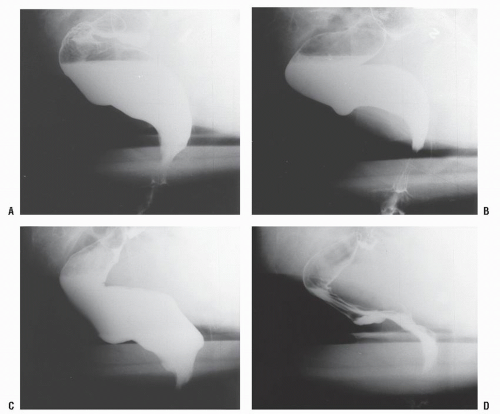
Because most of our patients have several physiologic investigations performed on the same day, bowel preparation by means of a phosphate enema is usually performed before manometry. With the patient in the left lateral decubitus position, a digital rectal examination is performed to lubricate the anal canal, to ascertain the presence of any pathologic abnormality, and to ensure that the patient understands the instructions of how to squeeze and to push. Through an enema-tipped catheter, 50 mL of liquid barium (58% wt/wt barium sulfate suspension, Polybar) is introduced into the rectum followed by the insufflation of a small quantity of air to coat the mucosa of the sigmoid colon. Thickened barium paste is then introduced into the rectum until the patient experiences a feeling of fullness. A small quantity of paste is inserted into the anus in order to outline the anal canal, after which the vagina is opacified with Gastrografin. The patient is then turned to the right lateral side, and the table is slowly brought to the erect position. This enables one to slide down the table and to sit on the commode. The fluoroscopy tube is positioned so that the coccyx and the symphysis pubis are both visible on screening. Still pictures (proctograms) are taken at rest and at maximal squeeze prior to starting the video recording (Figure 7-14).
At the commencement of the video, the patients are asked to squeeze, to relax, and then to try to commence evacuation. As elimination starts, a further still picture is taken. Following evacuation, the recording is recommenced while the patient squeezes and pushes in order to check for the presence of an intussusception or nonemptying rectocele. A single still image of the evacuated rectum at rest is also obtained (Figure 7-15B).


FIGURE 7-14. Proctogram series. A: Rest. B: Squeeze. C: Push. D: Postevacuation.

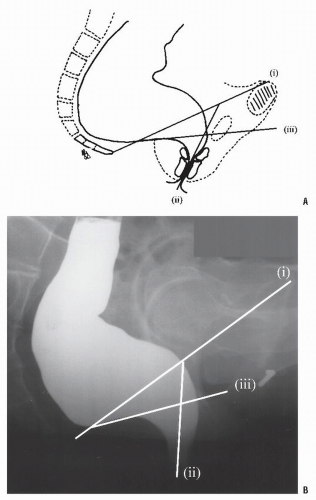
With completion of the study, three lines of reference are drawn, so that the anorectal angle, puborectalis length, and extent of perineal descent can be assessed on the proctograms. First, a line is drawn between the tip of the coccyx and the anterosuperior surface of the symphysis pubis. Lines are also drawn along the axis of the mid-anal canal and along the posterior rectal wall (Figure 7-15A).86 Whereas some have proposed using the dynamic images alone,156 the authors prefer to rely on the combination of the dynamic and still images. This has been shown to produce reliable and reproducible measurements.35


Anorectal Angle

Much attention has been directed to the anorectal angle since Parks (see Biography, Chapter 29) first proposed its role in maintaining fecal continence.129 The anorectal angle
is the angle formed by the axis of the posterior rectal wall and the axis formed by the anal canal; it is usually 70 to 140 degrees at rest.59,110 This angle becomes more acute during the squeeze phase (75 to 90 degrees) and becomes more obtuse during evacuation (100 to 180 degrees at maximum strain).86 Some investigators have reported greater anorectal angles in men,153 but most clinicians believe that it is not affected by gender.48 The wide variation in observed normal values is probably due to differences in interpretation.49,59 It has even been suggested that a normal range is impossible to define.153 Therefore, the change in angle seen during the squeeze and push phases is more important than the actual measurement.


FIGURE 7-15. Lines of reference for analysis of proctograms. A: Diagrammatic representation of (i) pubococcygeal line, (ii) midanal line, and (Hi) posterior rectal line. B: Resting proctogram with lines demonstrated.


Puborectalis Length and Perineal Descent

The puborectalis length is measured as the minimal distance between the anterosuperior aspect of the symphysis pubis and the puborectalis notch. This ranges between 14 and 16 cm in the rest position, shortens to between 12 and 15 cm during the squeezing phase, and is elongated to 15 to 18 cm during the push phase.88 As with the anorectal angle determination, the change in these parameters is more important than their definitive value.

Perineal descent is assessed as the length of a perpendicular line drawn from the pubococcygeal line to the anorectal junction. Perineal descent of either more than 3 cm in the rest phase or a further increase of 3 cm in the push phase is considered abnormal.


Interpretation

As with all of the studies performed in the physiology laboratory, interpretation of the results of defecography should be combined with a thorough assessment of the patient’s complaints through history and physical examination to ascertain the clinical significance of the findings. A normal defecogram should demonstrate relaxation of the puborectalis as indicated by (1) an increase in the anorectal angle, (2) lengthening of the puborectalis, and (3) a blunting of the puborectalis notch. These changes should be accompanied by symmetric opening of the anal canal to form a cone that is wider cephalad than it is caudad. This process takes ap- proximately 4 to 5 seconds. Contrast material in the upper rectum should subsequently be passed into the lower rectum and out the anus by rectal contraction, a process that may be facilitated by Valsalva’s maneuver, squeezing the rectum onto the levator plate.108 Complete evacuation takes about 10 to 12 seconds when thickened contrast medium is used but is more rapid (8 to 9 seconds) when the mixture is more fluid.97 Failure to relax the puborectalis is noted by the persistence of the puborectalis notch, failure of the anorectal angle to increase, and difficulty in emptying the rectum. Although these findings have been noted in normal individuals,81 patients with complaints of difficult evacuation who have these radiologic findings should be treated for paradoxical puborectalis contraction.

Jul 17, 2016 | Posted by in GASTROENTEROLOGY | Comments Off on Setting Up a Colorectal Physiology Laboratory

Full access? Get Clinical Tree
Get Clinical Tree app for offline access