Company
Stryker
Storz
Olympus
Olympus
Wolf
Wolf
Gyrus/ACMI
Gyrus/ACMI
Scope name/series
SRU-6/SRU6X
27001K/L
OES 4000 dual
OES pro single
E-Line offset/ultrathin
Needle
Bagley
Slimline
Working length
33/43 cm
34/43 cm
33/43 cm
33/43 cm
31/43 cm
31 /43 cm
33/43 cm
33/43 cm
Tapered shaft
Yes
Yes
Yes
Yes
Yes
Yes
Yes
Yes
Tip size
6.9Fr
7/8Fr
7.5Fr
6.4Fr
6/8Fr
4.5Fr
6.9Fr
7/7.7Fr
Pixel quantity
30,000
N/A
N/A
50,000
20,000/50,000
N/A
N/A
N/A
Eyepiece
Straight
Offers both
Offers both
Offers both
Angled
Angled
Straight
Angled
Single/dual channel
Dual
Single
Dual
Single
Single
Single
Dual
Single
Working channel size(s)
3.4/2.5Fr
5Fr
N/A
6.4Fr
4/5Fr
3.4Fr
3.4/2.3Fr
5.4Fr
Direction of view
12°
6°
7°
7°
5°/12°
5°
5°
5°
Consider: A semirigid ureteroscope is self-supporting due to a strong, thin-walled, metal outer tube that encloses the components of the telescope. It only needs to include a working channel and fiber bundles (to transmit light and an image). However, a flexible ureteroscope must in addition contain a deflection mechanism (whose cable occupies space within the instrument). Also, the flexible jacket of the device requires bendable, reinforcing layers that tend to be thicker than the metal comprising a semirigid telescope; without reinforcing layers, the flexible ureteroscope might wear out (fatigue) too quickly from repeated flexing and extending. In this vein, a semirigid instrument might be expected to have a narrower outer diameter than a flexible one. In reality, most are at least as thick, and they utilize their additional space within to contain a larger working channel, a greater number of optical fibers, or both.
While smaller sizes and newer designs are continually being developed and might be in production at the time of publication of this treatise, flexible ureteroscopes today are between 5.3 and 7.5 F (outer diameter of the tip), while adult semirigid ureteroscopes are between 4.5 and 8.4 F tip diameter [5].
But the above sizes are somewhat misleading; the tip of the smallest currently available semirigid ureteroscope (the Richard Wolf Model 8701 “Needle Ureteroscope”) actually describes the size of the absolute tip of the scope, including the lens, but excluding the bevel (beak) which contains the working/irrigation 3 F channel; just a few millimeters from the tip, the slope of the beak augments the total diameter to 6.5 F [30]. In contrast, the Olympus flexible ureteroscope (URF-P5) is truly 5.3 F diameter for a significant portion of the instrument’s length [31]. Both designs become thicker, as one progresses from the tip to the ocular, in order to provide more support and durability for the instrument. In addition, the semirigid scopes usually have a 5–12° look-down angle at the usually beveled tip of the scope, while the flexible scope tends to view straight ahead (0° angle).
Most manufacturers of semirigid ureteroscopes provide a choice of two lengths, 33 and 43 cm, enabling the urologist to pick the scope most appropriate to the task at hand. A short, semirigid scope might be better in a female; when one combines the length of the urethra with the distance from the vesical neck to the ureteral orifice, and then the ureteral length itself (up to the uretero-pelvic junction), it would be rare for the total distance to exceed 33 cm. However, for the male patient, the length of the urethra, plus the distance from the vesical neck to the ureteral orifice, and of course adding the length of the ureter, one might need a 43 cm long ureteroscope unless one is only working in the distal portion of the ureter. Figure 22.1 depicts a semirigid ureteroscope that was easily advanced to the patient’s right uretero-pelvic junction (UPJ); such is usually possible in patients with normal anatomy and whose psoas muscles are not unduly bulky.
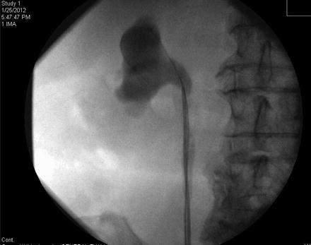
Fig. 22.1
A semirigid ureteroscope was easily passed alongside a safety guide wire in order to treat an acquired uretero-pelvic junction stricture in this patient. The instrument can be seen in the lower renal pelvis after the stricture was opened with a laser; contrast was instilled through the scope and there was only slight extravasation as contrast passed down the ureter alongside the ureteroscope
Flexible ureteroscopes are approximately 54–70 cm long, and this enables the surgeon to reach fully into all branches of the renal collecting system, even in a male patient with morbid obesity. But the surgeon usually sees better with a semirigid ureteroscope than with a flexible scope under most conditions. The optical resolution is nearly always better (30–50,000 pixels vs. 3–6,000 pixels, although the “chip on the tip” flexible scopes may correct this discrepancy in the next 3–5 years) [28, 32, 33], and the saline irrigant tends to flow more quickly, due to larger working channels (3.6 F for most flexible ureteroscopes vs. 4.5 F for most semirigid ureteroscopes); both factors can help with difficult situations such as active bleeding, compared to a flexible scope.
Introduction of a flexible ureteroscope is possible, even without a wire, but it tends to be difficult for those surgeons not practiced with such techniques. If a stone is fairly distal in the ureter, manipulation of a flexible ureteroscope can be difficult due to lack of purchase; i.e., the very nature of the flexible scope can allow buckling to occur, with subsequent loss of access into the ureter (Fig. 22.2). A semirigid ureteroscope provides its own inherent stability, and one thus has greater control over the tip, and consequent treatment of a stone or other lesion, while working in the lower ureter (Fig. 22.3).
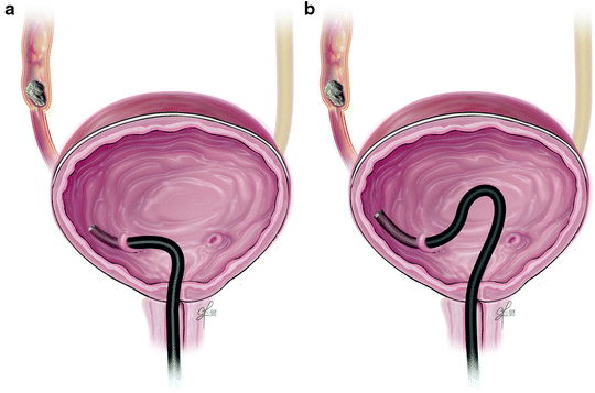
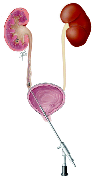
Fig. 22.2
This illustrates the sometimes encountered difficulty stabilizing a flexible ureteroscope when treating a distal ureteral calculus; pushing on the instrument may result in curling within the bladder rather than advancement of the device up the ureter, so control is thus limited in these instances
Fig. 22.3
A semirigid ureteroscope is self-stabilizing when used to access a lower third ureteral stone, and thus the tip can be readily controlled, in contrast to a flexible ureteroscope (previous illustration)
This author has had several instances performing laser lithotripsy where the unexpected happened: A Holmium:YAG laser failed in the midst of ureteral stone fragmentation during semirigid ureteroscopy, and no substitute laser device was immediately available. One option would have been to place a stent and complete the procedure another day, or keep the patient under anesthesia until another laser could be secured (>1 h delay). However, a Lithoclast was available and the procedure was safely completed using that device. Though laser is sometimes more effective and has a lower complication rate, pneumatic lithotripsy is still a reasonably safe alternative [12–17]. Due to limited flexibility of the pneumatic wire, the Lithoclast cannot currently be used with flexible ureteroscopes or with semirigid ureteroscopes lacking a straight-entry working channel.
A semirigid ureteroscope becomes difficult to use in a patient whose pelvic floor lacks elasticity. A patient with advanced cancer in the pelvis, one who has scar tissue from previous pelvic surgery, or one who has received previous pelvic radiation with subsequent fixation of organs and tissues, can prove more challenging for insertion of a semirigid ureteroscope. If one cannot pass the instrument initially, but the surgeon still wishes to utilize a semirigid rather than flexible ureteroscope, there are two “tricks” this author has found helpful at times. The first is to insert the semirigid instrument over a stiff glide wire, or even over an Amplatz Super-Stiff Guidewire. The wire helps direct the tip of the instrument and the stiffer wires tend to “straighten out” the angles and thus facilitate passage. One can also adjust the angle and/or position of one or both legs of the patient; sometimes this “straightens” the ureter as it relaxes the psoas and other muscles [34].
Interestingly, a semirigid ureteroscope can be utilized successfully in situations where angulation and/or tortuosity might be thought to totally preclude ureteral access. For example, a patient with prior surgery to correct vesico-ureteral reflux, as with the Cohen cross-trigonal technique, can usually undergo successful semirigid ureteroscopy despite the altered anatomy, if one uses a ureteral balloon dilating catheter (Uromax®, Boston Scientific, Inc.), the controlled expansion, stiff (when inflated under pressure) balloon usually eliminates curves and angles, thus permitting a straighter path even after deflation, and thus enabling passage of a semirigid ureteroscope [4, 35]. This technique can be applied to renal allograft transplant ureters (i.e., ones implanted in the dome or lateral bladder wall) as well [36, 37].
Semirigid ureteroscopy would be difficult (but not always impossible) when accessing the lower ureter through a bladder substitute (ileal conduit, Studor or Indiana Pouch, etc.). Such access is easier with a flexible ureteroscope and in many instances, requires antegrade insertion of a guidewire through a nephrostomy puncture, in order to instrument the ureter in a retrograde manner in patients with these types of anatomic alterations [38].
Patient Preparation, Set-Up, and Equipment
Patients should be prepared for ureteroscopy as for any surgical procedure that requires anesthesia. A complete history and physical examination should be obtained, and pertinent laboratory studies are usually ordered (a complete blood count and at least a basic chemistry, plus urinalysis). If the urinalysis shows pyuria, bacteriuria, positive nitrite, or otherwise is suggestive of a urinary tract infection, then a urine culture should be requested reporting any growth of uropathogens. Standard informed consent is obtained as per any surgical procedure, commensurate with the rules for the institution where the ureteroscopy is to be performed.
In select cases (patients with symptoms and/or comorbid medical conditions that increase the likelihood of heart disease), an electrocardiogram (EKG) should be ordered if one was not done within the preceding 6 months, and any major abnormalities should be addressed by the patient’s primary care physician, or by a cardiologist, to minimize any cardiac risk for surgery.
A preexisting urinary tract infection should be treated 24–48 h before surgery, or if bacteriuria cannot be completely eliminated, it should be suppressed. If the urine is sterile preoperatively, then prophylactic antibiotics should be administered before instrumentation of the upper urinary tract, according to the current American Urological Association (AUA) Guidelines [39]. In most cases, such will consist of a fluoroquinolone (FQL) or trimethoprim-sulfa (TMP-SMX), given with a sip of water an hour before surgery, or intravenously, so-as to enable adequate blood and urine levels before instrumentation. For patients allergic to these antibiotics, an aminoglycoside with a cell-wall inhibitor (i.e., a penicillin-type drug, a cephalosporin, or vancomycin) will provide good coverage of most uropathogens.
When a patient has had a joint replacement within the previous 2 years, when a patient is immunosuppressed (i.e., on chemotherapy, HIV positive, etc.), or when a patient has a prosthetic heart valve, he/she may be more susceptible to infection either of their implanted prosthetic device, or to urosepsis, so attention to the AUA Guidelines for Antimicrobial Prophylaxis is strongly recommended [39]. Anticoagulants are not routinely stopped prior to diagnostic ureteroscopy or laser lithotripsy of ureteral stones at our institution, but they might require temporary cessation if tissue is to be incised [40, 41].
Except in cases of pregnancy, fluoroscopy is routinely used for guidance of guidewires, ureteroscopic instruments, and stents during the procedure. Personnel should wear adequate protective coverage, including lead aprons, and the operating room should contain lead shielding in its walls and door(s). The operating surgeon(s) should additionally use protective eyewear (leaded glasses) if performing a high-volume of fluoroscopic procedures, as cataracts are a long-term risk after recurrent radiation exposure to one’s eyes [42, 43].
The patient should normally be in the dorsal lithotomy position (Fig. 22.4) or a modified dorsal lithotomy position where the ipsilateral leg (to the side being operated upon) is lowered and extended [44]. This latter position has been found by some to make the semirigid ureteroscopy easier and faster; it is thought that the ureter is better aligned for insertion of the instrument. The patient should not be able to move during insertion of a semirigid ureteroscope, as sudden patient movement could result in perforation or other complications. Thus, a good degree of muscle relaxation with general anesthesia or an adequate level of spinal or epidural anesthesia (with motor block) is preferred, although it has been reported that one can perform semirigid ureteroscopy under local anesthesia in very select patients [45].
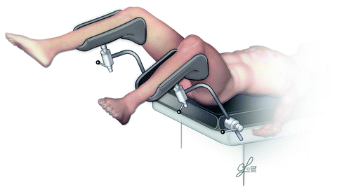
Fig. 22.4
The dorsal lithotomy position allows good access to the genitalia and facilitates semirigid ureteroscopy. This author finds nearly all patients can have suitable access with the semirigid instrument, without extending, lowering, or modifying leg position. Frozen hip and/or knee joints interfere with this patient position and would necessitate either a flexible ureteroscopic or percutaneous approach to the ureter
A routine cystoscopy/panendoscopy set-up (flexible or rigid) is arranged on a sterile field with standard drapes and the following additional items: The surgeon’s preferred guide wires, 50 ml or more of radiographic contrast, an open-ended ureteral catheter (usually 5 or 6 F size), a 12 or 14 F foley catheter, 10 and 20 ml syringes, and a pressure irrigation system with room temperature or body temperature saline. A single-action pump system or a pressure bag surrounding the saline for irrigation is the most commonly used methods for pressure irrigation through the ureteroscope. At our institution, we have not found any observable difference in outcome with room temperature saline vs. warmed (body temperature) saline for irrigation.
The length and size/type of ureteroscope should be appropriate to the location of the stone (or lesion) and the patient’s anatomy. A video camera imaging system is almost mandatory; otherwise, the surgeon must be a “contortionist” to keep his or her eye at the ocular end of the semirigid ureteroscope while advancing it into the lower ureter. Further, a camera system facilitates teaching, permits an assistant to better coordinate manipulation of a basket or other instrument, and keeps the operator’s face away from the ureteroscope’s working port, thus reducing the possibility of being sprayed in the face with a mixture of saline and the patient’s bodily fluids. It is more helpful if the video monitor and fluoroscopy monitor can be in close proximity, but as long as they are both easily visible for the urologic surgeon while performing the procedure, they will be adequate to the task at hand.
Technique
With the patient prepped and draped in the dorsal lithotomy position, standard cystourethroscopy is performed and the anatomic landmarks delineated. Assuming no preexisting ureteral stent is in place, the ureteral orifice of interest is cannulated with a 5 Fr open-ended ureteral catheter and retrograde pyelography is performed using radiographic contrast (Renografin-60®, Conray-60®, Ultravist-300®, Isovue-300®, or equivalent) diluted 50:50 with sterile water or normal saline. This will verify the location of the stone(s) and demonstrate the presence of stricture or other lesions that might affect the surgeon’s ability to safely insert the semirigid ureteroscope. If the patient is morbidly obese, sometimes it is necessary to dilute the contrast less, or even to use undiluted contrast, just to properly see the ureter on the fluoroscope (not usually an issue with newer, more powerful fluoroscopes that have improved resolution but sometimes necessary with older units).
A 0.035″ stiff-shaft glide wire is then advanced through the ureteral catheter into the renal collecting system using fluoroscopic guidance. The cysto-panendoscope and ureteral catheter are both removed over the wire, leaving the wire in place. Usually with semirigid ureteroscopy, only one wire is needed as a safety, so this wire is clipped to the drapes as a safety wire. Should a second wire be needed for the “ladder technique” or some other purpose, a dual lumen catheter may be placed over the wire and a second, identical wire advanced through the second lumen using fluoroscopic guidance, the dual lumen catheter is removed over the two wires, and one wire is clipped to the drapes as a safety wire. The second wire can be used as a “working wire” to pass a balloon dilating catheter or even to pass the ureteroscope. Or, it can also be clipped to the drapes as a second safety or stabilizing wire, permitting the ureteroscope to be passed between the two glide wires into the distal ureter, as shown in Fig. 22.5 (“ladder technique”).
Fig. 22.5
The semirigid ureteroscope is advanced between two wires into the ureteral orifice (“Ladder Technique”)
If a preexisting ureteral stent is present, then using graspers through the cystoscope, the stent is brought out to the urethral meatus and cannulated with a 0.035″ stiff-shaft glide wire, or other guide wire per surgeon’s preference. The stent is exchanged over the wire for a dual lumen ureteral catheter and a retrograde pyelo-ureterogram is performed via the second lumen. Once stone location is verified, a second identical glide wire, if needed, may be advanced through the dual lumen catheter as described above.
There are some surgeons who prefer to pass a safety wire alongside a preexisting stent before removing the stent, fearing possible loss of access if the stent is clogged with debris thus blocking advancement of a wire through the stent. The “alongside” technique usually works well, but on occasion a wire cannot be inserted alongside the stent and the stent must first be removed. This type of problem is sometimes encountered when the stent fits snugly through a tight ureteral stricture or alongside an impacted stone. Regardless, a safety wire is important in virtually all semirigid ureteroscopy cases, as it provides an exit strategy should one need to terminate the procedure prematurely, for any reason.
Related posts:
 Informed Consent and Perioperative Antibiotics
Complex Anatomy: Horseshoe, Pelvic, and Malrotated Kidneys
Informed Consent and Perioperative Antibiotics
Complex Anatomy: Horseshoe, Pelvic, and Malrotated Kidneys
 Ferromagnetics in Ureteroscopy
 Flexible Ureteroscopes: Fiberoptic and Digital
 Semirigid Ureteroscopy Step-by-Step: The Tulane Approach
 Upper Tract Urothelial Carcinoma: Ureteroscopic Biopsy and Specimen Preparation
Stay updated, free articles. Join our Telegram channel

Full access? Get Clinical Tree


