Trial
Magnetic removal (s)
Basket removal (s)
Difference (s)
Stones in ureteral orifice (magnetic, basket)
1
257
555a
298
1, 1
2
413
410
−3
2, 1
3
235
730
495
2, 2
4
174
298
124
0, 0
5
228
265
37
0, 0
6
46
250
204
0, 0
7
150
320
170
0, 0
Mean*
215
404
189
0, 0
One of the reasons for failure of complete stone extraction during PCNL and flexible ureteroscopy is of course the issue of poor visualization. This is often due to bleeding in the collecting system. Mir et al. [14] thus studied the effect of urine and blood on the binding of these novel microparticles to calcium oxalate stone fragments and the ability to attract the functionalized stones to the magnetic tool in these environments. In vitro urine concentrations of 5, 20, and 50 % were combined with microparticles (1 mg/ml) and stone fragments for a 10-min incubation period. Similarly blood concentrations of 0.5, 1, and 2 % (on visual inspection one cannot see through a test tube of 2 % concentration blood) were used. In both experiments, the investigators were able to achieve 100 % stone extraction, of fragment sizes of up to 3 mm, at all concentrations indicating a negligible interference of urine and blood in the clinical environment.
Mir et al. [14] also performed initial investigations of the in vitro toxicity of these microparticles on murine fibroblast, human urothelium, and human transitional cell carcinoma cell lines. Each cell line was plated at three different concentrations of cells and exposed uniformly to 1 mg/ml concentration of microparticles. No toxicity was seen in any of the three cell lines after 48 h of exposure except in urothelial cells at the lowest concentration of cells. Further biotoxicity tests have since been conducted in mice (data not published). The animals were exposed to these novel microparticles intravesically or intravenously. The systemic exposure was for a worst-case scenario where microparticles breached the urothelial membrane and entered the systemic circulation. Each group was further subdivided and given concentrations of the microparticles at 0.5, 1, and 5 mg/ml. We found that when particles were intravesically instilled, they did not translocate across the urothelial layer, regardless of the concentration used, with no associated systemic toxicity. In the mice that had intravenous injections of microparticles, there was a dose-dependent distribution of the particles to the liver, lung, and spleen. There was minimal inflammation seen on histology, and all the mice survived and gained weight appropriately indicating minimal toxicity.
Since the tests of the technology utilized an 8 F magnetic rod, new tool design is required for the intended use during flexible ureteroscope. As such, we have recently developed a ureteroscopic tool consisting of a magnet attached to a 0.038 in. guidewire (Fig. 40.1). This tool has undergone preliminary, non-published evaluation in an in vitro model of the pelvicollecting system. Access was gained via a ureteric access sheath and the magnetic tool was backloaded onto the flexible ureteroscope. Trials were performed comparing the magnetic tool against a standard 2.4 F nitinol basket in the extraction of stone fragments ranging from 1 to 2.5 mm in greatest diameter. Timed trials found that the magnetic tool outperformed the basket in the extraction of stones less than 2 mm in size but was of equal efficacy for fragments larger than 2 mm (Table 40.2). Nevertheless, there were a number of issues encountered with the current itineration of the tool. The size of the magnet affected the visibility of the system and it was easy to dislodge stones from the magnet as they were being trawled out of the pelvicollecting system (data unpublished).
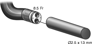
Fig. 40.1
Schematic of magnet mounted on guidewire and backloaded on flexible ureteroscope. Fernandez R, Tan YK, Kaberle W, et al. Determining a Performance Envelope for Capture of Kidney Stones. Journal of Endourology, in press
Table 40.2
Number of fragments extracted in 5 min in ureteroscopic model
Stone size | No. of stone fragments retrieved in 5 min | ||||||
|---|---|---|---|---|---|---|---|
Trial | 1 | 2 | 3 | 4 | Median | P-value | |
1–1.5 mm | Magnet
Related posts: Informed Consent and Perioperative Antibiotics
Complex Anatomy: Horseshoe, Pelvic, and Malrotated Kidneys Informed Consent and Perioperative Antibiotics
Complex Anatomy: Horseshoe, Pelvic, and Malrotated Kidneys
 Flexible Ureteroscopy: Holmium:YAG Laser and Optical Fibers Flexible Ureteroscopy: Holmium:YAG Laser and Optical Fibers
 Radiation Safety During Ureteroscopy Radiation Safety During Ureteroscopy
 Semirigid Ureteroscopy Step-by-Step: The Tulane Approach Semirigid Ureteroscopy Step-by-Step: The Tulane Approach
 Upper Tract Urothelial Carcinoma: Ureteroscopic Biopsy and Specimen Preparation Upper Tract Urothelial Carcinoma: Ureteroscopic Biopsy and Specimen Preparation
Stay updated, free articles. Join our Telegram channel
Full access? Get Clinical Tree
 Get Clinical Tree app for offline access

| ||||||