Manufacture
Model
Working length (cm)
Tip diameter (Fr)
Proximal shaft diameter
Deflection up/down degrees
Channel diameter (Fr)
Gyrus ACMI
DUR 8Ea
64
6.75
10.1
170/180
3.6
Gyrus ACMI
DUR-8 Ultraa
67.5
8.6
9.3 (avg)
270/270
3.6
Olympus
URF-P5b
70
8.4
8.4
180/275
3.6
Storz
Flex X2c
67.5
7.0
8.5
270/270
3.6
Stryker
Flex Vision U-500d
64
6.9
–
275/275
3.6
Wolf
Viper (7325. 076)e
68
6.0
8.8
270/270
3.6
Wolf
Cobra (7326.076)e
68
6.0
9.9
270/270
3.3/3.3
Many of the currently available fiberoptic flexible ureteroscopes are upgraded and enhanced version of prior models, and as such one must pay particular attention to the precise model in question. The most recent generation fiberoptic flexible ureteroscopes are described here. Though some prior generation ureteroscopes are still in use, they are not described in detail here, as most are no longer produced, though they still may be serviced by the manufacturer. One must be mindful that active deflection will be affected to a variable degree by working elements placed through the working channel. Though newer generations of flexible ureteroscopes generally prove to have better irrigant flow [13], this parameter will also be affected to a variable degree by introduced working elements.
At the current time a large number of the most recent generation of fiberoptic-based flexible ureteroscopes are still in use, and a large portion of the data available regarding flexible ureteroscopes involves these ureteroscopes. Though these ureteroscopes are more commonly being replaced with their digital counterparts, a thorough understanding of these ureteroscopes is important in that studies of these ureteroscopes reveal a number of issues that impact the use and durability of ureteroscopes in a large number of ways. It is likely that a number of these findings will also facilitate improvements in digital flexible ureteroscopes. Though digital flexible ureteroscopes have a different operative paradigm in terms of optics and image production, many mechanical and structural features of these ureteroscopes remain quite similar to their fiber- optic counterparts.
There are also a number of other qualities and parameters that most manufactures generally allude to in description of these ureteroscopes. For example, the various manufacturers state that the working shaft of their flexible ureteroscope is coated with a type of proprietary coating or polymer that allows for expeditious and easy passage of the scope. They also all state that the efforts have been made to make the shaft crush resistant and torque stable during rotation of the scope. Nonetheless, some notable specific points about some of these flexible ureteroscopes are worthy of note.
The Gyrus ACMI (now a business division of Olympus) DUR 8 Elite ureteroscope has two points of active deflection, accomplished by use of a second lever that provides deflection of the scope more proximal on the shaft. The active secondary deflection adds 130° down. It has also been equipped with a moiré reduction filter. Moiré patterns are often an undesired artifact of images produced by various imaging techniques.
The Gyrus ACMI (now a business division of Olympus) DUR 8 Ultra has been created as a successor to the DUR 8 Elite. This ureteroscope is a hybrid of Olympus and ACMI technologies. The ureteroscope has been enhanced so that it has 270° up and down deflection. It has also been equipped with a moiré reduction filter. The manufacturer also claims that construction of this ureteroscope has also been improved such that it has improved endurance with commonly used sterilization methods.
The Olympus URF-P5 is also an enhanced version of prior Olympus fiberoptic flexible ureteroscopes. This ureteroscope has a 5.3 Fr beveled or bullet-shaped tip. This scope also has an incorporated moiré reduction filter to enhance image and provide a clearer picture. The deflective portion of the shaft is also covered with thicker rubber to resist potential puncturing (Fig. 9.1).
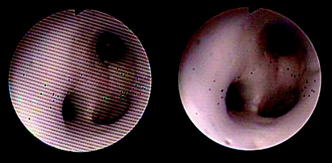
Fig. 9.1
Image through a fiberoptic ureteroscope showing moire effect and image after moire filtering. Also note that a number of fibers have been broken (courtesy of Olympus)
The Storz FLEX X2 ureteroscope is similar to its predecessor FLEX X scope in many aspects. Due to general concern regarding laser damage for all flexible ureteroscopes, this ureteroscope was enhanced with addition of a material laserite™ at its distal tip, in an attempt to make this portion of the ureteroscope more resistant to potential laser-induced damage.
Stryker offers the Flexvision U-500 fiberoptic flexible ureteroscope. Fiberoptic-based image quality relates in part to fiber number and density. This ureteroscope has been fitted with high-density fiber bundles in an effort to further improve image quality.
Richard Wolf produces the Wolf Viper and a more advanced flexible ureteroscope, the Wolf Cobra, which features dual channels for simultaneous use of two devices, such as a laser and a basket or a backstop. The dual 3.3 Fr. channels accommodate continuous irrigation capabilities with increased flow. The Cobra also includes an ergonomic laser dial with an integrated locking mechanism to advance the laser fiber and minimize laser damage, Figs. 9.2 and 9.3.
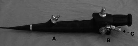
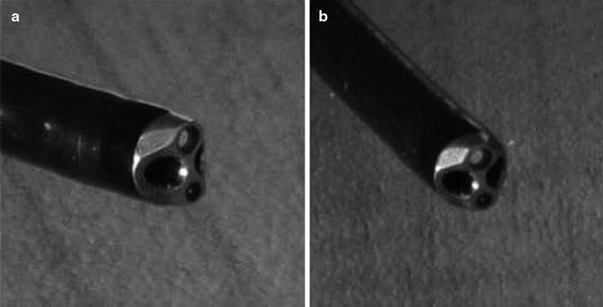
Fig. 9.2
Cobra dual-channel ureteroscope demonstrating two working channels (A, B) with laser advancement channel (B) being of longer length and optionally supported by thumb-operated laser advancement wheel (C) (thumb device not pictured). From [25], reprinted with permission from Mary Ann/Liebert Inc. Publishers
Fig. 9.3
Beveled tip of the dual-channel handle Cobra ureteroscope. From [25], reprinted with permission from Mary Ann/Liebert Inc. Publishers
Digital Ureteroscopes
Beyond doubt, the introduction of fiberoptic flexible ureteroscopes revolutionized urologic practice for management of upper urinary tract pathologies. Nonetheless, despite widespread use with these ureteroscopes, a number of concerns persist. Fiberoptic ureteroscopes tend to have a grainy image, water may leak into the lens, and fibers may burn out and fracture, resulting in loss of image quality [14]. The image produced by fiberoptic flexible ureteroscopes is not as sharp as that of a rod lens semirigid ureteroscope due to the smaller number of pixels in the flexible instrument’s image, when compared to (a) rod/lens semirigid ureteroscopes [15]. Image quality may be improved by increasing the diameter of the flexible ureteroscope, but this is likely to increase the risk of failure of access to the upper urinary tract and decreased flexibility [16, 17]. Thus, strong impetus was present for improvement of available flexible ureteroscopes.
Digital Imaging
In order to avoid the operation-related weaknesses of film cameras, digital imaging was developed in the 1960s and 1970s. The impetus at that time was primarily for scientific and military use. In 1970, Boyle and Smith developed the charge-coupled device (CCD). This chip could store data in the form of electrical charges within a grid for subsequent retrieval [18, 19]. These grids could be stored as pixels [20]. The complementary metal oxide semiconductor (CMOS), a lower cost alternative to the CCD chip, was developed during the same time period. In addition to lower cost, the CMOS chip had the advantage of reduced device size and suitability for mass production [21]. Subsequent improvements and modifications of this technology resulted in real-time digital imaging. Evolution of this technology also suggests that successive generations of these chips will be further miniaturized.
Working Paradigm
The key paradigm change with digital flexible ureteroscopes is the “chip on tip” design, where an image is picked up, at times processed, then transmitted by a digital sensor, and sent to a proximal point via a single wire, where further processing and transmission take place. This arrangement bypasses the fragile optical fiber system of conventional fiberoptic flexible ureteroscopes. This arrangement may also include a distal more luminous light source built into the ureteroscope itself.
The specific configuration and operative paradigm of available digital ureteroscopes vary in certain aspects. The DUR D and Storz Flex XC flexible ureteroscopes have distally located light-emitting diode (LED) light source and a distally located CMOS chip that picks up and processes images. The Olympus URF V digital ureteroscope has a light source that is transmitted from a proximal source and a CCD chip that picks up images and transmits the data to a proximal source for processing. At the current time, it is of unknown significance what these different arrangements will mean for long-term scope operation, durability, and chance for malfunction/catastrophic failure.
Currently Available Digital Ureteroscopes
The aforementioned digital ureteroscopes are now available for use. Their general specifications can be seen in Table 9.2. Due to design changes these ureteroscopes are all lighter than their fiberoptic counterparts. They generally retain the non-optical related improvements in ureteroscope structure and composition available in their fiberoptic counterparts. All available digital ureteroscopes provide high-quality digital imaging, autofocusing capabilities, and digital magnification, which improve the user experience. The overall experience with digital ureteroscopes is growing, but is still somewhat limited.
Table 9.2
Currently available digital flexible ureteroscopes
Manufacture | Model | Working length (cm) | Tip diameter (Fr) | Proximal shaft diameter | Deflection up/down degrees | Channel diameter (Fr) | Light source/chip |
|---|---|---|---|---|---|---|---|
Olympus-Gyrus ACMI | Invisio DUR Db | 65 | 8.7 | 9.3 | 250/250 | 3.6 | LED/CMOS chip |
Olympus | URF-Vb | 67 | 8.5 | 9.9 | 180/275 | 3.6 | Fiber/CCD chip |
Storz | FLEX XCa | 70 | 8.6 | 8.5 | 270/270 | 3.6 | LED/CMOS chip |
The Gyrus ACMI Invisio DUR D was the first digital flexible ureteroscope. The unique features of this ureteroscope are as aforementioned. The format of the hand piece, deflective mechanism, working port, and channels is relatively similar to its fiberoptic counterpart. The tip of the scope contains the dual LED light carriers and a 1-mm digital camera. The LED light can last for up to 10,000 h, which is significantly longer than more expensive xenon light sources. The ureteroscope has a configuration such that it is simply plugged in and ready for use. Of interest, the two aforementioned technological advances significantly reduce the weight of the scope (DUR D 505g, DUR 8E 1012g). Internalization of the light source also reduces risks of burning or fires. This ureteroscope also has a laser fiber detection system (IDC-1500-endoscope protection system) which can detect when the blue coating of a laser fiber is retracted within the ureteroscope and alert the operator. This option can be used to automatically disable (place on standby) the laser generator, with the goal of preventing laser-induced ureteroscope damage [14].
Though image quality is somewhat difficult to quantitate and qualify, the quality of the digital image that accompanies digital flexible ureteroscopy is notably improved. The DUR D allows digital magnification to 135%. Andonian et al. [14] compared the DUR 8 and DUR D ureteroscopes with test cards (USAF test pattern card), demonstrating a brighter image and higher resolution with the DUR D (Fig. 9.4).
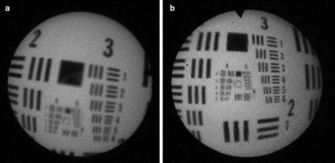
Fig. 9.4
(a) A photograph of the 1951 USAF test pattern card using the DUR-8 fiberoptic ureteroscope. (b) A photograph of the 1951 USAF test pattern card using the DUR-D digital ureteroscope. From [14], reprinted with permission from Mary Ann/Liebert Inc. Publishers
The Olympus digital flexible ureteroscope, URF-V, is also available and currently in use. As aforementioned, this digital ureteroscope contains a CCD chip and has light transmitted from a proximal source. This scope also is fitted with moiré reduction capability. This ureteroscope also has narrow band imaging (NBI) capability, which operates by alteration of the white light source to specific wavelength bands, which is useful for observation of mucosal morphology. This modality is currently being used for identification of urothelial carcinoma in a number of settings.
The Storz FLEX XC has also recently become available for use. This digital ureteroscope has a similar design to that of the DUR D in that it contains LED and CMOS technology. A notable difference with this digital ureteroscope is that it retains a relatively low caliber shaft along its working length. The FLEX XC, like its most recent fiberoptic counterpart, is equipped with laserite™.
Comparison of Flexible Ureteroscopes
A number of different parameters of flexible ureteroscopes have been compared in a variety of fashions. However, comparison of these ureteroscopes must be viewed with caution. Individual investigators generally investigate only what is available to them. Most manufacturers, as previously stated, have also produced a number of successive models of their fiberoptic flexible ureteroscopes, and comparison of available models, available to investigators in a time staggered fashion, is also quite variable. The availability of digital ureteroscopes has only further confounded this issue, with both fiberoptic and digital ureteroscopes included in some studies that do not necessarily focus on optics. As such, both fiberoptic and digital scopes are assessed together in many respects. However, differentiation of these two types of scopes will be noted. Investigators may also have different degrees of experience with the ureteroscopes available for study. Variability in results may also occur due to investigative methods used. In summary there are few all-inclusive direct comparison studies. Nonetheless, a brief survey of these investigations is worthwhile in that it lends further understanding into the features and limitations of these ureteroscopes.
Studies involving flexible ureteroscopes themselves (not clinical outcomes associated with their use) are generally one of the two types: a comparison of working parameters (optical/mechanical) of the ureteroscopes and overall durability of the ureteroscopes. A large number of studies have been performed on successive generations of fiberoptic flexible ureteroscopes. Representative studies to point out key features and differences, and studies including a large number or representative types of available ureteroscopes at any given time, are summarized here to give the reader an overall picture of how these ureteroscopes generally compare with one another. Studies relating to ureteroscopes still in use were also used in selection. The key findings of these studies are noted. The detailed methodologies used are beyond the purview of this review.
Experience in flexible ureteroscopy has made evident that irrigation flow and related quality of vision certainly are a critical issue, and as such, this specific issue has received due attention. Bach et al. [17], in 2008, evaluated the effects of tools and probes on deflection angle and irrigation flow in five unloaded and instrument-loaded (273 μm laser fiber, 2.4–3.0 Fr biopsy forceps, and tipless nitinol baskets 1.5–2.4 Fr) flexible ureteroscopes, Wolf Viper, Storz FLEX X2, ACMI DUR 8 Elite, ACMI DUR D, and Olympus URF P5, and noted that deflection had no influence on unloaded scope flow rates and baskets had no influence on the maximum angle of deflection. Laser fibers and biopsy forceps lead to loss of deflection (laser fiber 4.44–10.21%; biopsy forceps 30.7–57.8%). Flow rates depended on the size of the working device introduced. Loss of irrigation volume ranged from 62.24 (1.5 Fr) to 99.1% (3.0 Fr). The laser fiber reduced irrigation volume by 53.7%. This study summarizes and outlines well that irrigation flow, loss of deflection, and intraoperative visualization are dependent on the working devices used. Another very important point here is that improvements in ancillary devices, such as smaller nitinol baskets, can help minimize the impact of these devices on ureteroscope function and performance. Indeed, other studies also corroborate that newer smaller caliber baskets have significantly less effects on deflection or irrigation [22]. One must also bear in mind that as smaller diameter ancillary devices are created, working channel caliber of successive generations of ureteroscopes has also been somewhat improved (Figs. 9.5 and 9.6).
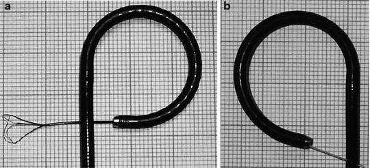
Fig. 9.5
 Informed Consent and Perioperative Antibiotics
Complex Anatomy: Horseshoe, Pelvic, and Malrotated Kidneys
Informed Consent and Perioperative Antibiotics
Complex Anatomy: Horseshoe, Pelvic, and Malrotated Kidneys
 Ferromagnetics in Ureteroscopy
 Radiation Safety During Ureteroscopy
 Semirigid Ureteroscopy Step-by-Step: The Tulane Approach
 Upper Tract Urothelial Carcinoma: Ureteroscopic Biopsy and Specimen Preparation


(a) Viper (Richard Wolf) with 1.5-Fr NGage basket (Cook). (b) Viper (Richard Wolf) with 273-μm laser fiber (FlexiFib, Lisa Laser): loss of deflection (7.41%) and increased diameter (+1 mm) can be seen. From [17], reprinted with permission from Mary Ann/Liebert Inc. Publishers
Related posts:
Informed Consent and Perioperative Antibiotics
Complex Anatomy: Horseshoe, Pelvic, and Malrotated Kidneys
Ferromagnetics in Ureteroscopy
Radiation Safety During Ureteroscopy
Semirigid Ureteroscopy Step-by-Step: The Tulane Approach
Upper Tract Urothelial Carcinoma: Ureteroscopic Biopsy and Specimen Preparation
Stay updated, free articles. Join our Telegram channel
Full access? Get Clinical Tree
