Fig. 34.1
Evolution of Roux-en-Y gastric bypass. (a) Mason and Ito (1966), (b) Griffin modification (1977), (c) Torres (1980)
Here we describe our technique for SITU-LRYGB.
34.3 Technique
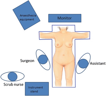
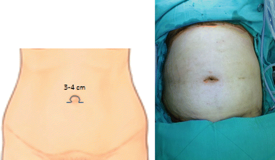
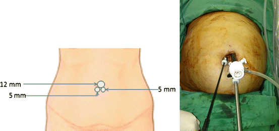
The patient is placed in the supine position with appropriate padding of the pressure points and strapped to the table to avoid slipping during the extremes of position. The arms are abducted laterally. The surgeon stands on the right side of the patient and the camera assistant on the left while the scrub nurse is behind the surgeon (Fig. 34.2). The incision is 4–6 cm transverse curvilinear (omega incision) [31] located at the superior border of the umbilicus and deepened up to the linea alba (Fig. 34.3). The subcutaneous fat is partly dissected to create the space for trocar placement. After creation of pneumoperitoneum, a 12-mm trocar is inserted at the 12 o’ clock position in the centre of the incision. Two other trocars (5 mm and 10 mm) are then placed under vision laterally in the bilateral ‘arms’ of the incision at the 4 o’ clock and 7 o’ clock positions (Fig. 34.4). A 10-mm, 30° long scope (45 cm) is employed for endovision which is placed through the right lateral trocar, while the two working instruments are placed through the central and left trocars. During the initial learning phase, we advise making a longer (6 cm) incision, allowing a sufficient space for manipulation between the trocars. The incision can then be reduced to 4 cm as the surgeon gains experience. The trocars are not placed perpendicularly but slightly obliquely, aiming towards the hiatus in order to avoid torque during instrument manipulation.


Fig. 34.2
Operation set-up

Fig. 34.3
Incision and port placement for SITU-LRYGB

Fig. 34.4
Port placement for SITU-LRYGB
The liver suspension tape [31] is prepared by cutting a 6 cm portion of Jackson–Pratt drain near the drainage hole. The drainage tube is then pierced with 2-0 Prolene suture (Monofilament Polypropylene Suture W8400™ (Ethicon, Cincinnati, OH, USA), according to the diameter of the hole. Needles are left in both sides for further liver puncture. The tube is placed inside the peritoneal cavity for retraction of the left lobe of the liver and for a clear view of the angle of His, the crura, and the gastroesophageal junction. Two or three tapes may be required, depending upon the bulkiness of the liver and the view obtained.
The dissection starts at the lesser curvature just below the first branch of the left gastric vessel. The tissue is cut using ultrasonic shears, and the retrogastric tunnel is entered. A 30 cc gastric pouch is created using laparoscopic linear staplers. The proximal jejunum is traced from the ligament of Treitz, and a length of 100 cm is measured. An enterotomy is created and a laparoscopic linear stapler is used to create 2 cm gastrojejunostomy which is placed in the antecolic and antegastric position. The proximal jejunum is then transected using a laparoscopic linear stapler adjacent to the gastrojejunostomy. One hundred centimeter of Roux limb is measured and side-to-side jejunojejunal anastomosis is performed using a laparoscopic linear stapler at this point. The entry holes of the staplers are closed by intra-corporeal continuous suturing using a 3-0 absorbable suture. The mesenteric, as well as the Petersen, defects are closed by continuous intracorporeal suturing with 2-0 non-absorbable sutures. The liver suspension tape is removed, and the puncture hole of the liver is cauterized for hemostasis.
After removing the trocars, all three fascial defects are closed individually with 2-0 Vicryl sutures. In cases of a long umbilical skin incision, the subcutaneous fat and the skin at the angles are removed, and an umbilicoplasty is performed. The final wound is circular and buried within the umbilicus. With increased experience, the initial skin incision made is smaller and an umbilicoplasty is unnecessary.
34.4 Surgical Results
The operation time reported has generally been longer than that of the conventional laparoscopic approach, mostly attributed to the technical difficulties encountered during the procedure. However, none of the groups reported any intra-operative or early postoperative complications related to the surgical technique. Problems related to loss of triangulation were managed by placement of an extra 5-mm trocar, as seen in 18 of 100 patients undergoing two-site LRYGB. Because of the surgeons’ sufficient experience, there was no need of conversion to conventional LRYGB or open procedure in any of the published reports. The two largest series compared their results with conventional LRYGB. The length of postoperative hospital stay, postoperative complications, and excess weight loss up to 12 months was not significantly different between the two groups. Although the SITU-LRYGB group in our study required more morphine administration for post-operative pain control, it did not reach statistical significance. There may be an increased risk of seroma formation due to wide dissection of the subcutaneous tissue, and the patients generally need to care for their wound for a longer time compared to conventional multiport laparoscopy [28]. Finally, the wound satisfaction score at 3 months was significantly higher for the SITU group, suggesting that they were more satisfied with the cosmetic outcome of the surgery.
34.5 Pitfalls and Their Management
34.5.1 Trocar Placement
The surgery may be performed using the commercially available single-port devices or conventional trocars [27–32]. The former devices provide less sufficient space between the instruments and hence necessitate the use of curved or articulating instruments [30, 32]. Because of their large size, they require the creation of an equivalent fascial defect which, if not closed properly, can increase the risk of incisional hernia. Moreover, these devices add considerable cost to the procedure. In SITU method, placement of all three operating trocars within a small umbilical incision would cause excessive crowding and hamper manipulation. This could be overcome by the technique we developed, omega umblicoplasty [31]. The initial umbilical incision was around 6 cm in length along the superior border of the umbilicus, allowing for comfortable placement of the three trocars with a gap of 4 cm between each trocar. The instrument clashing was overcome by using low profile trocars and instruments of different lengths, allowing adequate space between the surgeon’s hands. The trocars are all angled to the area of interest and not perpendicular to the abdominal wall, reducing the torque and shoulder strain for the surgeon.
The initial dissection of the subcutaneous space for trocar placement weakens the grip of the abdominal wall on the trocars and may result in gas leakage. The repeated instrument exchanges and excessive torque forces add to this problem and enlarge the existing fascial defect, necessitating the use of threaded trocars or skin suture for anchoring the ports. It is preferable to use a 30 ° 10 mm long telescope for creating good endovision. The light cable of the telescope is a frequent cause of interference. This is counteracted by using a light cable adapter that allows the light source to exit at an acute angle from the scope. Some authors have used semi-flexible scopes with integrated light and camera cables to make the procedure more comfortable, albeit at a higher cost.
34.5.2 Liver Retraction
Liver retraction is essential in bariatric surgery to perform dissection near the hiatus as the left lobe is usually bulky and friable and usually obscures this area if there is insufficient exposure. Earlier reports described the use of the conventional Nathanson liver retractor during RPL-RYGB [26]. Tacchino et al. performed the surgery without using any form of the liver retraction and managed to use the articulating stapler to push up the left liver lobe during pouch creation [30]. With this technique, the mean length of the gastric pouch was as large as 8 cm to avoid the technical difficulties that would have been encountered by big left liver, during the creation of a pouch and the subsequent higher gastrojejunal anastomosis. The liver suspension tape [31] that we developed can be easily prepared before the procedure, and multiple tapes can be used in the situation when one is not enough to retract liver well. The concern about the safety of liver suspension and its effect on postoperative liver function has been allayed by a prospective randomized trial wherein the three different methods of liver retraction were compared with respect to the time required for placement, postoperative pain, and liver function tests. Surprisingly, we found that the Nathanson liver retractor caused more pain and liver dysfunction compared to our liver suspension techniques [33].
34.5.3 Instruments and Surgical Technique
The successful performance of SITU-LRYGB requires that the dissection and anastomosis be confined to a single abdominal quadrant. Long instruments and harmonic shears (43 cm) are required to reach hiatus. Several authors have reported the use of articulating instruments like the endograsp for creating artificial triangulation within the abdomen. However, this requires crossing of the surgeon’s hands, creating confusion and sword fighting during the procedure. The success of using straight instruments mostly depends on the surgical steps we performed. After creating the pouch, we brought the uncut loop of jejunum to the gastric pouch for creation of the gastrojejunal anastomosis. Some authors then measure the foot of the Roux limb, create the jejunojejunostomy, and transect between the two anastomoses to finally separate the biliary and alimentary limbs [30]. In our technique, we transected the biliary limb after gastrojejunal anastomoses to avoid excessive tension. In both approaches, the entire manipulation and suturing is confined to the supracolic compartment and avoids excessive torque forces and change of position to the infracolic region which would make this step extremely difficult. We used instruments with curved opposing tips to create angles required for intracorporeal looping and knot tying. A to-and-fro movement of the instruments helps create loops especially using a monofilament suture material with a strong memory.
Stay updated, free articles. Join our Telegram channel

Full access? Get Clinical Tree


