Complications specific to right RDN
Liver
Duodenum
Complications specific to left RDN
Spleen
Pancreas
Nonside specific complications
Adrenal gland injury or removal
Pleural injury
Diaphragm injury
Bowel injury and injury to its mesentery
Major vessel injury – renal artery, renal vein, inferior vena cava, aorta
Minor vessel injury – adrenal vein, gonadal vein, lumber vein
Vascular stapler and hem-o-lok clip malfunction
Psoas sheath hematoma
Ureteric stricture and necrosis
Injuries during graft retrieval: bladder injury and injury to graft itself
Lymphatic injury and chylous ascites
Others: wound infection, orchalgia, epididymitis, medial thigh cutaneous paresthesia (entrapment of genitofemoral nerve)
Upper Pole Dissection and Injuries Specific to Right Side
Liver is the organ most commonly injured during right robotic donor nephrectomy while doing upper pole dissection or retracting the liver [1].
Prevention
Use of Self-retaining tooth grasper to elevate the liver, inserted via a 5 mm trocar under vision and clamped to the diaphragm or the sidewall.
Management
If there is liver laceration , most of the times it is self-limiting and stops by simple fulguration, packing alone, or by using Surgicel/flowseal. However, sometimes deeper lacerations may require suturing (horizontal mattress sutures).
After mobilization of colon medially, the duodenum can get injured. One should avoid cautery dissection to mobilize duodenum medially. If however duodenum is injured, primary closure is done and nasogastric tube is inserted. Patient should be closely followed in postoperative period and should be kept nil by mouth until gastrointestinal function returns to normal [1].
Upper Pole Dissection and Injuries Specific to Left Side
The most common intraoperative injury specific to left side donor nephrectomy while doing upper pole dissection is splenic injury [1].
Reason
Too much traction applied before complete division of splenorenal ligament
Management
Low grade injury (mild to moderate lacerations) can be managed conservatively using Surgicel/flowseal (fibrin sealant) and/or by spleenorrhaphy, while for severe splenic injury (significant blood loss leading to hemodynamic instability/requiring blood transfusion) splenectomy is the preferred option.
Pancreatic tail may also get injured while doing dissection at renal hilum and medial aspect of left kidney, which can present in postoperative period as acute pancreatitis, paralytic ileus. It should be managed conservatively when not associated with complications. However, if laceration is recognized intraoperatively, it is always better to seek gastro surgeon’s opinion. Rule out any injury to pancreatic duct. Non absorbable sutures should be used to repair parenchymal lesions.
Upper Pole Dissection and Nonside Specific Complication [1–3]
Robotic donor nephrectomy is an adrenal sparing surgery, where adrenal gland is preserved and is dissected off superior pole of the kidney using variable amount of unipolar or bipolar energy source in order to achieve hemostasis. Still, the adrenal gland and adrenal vein are sources of bleeding during the surgery. The right adrenal vein, due to its short length and direct insertion into vena cava, is more prone to injury. Severity of adrenal gland injuries varies from mild bleed, which can be controlled using simple cauterization or clipping the bleeding tissue, to major injuries requiring ipsilateral adrenalectomy.
Prevention
On left side
The renal vein margin and its junction with the adrenal vein should be defined.
The dissection on the adrenal vein side should extend till the point the adrenal gland is seen.
Interlocking clips should be used for securing the adrenal vein.
Vein stump on the renal vein side should be longer
Pleural injuries arising during dissection of superior/posterior aspect of kidney are also not uncommon and can result in pneumothorax. These can be identified as a curling of diaphragm into operative field and can be tested by asking anesthesiologist to hyper expand the lungs, while surgeon is irrigating near the diaphragm. Small injuries can be repaired using 4-0 chromic suture. While for larger ones, low pressure pneumoperitoneum is created and infant feeding tube no 10 is used to evacuate air from pneumothorax, tear is repaired using purse string suture, and infant feeding tube is removed and purse string suture tightened as the anesthesiologist hyper expands the lungs.
Bowel injuries are also common during reflection of bowel medially from superior surface of the kidney.
Prevention
Avoid inadvertent traction, avoid use of cautery, properly identify plane superior to gerota to avoid such complications.
Mesenteric tears during bowel mobilization can occur and should be repaired to prevent internal herniation of bowel.
Lumbar Veins Dissection and Related Complications [1–3]
The lumbar veins are a common source of troublesome bleeding. Typically, the lumbar veins arise from the renal vein and enter the lumbar canal.
Reasons
Failure to recognize the location
Dissecting the lumbar vein to near to its confluence with the renal vein.
Injury to the posterior wall of the lumbar vein while circumferentially dissecting the lumbar vein.
Prevention
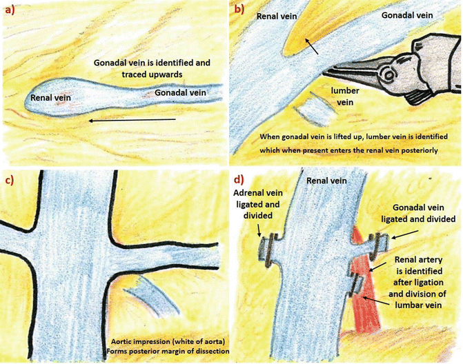
The exact location of the lumbar vein can be ascertained on the CT workstation. The number of lumbar veins and the spatial configuration can be made out if the surgeon views the same on a CT console. Whenever present, the lumber vein enters the renal vein posteriorly. Once the lumbar veins are secured the renal artery is visualized, which lies immediately posterior to it (Fig. 19.1a–d).


Fig. 19.1
Steps in lumber vein dissection (a) gonadal vein is traced upwards up to its insertion into renal vein. (b) Only when gonadal vein is lifted up surgeon can identify lumber vein, which enters renal vein posteriorly (c) diagram illustrates importance of recognizing glistening white layer over the aorta (which forms deep/posterior margin of dissection) in order to prevent major vessel injury (d) only after ligation of lumber vein (if present) the surgeon is able to identify and dissect renal artery accurately, which lies posterior to it
As a risk reduction strategy, the lumbar veins should be secured after the upper pole is dissected. In case of troublesome bleeding from these vessels, the surgeon can quickly secure the lumbar vein and retrieve the graft.
The lumbar veins can be secured with interlocking hem-o-lok clips. The controversy revolves around as to whether interlocking clips should be used or hem-o-lok clips should be used; the interlocking clips can be easily removed on the back bench. The lumbar veins should be secured keeping a cuff near the vein.
Management
The key and measures in the management of lumbar vein injury is decided
Is the upper pole is dissected?
Is the renal artery dissected?
Is the graft ready for retrieval?
Stay updated, free articles. Join our Telegram channel

Full access? Get Clinical Tree


