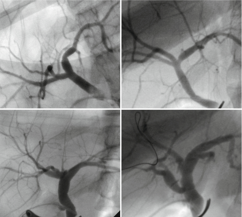
Fig. 30.1
Portal vein variations (including type 1, 2, 3)
30.2.2 The Classification of Hepatic Ducts in RPS Resection
Hepatic ducts can be classified into the four following types (Fig. 30.2). In the type I variant, the right hepatic duct (RHD) exhibits the usual bifurcation of the hilar bile duct and is found in 62.9 % of the population. In the type II RHD variant, the hilar bile duct trifurcates (12.2 %). Type III is the RHD variant with a Y-shaped union of the RAS and the left hepatic duct, with the RPS duct bent behind the RAS (13.2 %). Type IV is the RHD variant with a separate low-branching RPS duct (11.7 %) [5]. If the portal vein is suitable for RPS resection, the requirement for the hepatic duct is not strictly limited; however, the most suitable type of hepatic duct is type IV.


Fig. 30.2
Hepatic duct types (including type 1, 2, 3, 4)
30.2.3 Selection of the RPS Artery
Based on the origin of the RPS artery, the branching patterns of right hepatic artery are classified as extrahepatic and intrahepatic (Fig. 30.3). The extrahepatic type occurs when the RPS artery branches outside of the liver parenchyma. In this situation, the RPS is easy to liberate during a liver resection; therefore, it is the more suitable variant and is found in 78.7 % of the population. The other branching pattern occurs when the intrahepatic right hepatic artery is long and divides into a right posterior and anterior artery; in this case, liberation is difficult (21.3 %) [5].


Fig. 30.3
Type of RPS artery (including extrahepatic and intrahepatic)
30.2.4 Technical Difficulties in Resection and Implantation of RPS
Both RPS resection from the donor and implantation in the recipient demand a developed surgical technique; therefore, the RPS graft is an uncommon type of transplantation. First, the vessels of the RPS usually come from the secondary vessel and increase the complexity of the anatomy. The stump of the RPS would not be sufficient for anastomosis when the portal vein is the bifurcation variant. Thus, donors with this variant are not suitable for RPS. Second, variation in the hepatic duct, such as bifurcation in the RPS with the RHD running behind the portal vein, increases the difficulty of resection and anastomosis of the bile duct. Moreover, the split plane between the RPS and RAS is broad, and a tiny shift when dealing with the split plane during an RPS resection can lead to a dramatic deviation between the actual volume and the preoperative estimated volume of graft. Thus, the assessment of liver volume may be less accurate.
30.3 Preoperative Assessment
30.3.1 Routine Assessment Before Surgery
The safety of the donor is the most important principle in donor assessment. Additionally, the amount of hepatic issue should meet the metabolic needs of the recipient. The preoperative assessment should contain routine tests, including history collection, physical examination, assessment of the key organ function, imaging examination, mental condition assessment, determination of anesthetization risk, and an ethics review. Ages from approximately 18–55 years are most suitable, and the blood type should be compatible. Diabetic patients who are being treating with insulin and patients with unmanageable hypertension, physical diseases, or a history of malignancies and serious abdominal surgery should be excluded. Moreover, obese patients should be avoided due to the increased risk of cardiovascular disease, especially in patients with body mass indexes (BMIs) above 25, which significantly increases the operative risk during the perioperative period.
30.3.2 Measurement of the Liver Volume of RPS
A three-dimensional reconstruction using computer tomography angiography (CTA) of the liver can be used to measure the volume of the liver grafts. Experts from the University of Ulsan in Korea recommend that the residual liver without hepatic steatosis in donors who are 20–30 years of age should be more than 30 % of the whole liver volume. For those who are 30–50 years of age or exhibit mild or mid-stage hepatic steatosis, the residual liver volume should be more than 35 % [12]. In healthy adults, the right lobe of liver occupies an average of 67 % of the whole liver volume (Table 30.1). However, this figure is greater than 70 % in approximately one quarter of the donors [13]. In this situation, hepatectomy of the right lobe increases the risk to donors, whereas the left lobe is too small to transplant. RPS resection could be an approach for liver transplantation. The resection of the RPS requires that its volume be more than the sum volume of the left lobe and the caudate lobe, and the excess portion should be more than 3 % of the whole liver volume. At the same time, the estimated liver volume should be 40 % of a standard liver volume (SLT), or the graft-to-recipient weight ratio (GRWR) should be more than 0.8 %.
Lobe | Volume (cm3) | Percent (%) |
|---|---|---|
Left lobe | 395 ± 91 | 31 ± 4 |
Left lateral segment | 220 ± 57 | 17 ± 4 |
Left medial segment | 175 ± 65 | 14 ± 4 |
Caudate lobe | 25 ± 7 | 2 ± 1 |
Right lobe | 854 ± 151 | 67 ± 4 |
Right anterior segment | 473 ± 97 | 37 ± 5 |
Right posterior segment | 382 ± 84 | 30 ± 4 |
30.3.3 Assessment of the Anatomy of the Hepatic Vessels
The assessment of hepatic vessels includes the three-dimensional reconstruction of the artery, portal vein, and hepatic vein using hepatic CTA. The hepatic duct should be visualized using magnetic resonance cholangiopancreatography (MRCP). Most studies have reported that the choice of RPS should consider the anatomy of the vessels. As mentioned above, the most suitable variant types are type III for the portal vein and type IV for the hepatic duct and extrahepatic RPS artery branches. Due to the lack of an independent branch, type I is not suitable for RPS grafts. However, research from Korean experts in 2011 recommended that if the RPS volume is suitable for transplantation, the vessels should not be considered. These authors performed 13 cases of RPS graft resection in 65 LDLTs, showing excellent results [6]. Nevertheless, based on the experience of many transplantation centers, the anatomy of the portal vein is still an important factor in RPS resection [5, 7, 14].
Stay updated, free articles. Join our Telegram channel

Full access? Get Clinical Tree


