Transabdominal procedures
1. Repair of the pelvic floor
Abdominal repair of levator diastasis
Abdominoperineal levator repair
2. Suspension–fixation
Sigmoidopexy (Pemberton–Stalker)
Presacral rectopexy
Lateral strip rectopexy (Orr–Loygue)
Anterior sling rectopexy (Ripstein)
Posterior sling rectopexy (Wells)
Puborectal sling (Nigro)
3. Resection procedures proctopexy with sigmoid resection anterior resection
Perineal procedures
Perineal rectosigmoidectomy (Altemeier)
Rectal mucosal sleeve resection (Delorme)
Perineal suspension–fixation (Wyatt)
Anal encirclement (Thiersch + modification)
Choice of procedure is based upon patient and procedural factors. The key issues are gender, the patient’s overall medical condition, bowel function, and whether fecal incontinence is present.
There is a dearth of high-quality data regarding the optimum treatment method.
A comprehensive review, in 2008, of randomized trials found a few patterns:
The method of fixation during rectopexy did not change outcome.
Division of the lateral stalks was associated with a higher incidence of constipation.
Resection and rectopexy was associated with less constipation.
Laparoscopy was associated with a shorter hospitalization and less morbidity.
Patient Evaluation
Most patients present with complaints associated with the prolapse itself.
Constipation and/or fecal incontinence symptoms should be elucidated.
Physical examination may demonstrate a spontaneous prolapse (Fig. 33.1), while straining may be needed to demonstrate the prolapse in the squatting or sitting position.
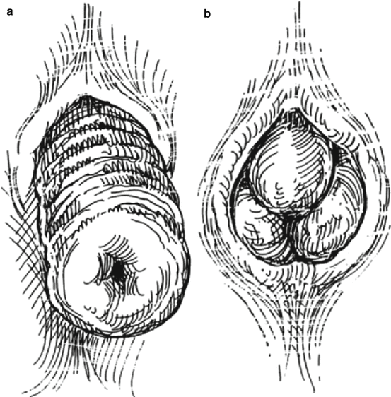
Fig. 33.1
Mucosal versus full-thickness prolapse. (a) Circumferential full-thickness prolapse with concentric mucosal folds. (b) Radial folds seen with hemorrhoidal prolapse (From Beck and Whitlow. Copyright 2003 by Taylor & Francis Group LLC. Reproduced with permission)
A differentiation should be made between full-thickness and mucosal prolapse.
Digital rectal examination detects concomitant anal pathology and evaluates adequacy of sphincter resting tone and squeeze pressure and function of the puborectalis muscle.
Colonoscopy or flexible sigmoidoscopy with air-contrast barium enema excludes and associated mucosal abnormalities.
Defecography adds little in full-thickness prolapse; however, it can be essential in the evaluation of internal or occult procidentia (rectorectal intussusception) or as part of pelvic floor musculature evaluation.
Anal manometry assesses sphincter function, as chronic prolapse typically damages the internal anal sphincter, resulting in poor resting pressures. A manometric study by Spencer reported that the anorectal inhibitory reflex was frequently absent or abnormal, that resting anal pressures were abnormally low, and that squeeze pressures were normal.
Surgical Procedures
There are two general approaches: abdominal and perineal operations.
The most common abdominal operations are rectopexy with or without concomitant sigmoid resection.
The typical perineal procedures are perineal rectosigmoidectomy (Altemeier) or a mucosal sleeve resection (Delorme).
The specific operation must be tailored to the condition and pathology of each patient, but some generalizations can be made.
Elderly, high-risk patients are best treated with perineal procedures (possibly with regional anesthesia).
An abdominal resection/rectopexy should be considered for a healthy patient with constipation and no incontinence.
The risk of impotence for abdominal rectopexy should approach 1–2 % in skilled hands.
A rectopexy with or without levatorplasty can be performed in patients without constipation symptoms
Perineal Procedures
Rectosigmoidectomy
Rectosigmoidectomy (Altemeier procedure) can be performed under a general or spinal anesthetic in either the prone, left lateral, or lithotomy position. A circumferential incision is made in the rectal wall approximately 1–2 cm above the dentate line (Fig. 33.2). The incision is deepened until the full thickness of the rectal wall has been divided. The rectum is withdrawn out of the body while progressively dividing and ligating the mesorectum, advancing more cephalad.
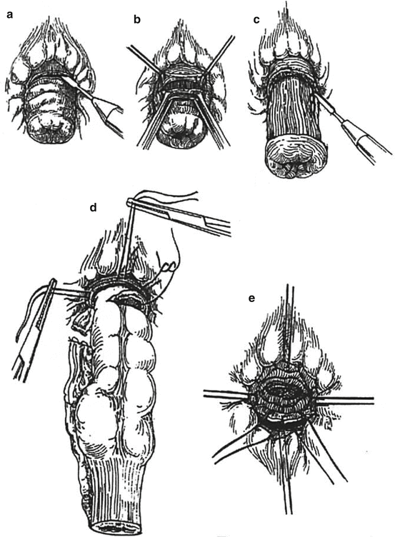
Fig. 33.2
Perineal rectosigmoidectomy. (a, b) Incision of rectal wall. (c) Division of vessel adjacent to bowel wall. (d) The prolapsed segment is amputated. Stay sutures previously placed in distal edge of outer cylinder are placed in cut edge of inner cylinder. (e) Anastomosis of distal aspect of remaining colon to the short rectal stump (From Beck and Whitlow. Copyright 2003 by Taylor & Francis Group LLC. Reproduced with permission)
Anteriorly, the peritoneal reflection (hernia sac) is opened. The dissection continues until there is no further redundancy remaining in the rectum/sigmoid colon. A hand-sutured or circular-stapled coloanal anastomosis is performed. A levator plication can be performed prior to the coloanal anastomosis, which has been reported to improve continence in two-thirds of patients.
Several studies have been reported on perineal rectosigmoidectomy, and clinical outcomes are summarized. An improvement in incontinence is reported in the majority of patients in whom levatorplasty was performed.
Mucosal Sleeve Resection (Delorme Procedure)
The Delorme procedure is ideally suited to those patients with a less extensive prolapse (e.g., about 5 cm in length) or with full-thickness prolapse limited to partial circumference (e.g., anterior wall).
In Delorme procedure, only the mucosa and submucosa are excised from the prolapsed segment (Fig. 33.3).
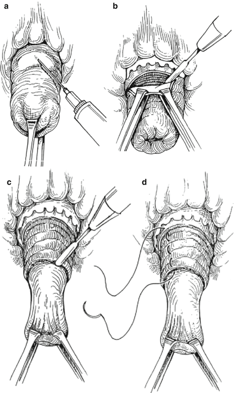
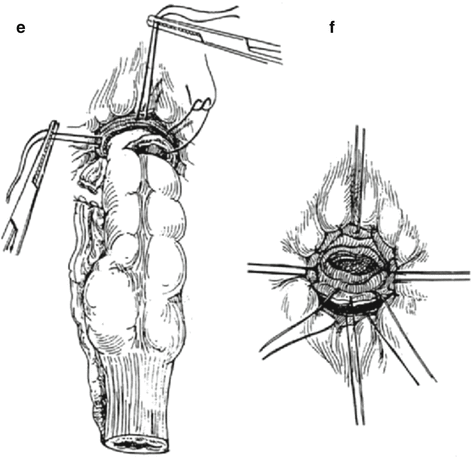
Fig. 33.3
Delorme procedure. (a) Subcutaneous infiltration of dilute epinephrine solution. (b) Circumferential mucosal incision. (c) Dissection of mucosa off muscular layer. (d) Plicating stitch approximating cut edge of mucosa, muscular wall, and mucosa just proximal to dentate line. (e) Plicating stitch tied. (f) Completed anastomosis (From Beck and Whitlow. Copyright 2003 by Taylor & Francis Group LLC (B). Reproduced with permission of Taylor & Francis Group (B) in the format Textbook via Copyright Clearance Center)
It can be performed under general, spinal, or local anesthesia. Prone position is preferred, but left lateral or lithotomy position can be used.
Results of Delorme procedure are summarized in Table 33.2. Recurrence rates (6–26 % at 1–13 years postoperatively) are generally higher than with a perineal rectosigmoidectomy. Incontinence is improved in 40–50 % of patients.
Table 33.2
Results of Delorme procedure
Authors | Number of patients | Recurrence | Mortality | Morbidity |
|---|---|---|---|---|
n | (%) | (%) | (%) | |
Uhlig and Sullivan | 44 | 7 | 0 | 34 |
Monson et al. | 27 | 7 | 0 | 0 |
Senapati et al. | 32 | 13 | 0 | 6 |
Oliver et al. | 41 | 22 | 2 | 62 |
Tobin and Scott | 43 | 26 | 0 | 12 |
Graf et al. | 14 | 21 | 0 | – |
Watkins et al. | 52 | 6 | 0 | 77 |
Lieberth et al. | 76 | 14 | 0 | 25 |
An alternative to the mucosal resection with muscular plication is the mucosal plication procedure (Gant–Miwa procedure). The best results seem to be when the mucosal plication is combined with an anal encircling procedure (see section “Thiersch Procedure” below).
Thiersch Procedure
Anal encirclement (Thiersch procedure) was originally performed with a silver wire placed subcutaneously around the anus under local anesthesia. The goal of this procedure was to mechanically supplement or replace the anal sphincter and stimulate a foreign body reaction in the perianal area, thereby increasing resistance at the anus.
Related posts:


Stay updated, free articles. Join our Telegram channel

Full access? Get Clinical Tree


