and Andrea Bischoff1
(1)
Pediatric Surgery, Colorectal Center for Children Cincinnati Children’s Hospital, Cincinnati, OH, USA
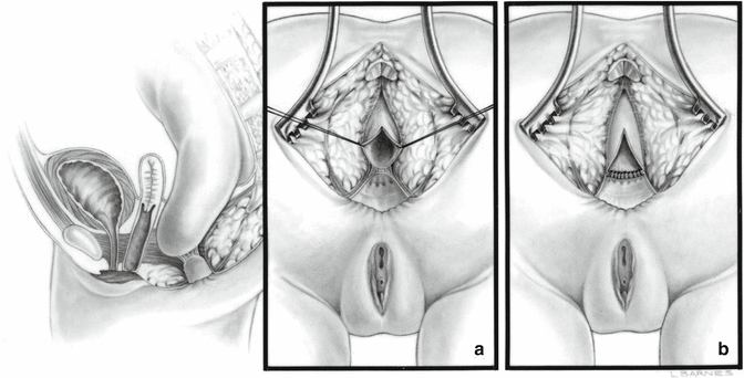
Rectal atresia is a very unique malformation that deserves a special description. It happens in our experience, in about 1 % of all cases of anorectal malformations. In this defect, the anus seems to be completely normal, including the quality of the sphincter and the location of the anal orifice. However, deep inside the anus, just at the junction of the anal canal with the rectum, there is an atresia or narrowing (stenosis) (Fig. 14.1). Occasionally, we see atresias or stenosis located at a different level. The space that separates the dilated blind rectum, from the anal canal, is represented by a septum that sometimes is extremely thin and can be perforated, and other times it is very thick. In some unusual cases, there is a significant separation between the blind upper rectum and the lower anal canal.
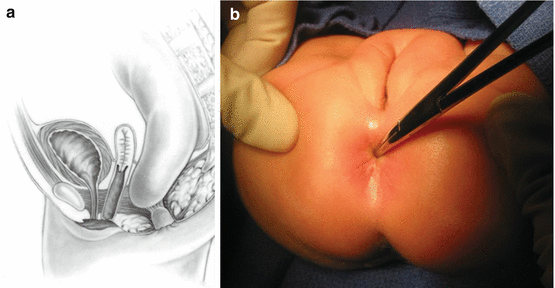
Fig. 14.1
Rectal Atresia. (a) Diagram. (b) External appearance
Interestingly, the sphincter mechanism is excellent in most cases. There is one particular malformation similar to this one that is represented by a stricture or by atresia of the rectum, associated to a presacral mass and a sacral defect (see Chap. 8, Sect. 8.2), which is a completely different type of defect. The only thing they have in common is the fact that the rectum is narrow or atretic.
We believe that rectal atresia with normal sacrum and no presacral mass is unique, because the sphincter mechanism is normal and also because these patients do not have the typical association with all the defects that we see in other anorectal malformations. As a consequence, the prognosis for these patients is excellent, in terms of bowel control. They have a significant tendency to suffer from severe constipation because they are born with a blind, very dilated rectum. These malformations have been previously described in the literature [1–5].
Rectal atresia has been traditionally described in the old textbooks. The baby is born with a normal-looking anus, and the nurse or the pediatrician tries to pass a thermometer through the anus and finds an obstruction. In fact, part of a routine examination of every “normal” newborn is to check the patency of the anus, unless the baby is already passing meconium.
14.1 Treatment
If one could think in an ideal indication for a posterior sagittal approach, this would be the malformation which seems to be more indicated. The defect is easily repaired through a posterior sagittal incision. In our initial cases, we simply remove the septum that separates the upper rectum from the anal canal and created an end-to-end anastomosis (Figs. 14.2 and 14.3). Subsequently, we found some cases in which the size discrepancy between the upper blind rectum and the small anal canal was very severe, and in order to expand the size of the anal canal, we introduced a technical modification maneuver [5] (Fig. 14.4). Most of the patients that we operated on came to us already with a colostomy in place. Since the patient has a colostomy, one can perform a distal colostogram and simultaneously introduce a metallic dilator in the anal canal to have a lateral image of the atresia and estimate the distance between the upper pouch and the anal canal. If we could make the diagnosis early in an otherwise healthy newborn baby, we would recommend to do the operation without a colostomy.