Fig. 6.1
The abdomen is entered via a midline xipho-umbilical incision
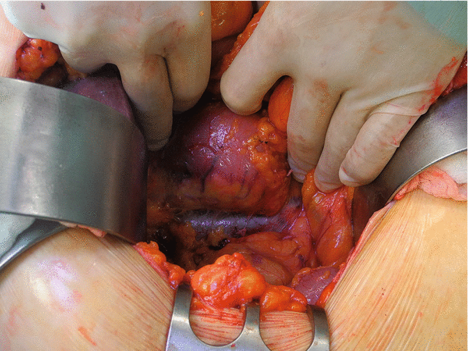
Fig. 6.2
After separation of the greater omentum, a Kocher maneuver is performed to ensure a better mobilization of the gastric tube
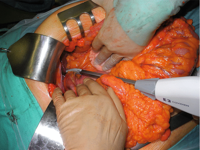
Fig. 6.3
Short gastric vessels are divided using a radiofrequency device (LigaSureTM), obtaining a complete mobilization of the greater curvature of the stomach
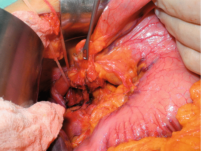
Fig. 6.4
The abdominal esophagus is completely exposed and encircled. The left gastric artery is dissected and lymphadenectomy at this level (station n. 7) is performed
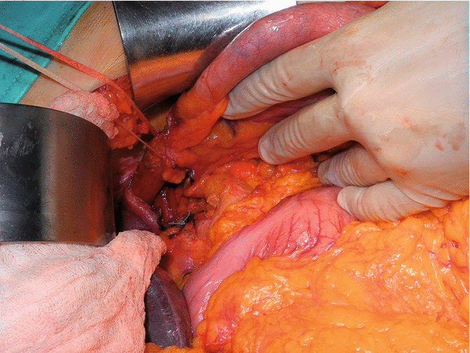
Fig. 6.5
The left gastric artery is divided at its origin
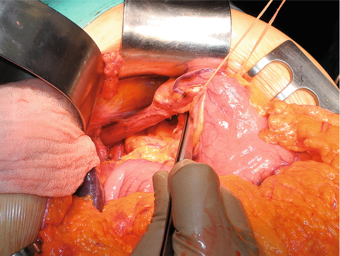
Fig. 6.6
Section of the anterior and posterior trunks of the vagus nerve with complete mobilization of the abdominal esophagus
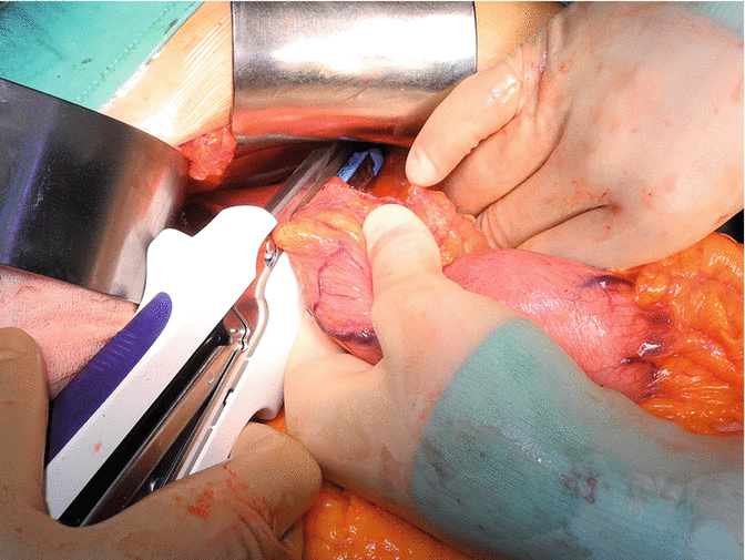
Fig. 6.7
The esophagus is divided with GIA 60 linear stapler above the level of the esophagogastric junction (EGJ), at least 2 cm above the upper pole of the EGJ tumor
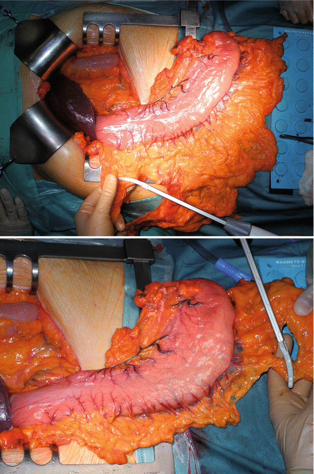
Figs. 6.8 and 6.9
The stomach is exposed outside the abdominal cavity and the omentum is resected preserving the gastroepiploic arch
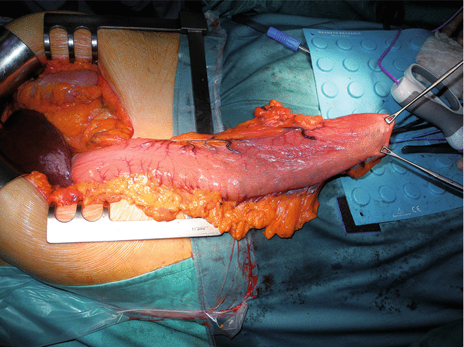
Fig. 6.10
Two Allis clamps are placed at the level of the gastric fundus to straighten the greater curvature of the stomach
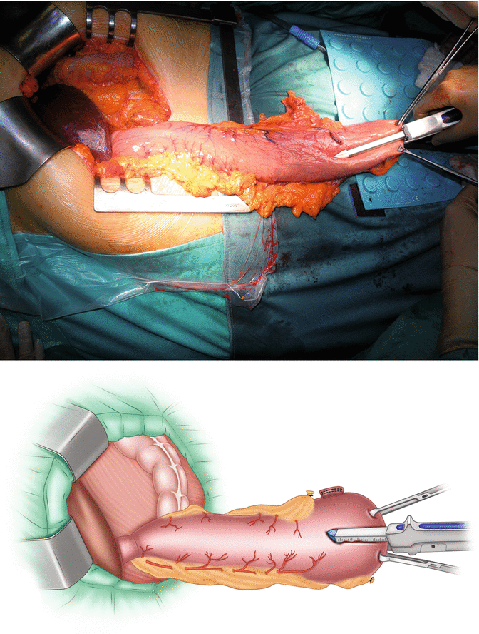
Figs. 6.11 and 6.12
The surgeon constructs the gastric tube along the greater curvature using multiple serial firings of linear staplers (GIA 60 and GIA 80). The procedure starts at the top, at the angle of His, with division of the stomach parallel to the greater curvature. The transverse diameter of the gastric conduit should not be more than 4 cm to ensure a good vascularization of the organ
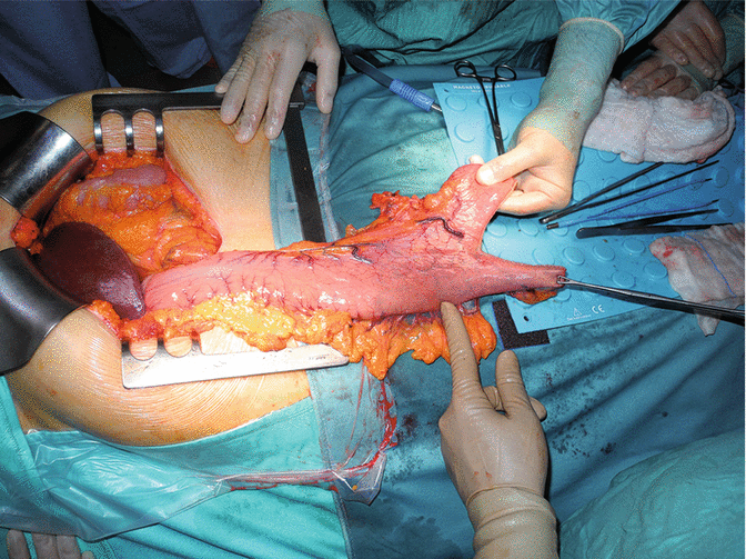
Fig. 6.13
The appearance after the first firing
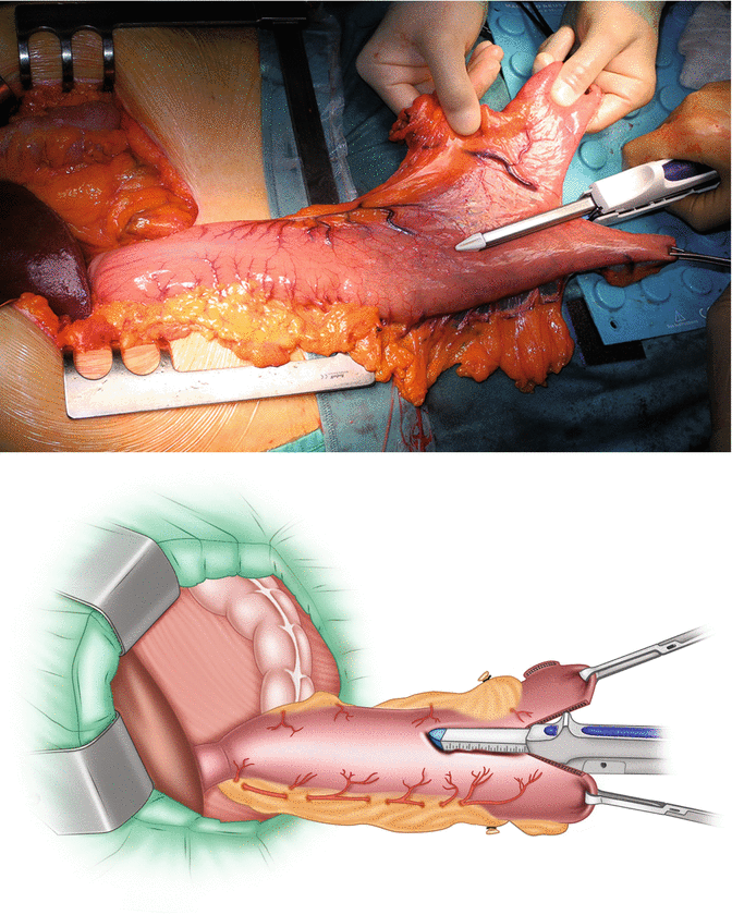
Figs. 6.14 and 6.15
The second firing with GIA 80 follows the same direction of the previous firing, maintaining a 4 cm distance from the greater curvature
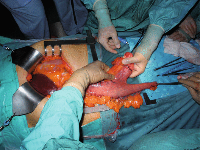
Fig. 6.16
The appearance after the second firing
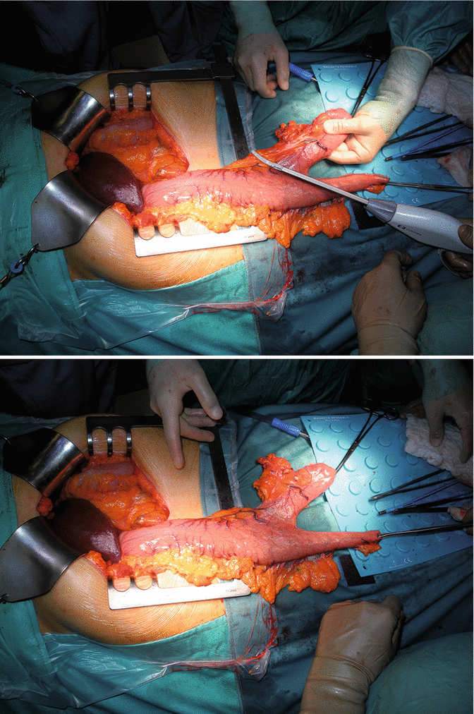
Figs. 6.17 and 6.18
Preparation of the lesser curvature of the stomach with radiofrequency device
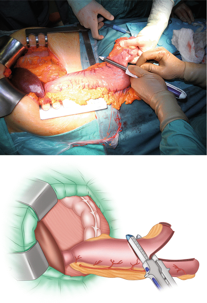
Figs. 6.19 and 6.20
The third firing is performed moving toward the lesser curvature. The specimen is removed; it consists of the esophagogastric junction, the gastric fundus, the upper portion of the lesser gastric curvature with right paracardial and proximal lesser curvature lymph nodes
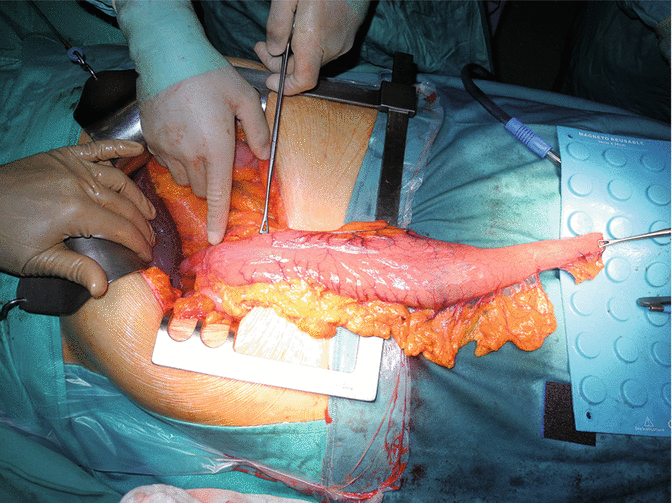
Fig. 6.21
The appearance after the third firing. The surgeon’s finger indicates the pylorus while the Duval clamp is applied 3 cm above. At this level the second step of gastric tube creation will begin
< div class='tao-gold-member'>
Only gold members can continue reading. Log In or Register to continue
Stay updated, free articles. Join our Telegram channel

Full access? Get Clinical Tree


